



Prostaglandin A2

Based on 1 Customer Validation
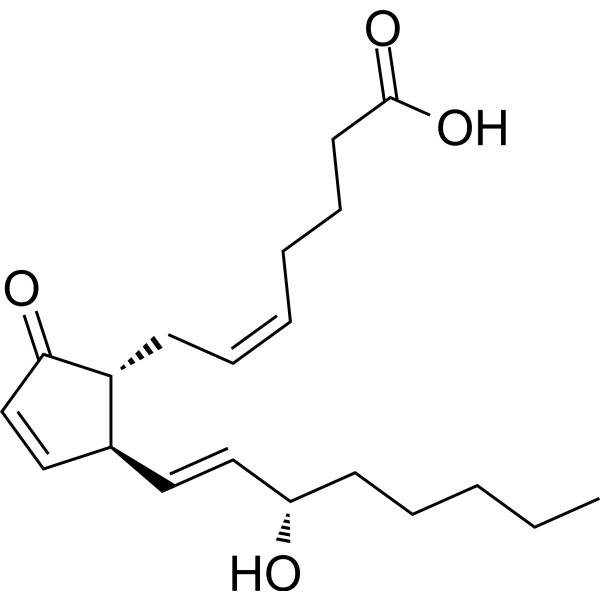
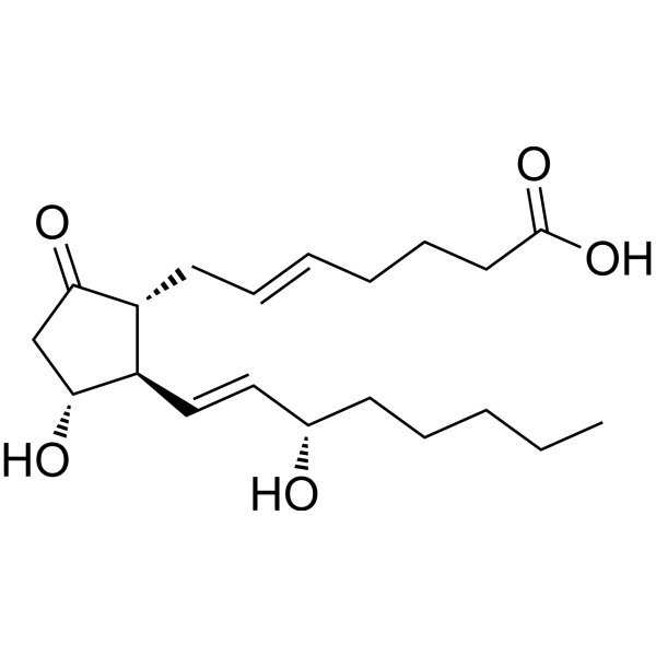
Prostaglandin A2 (PGA2) is a Cyclopentenone prostaglandin. Prostaglandin A2 induces Caspase-dependent Apoptosis, activates p53. Prostaglandin A2 activates ERK2 and JNK1/SAPK. Prostaglandin A2 shows antiviral activity against HSV-1. Prostaglandin A2 has anti-tumor effects. Prostaglandin A2 can be used for the research of colorectal cancer, colorectal carcinoma, breast carcinoma, and herpetic keratitis.
For research use only. We do not sell to patients.
- Purity : 99.9%
- CAS No.: 13345-50-1
- Formula: C20H30O4
- Molecular Weight:334.45
-
Storage:
Solution, -20°C, protect from light, 2 years

To place orders, for customer services and technical support, please contact: MedChemExpress USA
Tel: 609-228-6898 E-mail: [email protected] [email protected]


-
 Biological Activity
-
Chemical Information
- Protocols
- Purity & Documentation
- References
-
 Help & FAQs


-
HY-101952
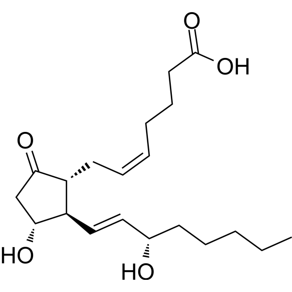
Prostaglandin E2Prostaglandin Receptor Agonist -
HY-12956
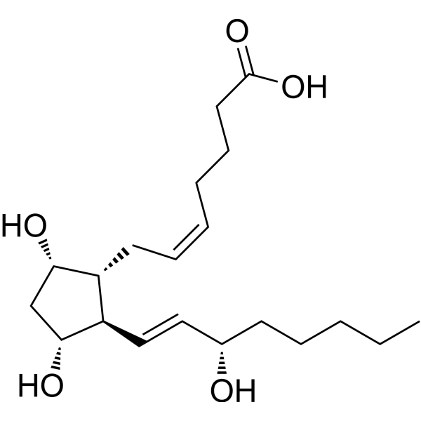
DinoprostFP Receptor Agonist -
HY-B0223S2
Albendazole-d7Stable Isotope -
HY-W010995
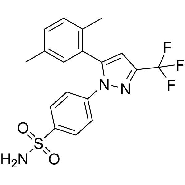
2,5-DimethylcelecoxibWnt/β-catenin inhibitor -
HY-P2588
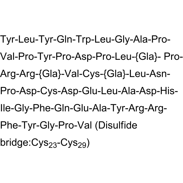
Osteocalcin (human)Bone Specific Protein -
HY-134135
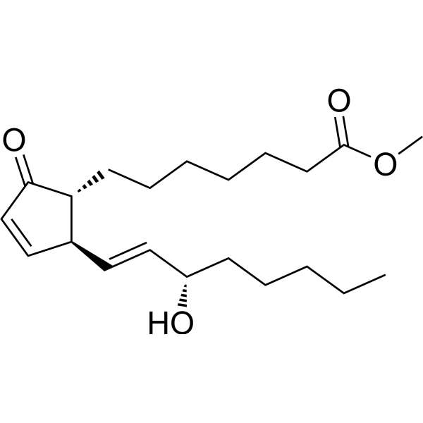
Prostaglandin A1 methyl esterCyclopentenone Analog -

HY-124273
C2 L-threo Ceramide (d18:1/2:0)Sphingolipid -
HY-137591
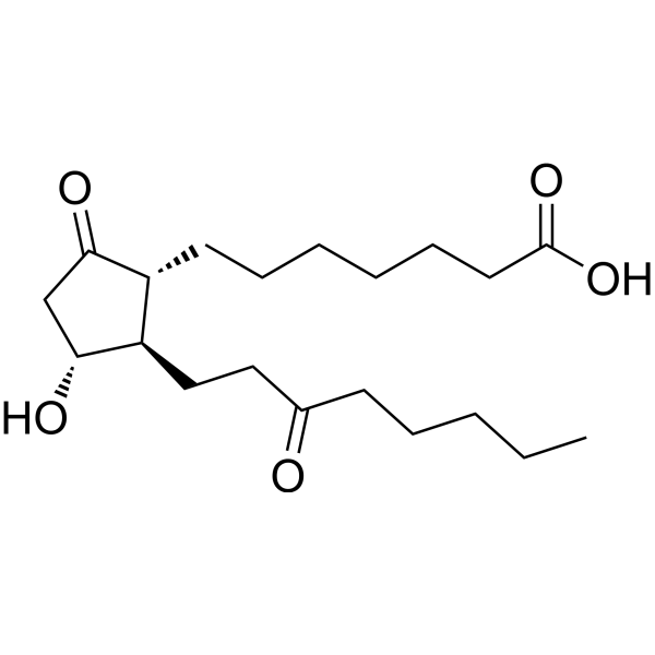
13,14-Dihydro-15-keto-PGE1PGE1 Metabolite -
HY-135023
5-trans-PGE2Aromatase Activator -

HY-135023R
5-trans-PGE2 (Standard)Aromatase Activator
All Caspase Isoforms
More

Biological Activity
Description
|
JNK1 |
ERK2 |
PARP-1 |
Caspase 3 |
HSV-1 |
In Vitro
Prostaglandin A2 (5-15 μg/mL; 12 h) induces caspase-dependent apoptosis in HCT116 (wild-type p53) cells, but exerts no such effect in HCT116 p53-/- cells[1].
Prostaglandin A2 (36 μM; 48 h) induces growth arrest in MCF-7 human breast cancer cells, and triggers apoptotic death in RKOneo and RKOE6 human colorectal cancer cells[2].
Prostaglandin A2 (36 μM; 30-120 min) potently activates ERK2 and JNK1/SAPK kinases in MCF-7 cells, but exerts no such effect in RKO cells[2].
Prostaglandin A2 inhibits the replication of HSV-1 strain McKrae in rabbit stromal cells, with an ED50 of 1.2 μM[3].
MedChemExpress (MCE) has not independently confirmed the accuracy of these methods. They are for reference only.
-
Cell Line:human colorectal HCT116 (wild-type p53) cells, HCT116 p53-/- cells
-
Concentration:5, 10, 15 μg/mL
-
Incubation Time:12 h
-
Result:Induced cleavage of PARP1 and caspase-3 in HCT116 cells in a dose-dependent manner, but not in HCT116 p53-/- cells.
Increased p53 protein levels and p53 phosphorylation at Ser-15 in HCT116 cells in a dose-dependent manner.
In Vivo
MedChemExpress (MCE) has not independently confirmed the accuracy of these methods. They are for reference only.
-
Animal Model:New Zealand White rabbits (2.3 to 2.7 kg, free of preexisting corneal defects)[3]
-
Dosage:0.5 mg/mL; 1.0 mg/mL; 2.0 mg/mL
-
Administration:topical eyedrops; five doses per day every 2 hours; days 3 to 7 post-infection
-
Result:Showed only one significant epithelial lesion score reduction vs.
placebo at a single dose and time point; overall, epithelial disease did not respond in a dose-dependent manner.
Recorded mean lesion scores on day 8 post-infection of 1.7 (1 mg/mL) and 2.1 (2 mg/mL).
Observed no significant differences in corneal thickness between treated and placebo groups at peak stromal involvement (12 to 14 days post-infection), with mean corneal thicknesses at day 12 post-infection of 473 ± 23 μm (0.5 mg/mL), 582 ± 76 μm (1.0 mg/mL), and 541 ± 42 μm (2.0 mg/mL).
Detected significantly higher anterior chamber flare scores in treated groups vs.
placebo in a dose-dependent manner, with mean flare scores after 1 day of therapy of 0.44 (0.5 mg/mL), 0.88 (1 mg/mL), and 1.03 (2 mg/mL).
Caused worse long-term sequelae in treated rabbits, with mean corneal thickness at 67 days post-infection of 569 ± 120 μm (1 mg/mL) and 694 ± 124 μm (2 mg/mL) vs.
480 ± 70 μm for placebo, and 14 of 28 treated eyes developing necrotizing stromal keratitis vs.
3 of 10 placebo eyes.
Observed no differences in virus recovery from trigeminal ganglia or corneas between treated and placebo groups, with 15 of 18 trigeminal ganglia from treated rabbits yielding infectious virus upon cocultivation, and 4 of 18 or 5 of 12 corneas from treated rabbits yielding infectious virus.


Chemical Information
-
CAS No. 13345-50-1
-
Appearance Liquid
-
Molecular Weight 334.45
-
Formula C20H30O4
-
Color Colorless to light yellow
-
SMILES
CCCCC[C@H](O)/C=C/[C@@H]1[C@H](C(C=C1)=O)C/C=C\CCCC(O)=O
-
Synonyms
PGA2; Medullin
-
Shipping
Room temperature in continental US; may vary elsewhere.
-
Storage
Solution, -20°C, protect from light, 2 years


Purity & Documentation
-
Data Sheet (278 KB)
-
SDS (558 KB)
- English - EN (558 KB)
- Français - FR (558 KB)
- Deutsch - DE (558 KB)
- Norwegian - NO (558 KB)
- Español - ES (558 KB)
- Swedish - SV (558 KB)
- Italian - IT (558 KB)
- Korean - KR (558 KB)
- Portuguese - PT (558 KB)
-
Handling Instructions (2659 KB)
References
[1]. Lee SB, et al. Induction of p53-Dependent Apoptosis by Prostaglandin A2. Biomolecules. 2020;10(3):492. Published 2020 Mar 24. [Content Brief]
[2]. Gorospe M, et al. Protective role of p21(Waf1/Cip1) against prostaglandin A2-mediated apoptosis of human colorectal carcinoma cells. Mol Cell Biol. 1996 Dec;16(12):6654-60. [Content Brief]
[3]. O'Brien WJ, et al. Assessment of antiviral activity, efficacy, and toxicity of prostaglandin A2 in a rabbit model of herpetic keratitis. Antimicrob Agents Chemother. 1996;40(10):2327-2331. [Content Brief]

Calculators
Concentration (start) × Volume (start) = Concentration (final) × Volume (final)


Prostaglandin A2 Related Classifications
HY-113041 Related Classifications
 Powered by Bioz
Keywords
- Prostaglandin A2
- 13345-50-1
- PGA2
- Medullin
- Prostaglandin A 2
- Prostaglandin A-2
- PGA 2
- PGA-2
- Apoptosis
- Caspase
- PARP
- ERK
- MDM-2/p53
- JNK
- HSV
- p53
- cyclin-dependent kinases
- DNA-activated protein kinase
- poly(ADP-ribose) polymerase 1
- histone H2AX
- DR5
- extracellular signal-regulated kinase
- c-Jun terminal kinase 1/stress-activated protein kinase
- caspase-3
- colorectal cancer
- Inhibitor
- inhibitor
- inhibit