



PHA-793887
Based on 4 publication(s) in Google Scholar
PHA-793887 is a CDK inhibitor (with IC50 values of 8 nM for Cdk2, 60 nM for Cdk1, 62 nM for Cdk4, 138 nM for Cdk9). PHA-793887 inhibits purified GSK3β (IC50 79 nM). PHA-793887 reduces the phosphorylation level of nucleophosmin/cdc6, induces G1/G2/M phase arrest, and triggers Apoptosis by activating Caspase-3. PHA-793887 exhibits anticancer activity against leukemia. PHA-793887 can be used in research related to acute leukemia, chronic myeloid leukemia, and advanced/metastatic solid tumors.
For research use only. We do not sell to patients.
- Purity : 99.25%
- CAS No.: 718630-59-2
- Formula: C19H31N5O2
- Molecular Weight:361.48
-
Storage:Powder -20°C, 3 years , 4°C, 2 years ; In solvent -80°C, 2 years , -20°C, 1 year

To place orders, for customer services and technical support, please contact: MedChemExpress USA
Tel: 609-228-6898 E-mail: [email protected] [email protected]


-
 Biological Activity
-
Chemical Information
-
Solvent & Solubility
- Protocols
- Purity & Documentation
- References
-
 Help & FAQs


-
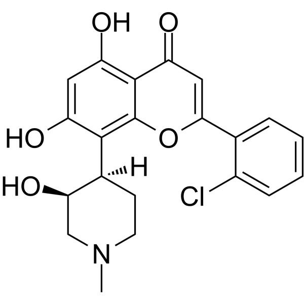
HY-10005
FlavopiridolCDK Inhibitor -
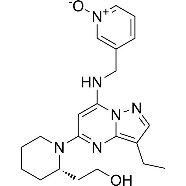
HY-10492
DinaciclibCDK2/5/9 Inhibitor -
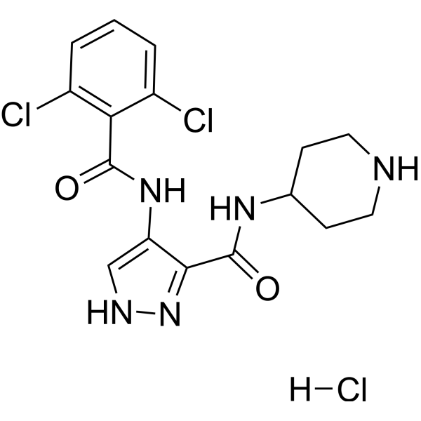
HY-50943
AT7519 HydrochlorideCDK Inhibitor -
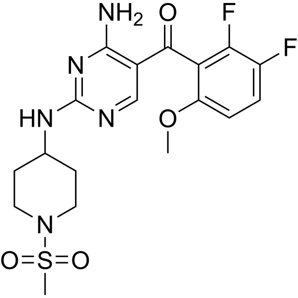
HY-10014
R547CDK Inhibitor -
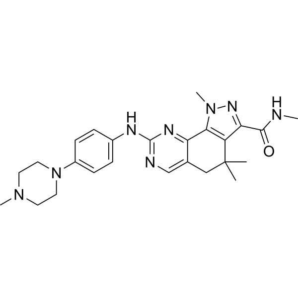
HY-10424
MilciclibCDK/TRK Inhibitor -
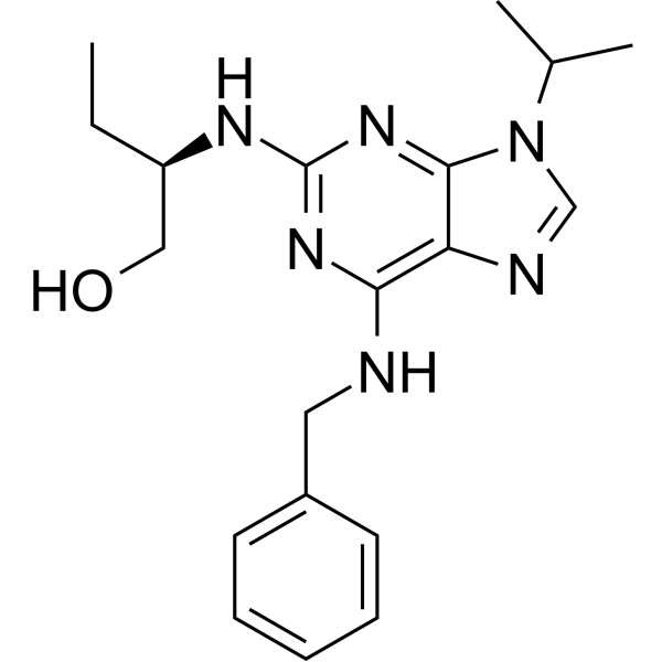
HY-30237
(R)-RoscovitineCDKs Inhibitor -
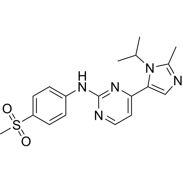
HY-10012
AZD-5438CDK1/2/9 Inhibitor -
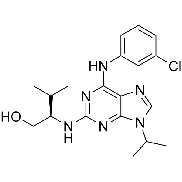
HY-18299A
Purvalanol ACDK Inhibitor -
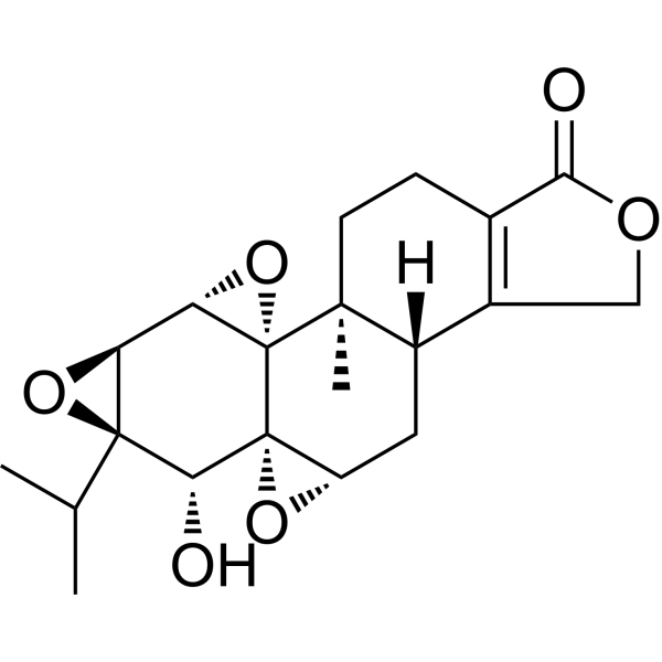
HY-32735
TriptolideImmunosuppressive Agent -
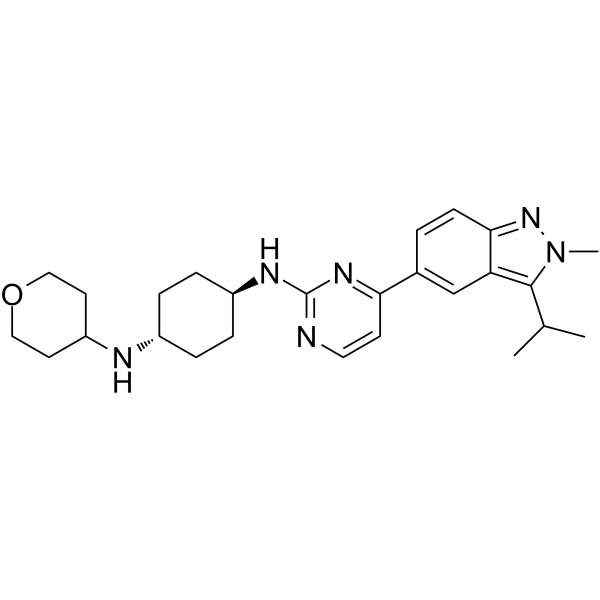
HY-12293
LY2857785CDK9/8/7 Inhibitor -
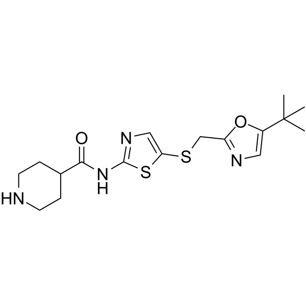
HY-10008
SNS-032CDK Inhibitor -
HY-13266A
BS-181 hydrochlorideCDK7 Inhibitor
Publications Citing Use of MedChemExpress (MCE) PHA-793887
MoreAll Caspase Isoforms
More

Biological Activity
Description
IC50 & Target
[4]|
CDK2 8 nM (IC50) |
CDK1 60 nM (IC50) |
CDK4 62 nM (IC50) |
CDK9 138 nM (IC50) |
Caspase 3 |
GSK-3β 79 nM (IC50) |
Cellular Effect
|
Cell Line

|
Type | Value | Description | References |
|---|---|---|---|---|
| A2780 | IC50 |
0.09 μM
Compound: 31
|
Antiproliferative activity against human A2780 cells after 72 hrs by fluorescence assay
Antiproliferative activity against human A2780 cells after 72 hrs by fluorescence assay
|
[PMID: 20153204] |
| A-375 | IC50 |
0.396 μM
Compound: 31
|
Antiproliferative activity against human A375 cells after 72 hrs by SRB assay
Antiproliferative activity against human A375 cells after 72 hrs by SRB assay
|
[PMID: 20153204] |
| BXPC-3 | IC50 |
3.444 μM
Compound: 31
|
Antiproliferative activity against human BxPC3 cells after 72 hrs by SRB assay
Antiproliferative activity against human BxPC3 cells after 72 hrs by SRB assay
|
[PMID: 20153204] |
| COLO 205 | IC50 |
0.188 μM
Compound: 31
|
Antiproliferative activity against human COLO205 cells after 72 hrs by SRB assay
Antiproliferative activity against human COLO205 cells after 72 hrs by SRB assay
|
[PMID: 20153204] |
| DU-145 | IC50 |
0.303 μM
Compound: 31
|
Antiproliferative activity against human DU145 cells after 72 hrs by SRB assay
Antiproliferative activity against human DU145 cells after 72 hrs by SRB assay
|
[PMID: 20153204] |
| HCT-116 | IC50 |
0.163 μM
Compound: 31
|
Antiproliferative activity against human HCT116 cells after 72 hrs by SRB assay
Antiproliferative activity against human HCT116 cells after 72 hrs by SRB assay
|
[PMID: 20153204] |
| MCF7 | IC50 |
1.284 μM
Compound: 31
|
Antiproliferative activity against human MCF7 cells after 72 hrs by SRB assay
Antiproliferative activity against human MCF7 cells after 72 hrs by SRB assay
|
[PMID: 20153204] |
| PC-3 | IC50 |
0.601 μM
Compound: 31
|
Antiproliferative activity against human PC3 cells after 72 hrs by SRB assay
Antiproliferative activity against human PC3 cells after 72 hrs by SRB assay
|
[PMID: 20153204] |
In Vitro
PHA-793887 potently inhibits multiple cyclin-dependent kinases with IC50 values between 5 and 140 nM, and is inactive against non-cdk kinases including c-abl, c-kit, lck, and TRKA[1].
PHA-793887 (0.01-10 μM; 48 h) is cytotoxic to a panel of leukemic cell lines with IC50 values from 0.04 μM to >10 μM, is not cytotoxic to unstimulated normal PBMC and CD34+ cells, and inhibits proliferation of mitogen- or growth factor-stimulated normal hematopoietic cells with IC50 values of 0.75 μM and 1.85 μM, respectively[1].
PHA-793887 (0.04-5 μM; 48 h; 24 h) induces G1 phase cell cycle arrest at 0.04 to 1 μM and apoptosis at 5 μM in sensitive leukemic cell lines (697, KCL22, TOM1), but has no effect on resistant KG1 cells; it activates caspase-3 in KCL22 cells at 5 μM[1].
PHA-793887 potently inhibits multiple CDKs, including CDK2/cyclin A (IC50 = 0.008 μM), and shows selective activity against GSK3β among a panel of non-CDK kinases[3].
PHA-793887 inhibits proliferation of a broad range of human cancer cell lines, with the highest potency against colon carcinoma HCT-116 cells (IC50 = 0.163 μM) and lowest potency against pancreatic carcinoma BX-PC3 cells (IC50 = 3.444 μM)[3].
PHA-793887 (1-3 μM; 24 h) disrupts cell cycle progression and inhibits DNA synthesis in human ovarian carcinoma A2780 cells, inducing G1 arrest at 1 μM and G2/M arrest with apoptosis at 3 μM[3].
PHA-793887 potently inhibits purified Cdk2 (IC50 8 nmol/L), Cdk1 (IC50 60 nmol/L), Cdk4 (IC50 62 nmol/L), Cdk9 (IC50 138 nmol/L), and GSK3β (IC50 79 nmol/L) in cell-free biochemical assays, while sparing 35 other tested kinases[4].
MedChemExpress (MCE) has not independently confirmed the accuracy of these methods. They are for reference only.
-
Cell Line:13 leukemic cell lines (K562, KCL22, KU812, TOM1, SUP-B15, REH, 697, RS4;11, HL60, KG1, RPMI8226, ALL-2, AML-PS), normal unstimulated peripheral blood mononuclear cells (PBMC), phytohemagglutinin-stimulated PBMC, normal unstimulated CD34+ hematopoietic stem cells, growth factor cocktail-stimulated CD34+ hematopoietic stem cells
-
Concentration:0.01-10 μM
-
Incubation Time:48 h
-
Result:Showed variable cytotoxicity across leukemic cell lines, with IC50 values ranging from 0.04 μM (most sensitive lines: TOM1, 697, RS4;11, HL60) to >10 μM (least sensitive lines: SUP-B15, KG1).
Primary leukemic cells ALL-2 and AML-PS had IC50 values of 3.4 μM and 6.2 μM, respectively.
Was not cytotoxic to unstimulated normal PBMC or CD34+ cells (IC50 >10 μM), but inhibited proliferation of stimulated PBMC (IC50 0.75 μM) and stimulated CD34+ cells (IC50 1.85 μM).
-
Cell Line:697, KCL22, TOM1, and KG1 leukemic cell lines
-
Concentration:0.04-5 μM
-
Incubation Time:48 h (cell cycle analysis); 24 h (apoptosis analysis)
-
Result:Induced G1 phase arrest (14-40% increase compared to control) and reduced S-phase cell counts at doses of 0.04 to 1 μM in sensitive cell lines (697, KCL22, TOM1).
Induced a G2/M phase block (34% increase) in TOM1 cells at 1 μM.
Induced apoptosis (15-35% sub-G1 population) in sensitive cell lines at 5 μM.
Showed no significant cell cycle arrest or apoptosis in resistant KG1 cells at any dose.
Induced caspase-3 activation (43% positive cells, a 26% increase compared to control) in KCL22 cells at 5 μM, while 0.2 μM had no effect.
-
Cell Line:human ovarian carcinoma A2780 cells
-
Concentration:1-3 μM
-
Incubation Time:24 h
-
Result:Caused a 25% increase in G0/G1 population, 76% decrease in S phase population, 62% increase in G2/M population, and 85% reduction in BrdU incorporation relative to control at 1 μM.
Caused an 11% decrease in G0/G1 population, 48% decrease in S phase population, 263% increase in G2/M population, 100% reduction in BrdU incorporation, and an increase in sub-G1 cells (from 2.6% to 26.2%) indicative of apoptosis at 3 μM.
In Vivo
PHA-793887 (20 mg/kg; i.v.; once daily; two 5-day cycles with 3-day rest between cycles) significantly reduces tumor growth and causes a 4.7-day growth delay in K562 subcutaneous CML xenografts in SCID mice[1].
PHA-793887 (20 mg/kg; i.v.; once daily; three 5-day cycles with 3-day rests between cycles) significantly increases median survival by 7 days in the preemptive disseminated AML-PS xenograft model in SCID mice[1].
PHA-793887 (20 mg/kg; i.v.; once daily; three 5-day cycles with 3-day rests between cycles) significantly increases median survival by 22.5 days in the preemptive disseminated ALL-2 xenograft model in SCID mice[1].
PHA-793887 (10-30 mg/kg; i.v.; daily; 10 days) exhibits 76% tumor growth inhibition in human A2780 ovarian carcinoma xenografts when administered at 30 mg/kg i.v. daily for 10 days, with confirmed in vivo target engagement via reduced tumor cell proliferation and pRb phosphorylation[3].
PHA-793887 (10-20 mg/kg; i.v.; daily; 10 days) exhibits 81% tumor growth inhibition in human HCT-116 colon carcinoma xenografts when administered at 20 mg/kg i.v. daily for 10 days, with good tolerability[3].
PHA-793887 (20 mg/kg; i.v.; daily; 10 days) exhibits 84% tumor growth inhibition in human BX-PC3 pancreatic carcinoma xenografts when administered at 20 mg/kg i.v. daily for 10 days, with good tolerability[3].
PHA-793887 (15-30 mg/kg; i.v.; daily; 2-10 days) induces dose-dependent tumor growth inhibition (50% at 15 mg/kg, 75% at 30 mg/kg) and downregulation of an E2F-dependent gene signature in A2780 ovarian carcinoma xenografts in CD-1 nude mice[4].
MedChemExpress (MCE) has not independently confirmed the accuracy of these methods. They are for reference only.
-
Animal Model:SCID mice (unspecified gender, age not specified)[1]
-
Dosage:20 mg/kg
-
Administration:i.v.; once daily; 10 consecutive days
-
Result:Induced tumor regression.
Achieved a tumor growth delay of 23.3 days.
Left all treated mice tumor-free one week after treatment ended.
Caused no body weight loss or toxic effects.
-
Animal Model:SCID mice (unspecified gender, age not specified)[1]
-
Dosage:20 mg/kg
-
Administration:i.v.; once daily; two 5-day cycles with 3-day rest between cycles
-
Result:Significantly reduced tumor growth from day 14 compared to vehicle controls (p < 0.01).
Achieved a tumor growth delay of 4.7 days.
Caused no body weight loss or toxic effects.
-
Animal Model:SCID mice (5-week-old, unspecified gender)[1]
-
Dosage:20 mg/kg
-
Administration:i.v.; once daily; three 5-day cycles with 3-day rests between cycles
-
Result:Significantly increased median survival time by 7 days compared to vehicle controls (p < 0.0001).
-
Animal Model:SCID mice (5-week-old, unspecified gender)[1]
-
Dosage:20 mg/kg
-
Administration:i.v.; once daily; three 5-day cycles with 3-day rests between cycles
-
Result:Significantly increased median survival time by 22.5 days compared to vehicle controls (p < 0.0001).\nSignificantly increased median survival time by 22.5 days compared to vehicle controls (p < 0.0001).
Reduced tumor infiltration from 22.5% to 1.6% in peripheral blood after two treatment cycles.
Reduced tumor infiltration from 97.7% to 24.8% in bone marrow after two treatment cycles.
Reduced tumor infiltration from 91.6% to 44.7% in spleen after two treatment cycles.
Caused no body weight loss or toxic effects.
-
Animal Model:Nu/Nu (male)[3]
-
Dosage:10 mg/kg; 20 mg/kg; 30 mg/kg
-
Administration:i.v.; daily; 10 days
-
Result:Caused dose-dependent inhibition of A2780 tumor growth, reaching 76% tumor growth inhibition at the 30 mg/kg dose at the end of treatment.
Showed marginal body weight reduction (<10% vs control mice) and no toxic effects observed at gross autopsy.
Reduced tumor BrdU incorporation (p = 0.0022) and phosphorylated retinoblastoma protein (pRb) levels (p = 0.005) in mice treated with 30 mg/kg for 5 days compared to vehicle controls.
-
Animal Model:Nu/Nu (male)[3]
-
Dosage:10 mg/kg; 20 mg/kg
-
Administration:i.v.; daily; 10 days
-
Result:Caused dose-dependent inhibition of HCT-116 tumor growth, reaching 81% tumor growth inhibition at the 20 mg/kg dose at the end of treatment.
Showed marginal body weight reduction (<10% vs control mice) and no toxic effects observed at gross autopsy.
-
Animal Model:Nu/Nu (male)[3]
-
Dosage:20 mg/kg
-
Administration:i.v.; daily; 10 days
-
Result:Caused 84% tumor growth inhibition in BX-PC3 xenografts at the end of treatment.
Showed marginal body weight reduction (<10% vs control mice) and no toxic effects observed at gross autopsy.
- Kinase activity and phosphorylation assays
- Western Blot
- Detection of Apoptosis
- TUNEL staining for apoptotic DNA fragmentation
- Flow cytometric DNA-content cell-cycle staining
- Annexin V plus membrane-impermeant dye apoptosis staining
- Apoptosis Solutions
- Protocol for Cell Cycle
- Protocol for Kinase activity and phosphorylation assays
Clinical Trial
| NCT Number | Sponsor | Condition | Start Date |
Phase
|
|---|---|---|---|---|
| NCT01329991 | Plexxikon| | 2011-05 | PHASE1 |


Chemical Information
-
CAS No. 718630-59-2
-
Appearance Solid
-
Molecular Weight 361.48
-
Formula C19H31N5O2
-
Color White to yellow
-
SMILES
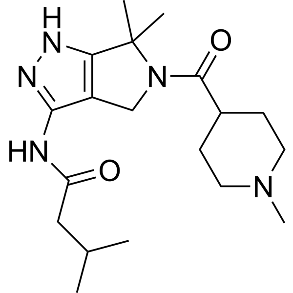
CC(C)CC(NC1=NNC2=C1CN(C(C3CCN(C)CC3)=O)C2(C)C)=O
-
Shipping
Room temperature in continental US; may vary elsewhere.
-
Storage
Powder -20°C 3 years 4°C 2 years In solvent -80°C 2 years -20°C 1 year


Publications (4)
-
Journal Impact Factor
-
Most Recent
-
Sci Transl Med
PP2A inhibition is a druggable MEK inhibitor resistance mechanism in KRAS-mutant lung cancer cells. [Abstract]2018 Jul 18;10(450):eaaq1093. PMID: 30021885 -
Sci Data
High-throughput drug screening identifies novel therapeutics for Low Grade Serous Ovarian Carcinoma. [Abstract]2024 Sep 19;11(1):1024. PMID: 39300112 -
Bioinform Biol Insights
Integrative Analysis for Identification of Therapeutic Targets and Prognostic Signatures in Non-Small Cell Lung Cancer. [Abstract]2022 Apr 6;16:11779322221088796. PMID: 35422618 -


Solvent & Solubility
In Vitro:
DMSO : ≥ 50 mg/mL (138.32 mM; Hygroscopic DMSO has a significant impact on the solubility of product, please use newly opened DMSO)
* "≥" means soluble, but saturation unknown.
Please refer to the solubility information to select the appropriate solvent. Once prepared, please aliquot and store the solution to prevent product inactivation from repeated freeze-thaw cycles.
Storage method and period of stock solution: -80°C, 2 years; -20°C, 1 year. When stored at -80°C, please use it within 2 years. When stored at -20°C, please use it within 1 year.
Please refer to the solubility information to select the appropriate solvent. Once prepared, please aliquot and store the solution to prevent product inactivation from repeated freeze-thaw cycles.
Storage method and period of stock solution: -80°C, 2 years; -20°C, 1 year. When stored at -80°C, please use it within 2 years. When stored at -20°C, please use it within 1 year.
Concentration (start) × Volume (start) = Concentration (final) × Volume (final)
In Vivo:
Select the appropriate dissolution method based on your experimental animal and administration route.
- For the following dissolution methods, please ensure to first prepare a clear stock solution using an In Vitro approach and then sequentially add co-solvents:
- To ensure reliable experimental results, the clarified stock solution can be appropriately stored based on storage conditions. As for the working solution for In Vivo experiments, it is recommended to prepare freshly and use it on the same day.
- The percentages shown for the solvents indicate their volumetric ratio in the final prepared solution. If precipitation or phase separation occurs during preparation, heat and/or sonication can be used to aid dissolution.
Add each solvent one by one: 10% DMSO 90% (20% SBE-β-CD in Saline)
Solubility: ≥ 2.5 mg/mL (6.92 mM); Clear solution
This protocol yields a clear solution of ≥ 2.5 mg/mL (saturation unknown).
Taking 1 mL working solution as an example, add 100 μL DMSO stock solution (25.0 mg/mL) to 900 μL 20% SBE-β-CD in Saline, and mix evenly.
Preparation of 20% SBE-β-CD in Saline (4°C, storage for one week): 2 g SBE-β-CD powder is dissolved in 10 mL Saline, completely dissolve until clear.
In Vivo Dissolution Calculator
Please enter the basic information of animal experiments:
-
-
-
-
Recommended: Prepare an additional quantity of animals to account for potential losses during experiments.
Please enter your animal formula composition:
-
%DMSO +
Recommended: Keep the proportion of DMSO in working solution below 2% if your animal is weak.
-
%+
-
+%Tween-80 + +
-
%Saline +
The co-solvents required include: DMSO, . All of co-solvents are available by MedChemExpress (MCE). , Tween 80. All of co-solvents are available by MedChemExpress (MCE).
Working solution concentration: 0.22 mg/mL
Method for preparing stock solution: mg drug dissolved in μL DMSO. Stock solution concentration: mg/mL.
1. Take μL DMSO stock solution;
2. Add μL .
μL , mix evenly;
3. Then add μL Tween 80, mix evenly;
4. Then add μL
Please ensure that the stock solution in the first step is dissolved to a clear state, and add co-solvents in sequence. You can use ultrasonic heating (ultrasonic cleaner, recommended frequency 20-40 kHz), vortexing, etc. to assist dissolution.


Purity & Documentation
-
Data Sheet (297 KB)
-
SDS (393 KB)
- English - EN (393 KB)
- Français - FR (393 KB)
- Deutsch - DE (393 KB)
- Norwegian - NO (393 KB)
- Español - ES (393 KB)
- Swedish - SV (393 KB)
- Italian - IT (393 KB)
- Korean - KR (393 KB)
- Portuguese - PT (393 KB)
-
Handling Instructions (2659 KB)
References
[1]. Alzani R, et al. Therapeutic efficacy of the pan-cdk inhibitor PHA-793887 in vitro and in vivo in engraftment and high-burden leukemia models. Experimental hematology. 2010 Apr;38(4):259-269.e2. [Content Brief]
[2]. Massard C, et al. A first in man, phase I dose-escalation study of PHA-793887, an inhibitor of multiple cyclin-dependent kinases (CDK2, 1 and 4) reveals unexpected hepatotoxicity in patients with solid tumors. Cell cycle (Georgetown, Tex.). 2011 Mar 15;10(6):963-70. [Content Brief]
[3]. Brasca MG, et al. Optimization of 6,6-dimethyl pyrrolo[3,4-c]pyrazoles: Identification of PHA-793887, a potent CDK inhibitor suitable for intravenous dosing. Bioorganic & medicinal chemistry. 2010 Mar 01;18(5):1844-53. [Content Brief]
[4]. Locatelli G, et al. Transcriptional analysis of an E2F gene signature as a biomarker of activity of the cyclin-dependent kinase inhibitor PHA-793887 in tumor and skin biopsies from a phase I clinical study. Molecular cancer therapeutics. 2010 May;9(5):1265-73. [Content Brief]


Complete Stock Solution Preparation Table
Please refer to the solubility information to select the appropriate solvent. Once prepared, please aliquot and store the solution to prevent product inactivation from repeated freeze-thaw cycles.
Storage method and period of stock solution: -80°C, 2 years; -20°C, 1 year. When stored at -80°C, please use it within 2 years. When stored at -20°C, please use it within 1 year.
| Optional Solvent | Concentration Solvent Mass | 1 mg | 5 mg | 10 mg | 25 mg |
|---|---|---|---|---|---|
| DMSO | 1 mM | 2.7664 mL | 13.8320 mL | 27.6640 mL | 69.1601 mL |
| 5 mM | 0.5533 mL | 2.7664 mL | 5.5328 mL | 13.8320 mL | |
| 10 mM | 0.2766 mL | 1.3832 mL | 2.7664 mL | 6.9160 mL | |
| 15 mM | 0.1844 mL | 0.9221 mL | 1.8443 mL | 4.6107 mL | |
| 20 mM | 0.1383 mL | 0.6916 mL | 1.3832 mL | 3.4580 mL | |
| 25 mM | 0.1107 mL | 0.5533 mL | 1.1066 mL | 2.7664 mL | |
| 30 mM | 0.0922 mL | 0.4611 mL | 0.9221 mL | 2.3053 mL | |
| 40 mM | 0.0692 mL | 0.3458 mL | 0.6916 mL | 1.7290 mL | |
| 50 mM | 0.0553 mL | 0.2766 mL | 0.5533 mL | 1.3832 mL | |
| 60 mM | 0.0461 mL | 0.2305 mL | 0.4611 mL | 1.1527 mL | |
| 80 mM | 0.0346 mL | 0.1729 mL | 0.3458 mL | 0.8645 mL | |
| 100 mM | 0.0277 mL | 0.1383 mL | 0.2766 mL | 0.6916 mL |


PHA-793887 Related Classifications
HY-11001 Related Classifications
 Powered by Bioz