



4,4'-Sulfonyldiphenol

Based on 1 Customer Validation
4,4'-Sulfonyldiphenol (Bisphenol S; Bis(4-hydroxyphenyl) sulfone), a substitute for Bisphenol A (HY-18260), is widely used in industrial and consumer products. 4,4'-Sulfonyldiphenol is an oally ative estrogen receptor (ER) agonist and can competitively bind to thyroid hormone receptors (TR) with IC50 values for TRα and TRβ are 2650 μM and 2294 μM respectively, thereby affecting breast development and reducing the expression of androgen receptor (AR) in fetal testes. 4,4'-Sulfonyldiphenol promotes the progression of glioblastoma by upregulating the EZH2 mediated PI3K/AKT/mTOR pathway. Under chronic exposure, 4,4'-Sulfonyldiphenol can cause significant lipid deposition and dyslipidemia in the mouse liver by upregulating JunB and Atf3, and has a role in causing obesity at low doses. 4,4'-Sulfonyldiphenol induces intestinal inflammation by altering the intestinal microbiome. 4,4'-Sulfonyldiphenol accelerates the progression of atherosclerosis in zebrafish embryo larvae.
For research use only. We do not sell to patients.
- Purity : 99.44%
- CAS No.: 80-09-1
- Formula: C12H10O4S
- Molecular Weight:250.28
-
Storage:
Store at room temperature 3 years.
In solvent -80°C, 2 years , -20°C, 1 year


To place orders, for customer services and technical support, please contact: MedChemExpress USA
Tel: 609-228-6898 E-mail: [email protected] [email protected]


-
 Biological Activity
-
Chemical Information
-
Solvent & Solubility
- Protocols
- Purity & Documentation
- References
-
 Help & FAQs


-
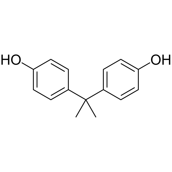
HY-18260
Bisphenol AEndocrine-Disrupting Compound -

HY-W011927R
4,4'-Sulfonyldiphenol (Standard)Standard of 4,4'-Sulfonyldiphenol -
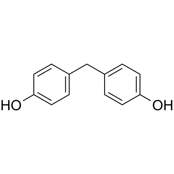
HY-W014901
Bisphenol FEndocrine Disruptor -
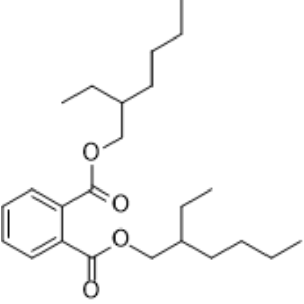
HY-B1945
DEHPEndogenous Metabolite -
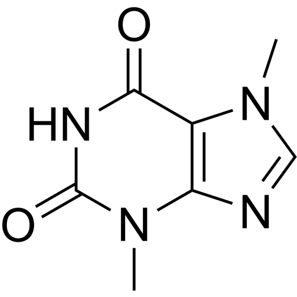
HY-N0138
TheobromineEndogenous Metabolite -
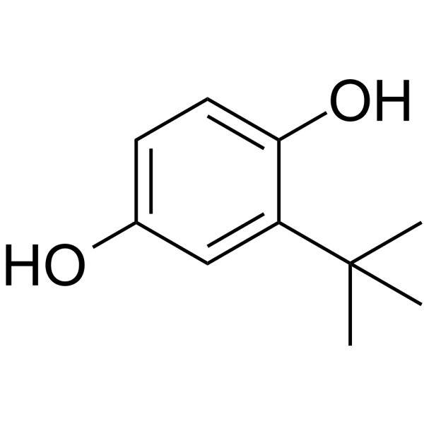
HY-100489
TBHQNrf2 Activator -
HY-W013712
GI-530159TREK1/2 Channels Opener -
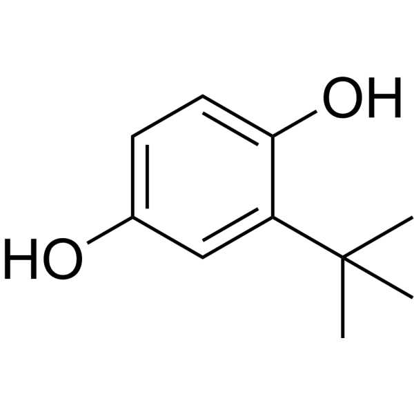
HY-100489R
TBHQ (Standard)Nrf2 Activator -
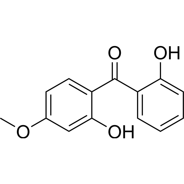
HY-B0966
DioxybenzoneUV Blocker -

HY-121254
GERI-BP002-AACAT Inhibitor -
HY-W013631
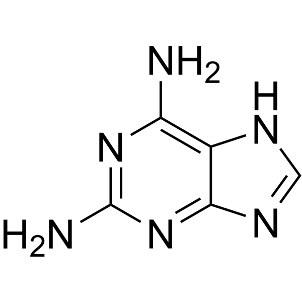
7H-Purine-2,6-diamineBiochemical Reagent -

HY-117007
Glycidyl methacrylateBifunctional Monomer
All Histone Methyltransferase Isoforms
More

Biological Activity
Description
In Vitro
4,4'-Sulfonyldiphenol (0-50 μM) induces thyroid hormone (TH)-dependent GH3 cell proliferation, and inhibits T3 (HY-A0070A) induction in the presence of T3[3].
4,4'-Sulfonyldiphenol (100 μM, 24 h) Significantly increases lipid deposition in AML12 and primary hepatocytes via the JunB/ATF3 axis[4].
4,4'-Sulfonyldiphenol (0.001-100 μM, 4-24 h) promotes the proliferation and migration of U-87 MG cells at a concentration of 0.1 µM and appears to be linked to the activation of the EZH2-mediated PI3K/ AKT/mTOR pathway[6].
4,4'-Sulfonyldiphenol (0.01-1 μM, 0-24 h) exhibits enhanced formation of foam cells with oxidized lowdensity lipoprotein (oxLDL) in macrophages[7].
MedChemExpress (MCE) has not independently confirmed the accuracy of these methods. They are for reference only. Further protocols information, click here.
-
Cell Line:U-87 MG cells
-
Concentration:0, 0.001, 0.01, 0.1, 1, 10 and 100 μM
-
Incubation Time:4 and 8 h
-
Result:Significantly increased the proliferation rate at 0.1 μM, and the effect was more significant after 8 hours of treatment than after 4 hours.
-
Cell Line:U-87 MG cells
-
Concentration:0.1 μM
-
Incubation Time:0, 2, 4, 6, 8, 10 and 12 h
-
Result:Significantly increased the mobility within 12 hours.
-
Cell Line:U-87 MG cells
-
Concentration:0.1, 1 and 10 μM
-
Incubation Time:24 h
-
Result:Induced glioblastoma invasion in U-87 MG cells.
-
Cell Line:U-87 MG cells
-
Concentration:0.1 μM
-
Incubation Time:4 and 8 h
-
Result:Increased the mRNA level of EZH2.
-
Cell Line:U-87 MG cells
-
Concentration:0.1 μM
-
Incubation Time:4 and 8 h
-
Result:Increased the expression of EZH2, p-PI3K, p-AKT, p-mTOR and p-4E-BP1.
Decreased the expression of PIK3IP1.
In Vivo
4,4'-Sulfonyldiphenol (0.2-50 μg/kg, p.o., once daily beginning at gestational day 0 and continuing in offspring up to 23-weeks old) can be an obesogen at low doses and after perinatal and chronic exposure for the male mice[2].
4,4'-Sulfonyldiphenol (0-10 μM, single dose) induces TH-response gene transcription in Pelophylax nigromaculatus tadpoles, but in the presence of T3 altered T3-induced gene transcription in a biphasic concentration-response manner[3].
4,4'-Sulfonyldiphenol (0.1-1 mg/kg, i.g., once daily for 3 months) induces mice to show significant lipid deposition in the liver and dyslipidemia and were predisposed to metabolic dysfunction-associated steatotic liver disease (MASLD)[4].
4,4'-Sulfonyldiphenol (0.1-1 mg/kg, i.g., once daily for 3 months) induces intestinal inflammation via altering gut microbiome in mice[5].
4,4'-Sulfonyldiphenol (0-100 μg/L, for 15-45 days) accelerates the progression of atherosclerotic cardiovascular disease (ASCVD) using zebrafish embryo-larvae as a model[7].
MedChemExpress (MCE) has not independently confirmed the accuracy of these methods. They are for reference only.
-
Animal Model:Long-term and short-term exposure effect assessment established in female CD-1 mice[1]
-
Dosage:2 and 200 μg/kg (long-term); 2, 200, and 2000 μg/kg (short-term)
-
Administration:Oral administration (p.o.), once daily starting on pregnancy day 9 through lactation day 20 (long-term) and starting on pregnancy day 9 through lactation day 2 (short-term)
-
Result:Showed signs of ductal formation in the mammary gland and significantly enlarged the intercellular spaces between the tissues.
Increased ERα expression in epithelial cells and dose-dependently decreased AR expression in the stroma.
Increased the right duct area in all groups, and the left duct area also increased significantly in the high-dose group.
Altered the response of male mice to a pre-pubertal estrogen challenge.
-
Animal Model:High fat diet model established in pregnant C57Bl/6J mice[2]
-
Dosage:0.2, 1.5, 50 µg/kg
-
Administration:Oral administration (p.o.) with water, once daily beginning at gestational day 0 and continuing in offspring up to 23-weeks old
-
Result:Significantly increased in body weight and fat mass on a high-fat diet.
Had no effect on the food intake and energy expenditure.
Exhibited a weak but significant hypercholesterolemia with a fold increase of 1.21 and 1.29 at 1.5 and 50 µg/kg, respectively.
Showed a significant decrease of the AUC (increase of blood TG clearance).
Significantly increased the calculated HOMA-IR index in mice exposed to 1.5 and 50 µg/kg.
Induced a significant overexpression of SOCS3 mRNA and a significant mRNA down-regulation of INSR and adiponectin.
Induced a weak but significant decrease in mRNA expression of HSL and PPARγ genes in adipose tissue of male mice fed with a HFD.
-
Animal Model:MASLD model established in 7-week-old C57BL/6 male mice[4]
-
Dosage:0.1 and 1 mg/kg
-
Administration:Oral gavage (i.g.), once daily for 3 months
-
Result:Induced MASLD phenotype: elevated LDL, increased NEFA, and elevated ALT.
Showed obvious lipid droplet accumulation.
-
Animal Model:Intestinal inflammation model established in 6-7 week old male C57BL/6 mice[5]
-
Dosage:0.1 and 1 mg/kg alone and 1 mg/kg with antibiotic cocktail (ABX)
-
Administration:Oral gavage (i.g.), once daily for 3 months alone and also for 3 months, during the exposure process, every two-week ABX treatment was followed by a two-week recovery in cycles
-
Result:Led to typical features of intestinal inflammation: significant shortening of colon length, tissue pathological damage, increased immune cell infiltration and apoptosis.
Induced bacterial imbalance, increasing pro-inflammatory bacteria and decreasing anti-inflammatory bacteria.
Caused an increase in arachidonic acid (pro-inflammatory) and a decrease in madecainic acid (anti-inflammatory).
Normalized intestinal inflammation, improved metabolism after antibiotic intervention.


Chemical Information
-
CAS No. 80-09-1
-
Appearance Solid
-
Molecular Weight 250.28
-
Formula C12H10O4S
-
Color White to yellow
-
SMILES
O=S(C1=CC=C(O)C=C1)(C2=CC=C(O)C=C2)=O
-
Synonyms
Bisphenol S; Bis(4-hydroxyphenyl) sulfone
-
Shipping
Room temperature in continental US; may vary elsewhere.
-
Storage
Store at room temperature 3 years
In solvent -80°C 2 years -20°C 1 year


Solvent & Solubility
In Vitro:
DMSO : 100 mg/mL (399.55 mM; Need ultrasonic; Hygroscopic DMSO has a significant impact on the solubility of product, please use newly opened DMSO)
Please refer to the solubility information to select the appropriate solvent. Once prepared, please aliquot and store the solution to prevent product inactivation from repeated freeze-thaw cycles.
Storage method and period of stock solution: -80°C, 2 years; -20°C, 1 year. When stored at -80°C, please use it within 2 years. When stored at -20°C, please use it within 1 year.
Please refer to the solubility information to select the appropriate solvent. Once prepared, please aliquot and store the solution to prevent product inactivation from repeated freeze-thaw cycles.
Storage method and period of stock solution: -80°C, 2 years; -20°C, 1 year. When stored at -80°C, please use it within 2 years. When stored at -20°C, please use it within 1 year.
Concentration (start) × Volume (start) = Concentration (final) × Volume (final)
In Vivo:
Select the appropriate dissolution method based on your experimental animal and administration route.
- For the following dissolution methods, please ensure to first prepare a clear stock solution using an In Vitro approach and then sequentially add co-solvents:
- To ensure reliable experimental results, the clarified stock solution can be appropriately stored based on storage conditions. As for the working solution for In Vivo experiments, it is recommended to prepare freshly and use it on the same day.
- The percentages shown for the solvents indicate their volumetric ratio in the final prepared solution. If precipitation or phase separation occurs during preparation, heat and/or sonication can be used to aid dissolution.
Add each solvent one by one: 10% DMSO 40% PEG300 5% Tween-80 45% Saline
Solubility: ≥ 2.5 mg/mL (9.99 mM); Clear solution
This protocol yields a clear solution of ≥ 2.5 mg/mL (saturation unknown).
Taking 1 mL working solution as an example, add 100 μL DMSO stock solution (25.0 mg/mL) to 400 μL PEG300, and mix evenly; then add 50 μL Tween-80 and mix evenly; then add 450 μL Saline to adjust the volume to 1 mL.
Preparation of Saline: Dissolve 0.9 g sodium chloride in ddH₂O and dilute to 100 mL to obtain a clear Saline solution.
Add each solvent one by one: 10% DMSO 90% (20% SBE-β-CD in Saline)
Solubility: ≥ 2.5 mg/mL (9.99 mM); Clear solution
This protocol yields a clear solution of ≥ 2.5 mg/mL (saturation unknown).
Taking 1 mL working solution as an example, add 100 μL DMSO stock solution (25.0 mg/mL) to 900 μL 20% SBE-β-CD in Saline, and mix evenly.
Preparation of 20% SBE-β-CD in Saline (4°C, storage for one week): 2 g SBE-β-CD powder is dissolved in 10 mL Saline, completely dissolve until clear.
In Vivo Dissolution Calculator
Please enter the basic information of animal experiments:
-
-
-
-
Recommended: Prepare an additional quantity of animals to account for potential losses during experiments.
Please enter your animal formula composition:
-
%DMSO +
Recommended: Keep the proportion of DMSO in working solution below 2% if your animal is weak.
-
%+
-
+%Tween-80 + +
-
%Saline +
The co-solvents required include: DMSO, . All of co-solvents are available by MedChemExpress (MCE). , Tween 80. All of co-solvents are available by MedChemExpress (MCE).
Working solution concentration: 0.22 mg/mL
Method for preparing stock solution: mg drug dissolved in μL DMSO. Stock solution concentration: mg/mL.
1. Take μL DMSO stock solution;
2. Add μL .
μL , mix evenly;
3. Then add μL Tween 80, mix evenly;
4. Then add μL
Please ensure that the stock solution in the first step is dissolved to a clear state, and add co-solvents in sequence. You can use ultrasonic heating (ultrasonic cleaner, recommended frequency 20-40 kHz), vortexing, etc. to assist dissolution.

Protocols
-
RNA extraction experimental
By lysing cells, releasing RNA, and removing impurities such as proteins and DNA, high-purity RNA products are finally obtained. The commonly used traditional method is the guanidine isothiocyanate/phenol/chloroform method (Trizol), which is suitable for a variety of animal materials including animal tissues, microorganisms, cultured cells, etc., and most plant materials.
-
Cell Cytotoxicity Assay
Cytotoxicity assays are usually based on the assessment of cell membrane damage, which can also be indirectly detected by measuring cell viability. Detection methods include MTT assay, CKK-8 assay, LDH assay and ATP assay, etc.
-
Research Protocol for Endocrine Diseases
Endocrine diseases often arise from disrupted hormone production, hormone signaling, or target-tissue responsiveness; for diabetes-focused endocrine disease models, insulin signaling regulates glucose uptake, hepatic glucose output, lipid metabolism, and β-cell compensation. Type 2 diabetes develops through interacting defects in insulin resistance, β-cell dysfunction, adipose inflammation, hepatic glucose overproduction, altered incretin signaling, and ectopic lipid metabolism. A major unresolved question is whether endocrine dysfunction is driven primarily by target-tissue insulin resistance, intrinsic β-cell failure, immune/inflammatory stress, or combined multi-organ failure that differs by disease stage.
-
DSS-Induced Colitis
Dextran sulfate sodium (DSS)-induced colitis is generated by administering DSS in mouse drinking water, producing epithelial injury, barrier disruption, weight loss, diarrhea, fecal blood, colon shortening, histologic mucosal damage, and inflammatory mediator changes; the model is mainly used to study acute or chronic intestinal inflammation resembling selected features of ulcerative colitis. DSS injury is interpreted through clinical and tissue readouts rather than a single molecular endpoint: daily body weight, stool consistency, and bleeding are combined into a disease activity index, while colon length, histology, cytokines, myeloperoxidase activity, intestinal permeability, and tight-junction markers provide complementary measures of inflammation and barrier damage.
-
TNBS-Induced Colitis
TNBS-induced colitis is produced by intrarectal delivery of 2,4,6-trinitrobenzene sulfonic acid in ethanol, where ethanol disrupts the mucosal barrier and TNBS haptenates colonic proteins, generating immune-mediated colonic inflammation with weight loss, diarrhea, ulceration, transmural injury, inflammatory-cell infiltration, and cytokine responses. The model is used as an experimental intestinal inflammation model with Crohn’s disease–like features, especially when Th1-type responses, IL-12–dependent inflammation, chronic relapsing inflammation, or fibrosis-related endpoints are studied.
-
Research Protocol for Microbiome Analysis
Microbiome analysis characterizes microbial communities in biological or environmental samples by measuring community composition, diversity, taxonomic structure, functional potential, and associations with host or environmental phenotypes. 16S rRNA gene amplicon sequencing is commonly used for bacterial and archaeal taxonomic profiling, while shotgun metagenomics provides higher taxonomic resolution and direct functional information, including microbial genes, pathways, viruses, fungi, and antimicrobial-resistance genes when sequencing depth and host-DNA contamination are adequately controlled. Microbiome results are strongly affected by sample collection, storage, DNA extraction, contamination, sequencing method, reference database, and bioinformatic pipeline; therefore, standardized protocols, negative controls, mock communities, and transparent analysis workflows are required. Unresolved issues include low-biomass contamination, compositional-data bias, inconsistent species-level c
-
Research Protocol for Cardiovascular Diseases
Cardiovascular disease can be modeled as maladaptive cardiac remodeling, where ischemic injury or pressure overload activates inflammatory signaling, fibroblast activation, extracellular-matrix deposition, cardiomyocyte hypertrophy, vascular remodeling, and progressive ventricular dysfunction. The TGF-β/SMAD axis is a central profibrotic pathway after myocardial injury and pressure overload, while innate immune and cytokine pathways regulate leukocyte recruitment, scar formation, and adverse remodeling. Key unresolved questions include which inflammatory signals are reparative versus harmful, when fibrosis is protective versus maladaptive, and whether pathway inhibition improves function without weakening necessary infarct healing or compensatory remodeling.
-
Research Protocol for Inflammation-related Diseases
The NLRP3 inflammasome is a cytosolic innate immune signaling platform that integrates priming signals and danger-signal activation to promote caspase-1 activation, maturation of IL-1β and IL-18, and gasdermin D-mediated pyroptotic cell death. The core experimental logic is to determine whether inflammatory disease phenotypes are driven by increased NLRP3 expression, ASC-containing inflammasome assembly, caspase-1 cleavage, GSDMD cleavage, and extracellular release of IL-1β/IL-18 rather than by nonspecific cell injury alone. The pathway is strongly linked to inflammation-related disease phenotypes because monosodium urate crystals activate NALP3/NLRP3 inflammasome signaling in gout-like crystal inflammation, cholesterol crystals activate NLRP3 inflammasomes in atherogenesis models, and DSS-induced intestinal inflammation has been reported to involve NLRP3 inflammasome activity. However, experimental colitis studies also show context-dependent protective effects of NLRP3 inflammasome co
-
Research Protocol for Metabolic Diseases
AMP-activated protein kinase, AMPK, is a conserved cellular energy sensor that responds to reduced cellular energy status and coordinates metabolism by increasing ATP-generating catabolic pathways while suppressing ATP-consuming anabolic processes. In metabolic disease research, the AMPK pathway is experimentally relevant because it regulates hepatic lipid synthesis, fatty acid oxidation, glucose production, skeletal-muscle glucose disposal, mTORC1-linked biosynthesis, autophagy, mitochondrial homeostasis, and whole-body energy balance. The central pathway logic is that energy stress, metformin, exercise-like stimulation, or direct AMPK activators increase AMPKα Thr172 phosphorylation and downstream substrate phosphorylation, including ACC and RAPTOR. Phosphorylation of ACC suppresses lipogenesis and supports fatty acid oxidation, whereas phosphorylation of RAPTOR suppresses mTORC1 signaling and links cellular energy status to growth and protein synthesis control. The pathway is linked


Purity & Documentation
-
Data Sheet (286 KB)
-
SDS (419 KB)
- English - EN (419 KB)
- Français - FR (419 KB)
- Deutsch - DE (419 KB)
- Norwegian - NO (419 KB)
- Español - ES (419 KB)
- Swedish - SV (419 KB)
- Italian - IT (419 KB)
- Korean - KR (419 KB)
- Portuguese - PT (419 KB)
-
Handling Instructions (2659 KB)
References
[1]. Kolla S, et al. Bisphenol S alters development of the male mouse mammary gland and sensitizes it to a peripubertal estrogen challenge. Toxicology. 2019 Aug 1;424:152234. [Content Brief]
[2]. Ivry Del Moral L, et al. Obesogen effects after perinatal exposure of 4,4'-sulfonyldiphenol (Bisphenol S) in C57BL/6 mice. Toxicology. 2016 May 16;357-358:11-20. [Content Brief]
[3]. Zhang YF, et al. Bisphenol A alternatives bisphenol S and bisphenol F interfere with thyroid hormone signaling pathway in vitro and in vivo. Environ Pollut. 2018 Jun;237:1072-1079. [Content Brief]
[4]. Li S, et al. Bisphenol S Exposure and MASLD: A Mechanistic Study in Mice. Environ Health Perspect. 2025 May;133(5):57009. [Content Brief]
[5]. Ko MY, et al. Bisphenol S (BPS) induces glioblastoma progression via regulation of EZH2-mediated PI3K/AKT/mTOR pathway in U87-MG cells. Toxicology. 2024 Sep;507:153898. [Content Brief]
[6]. Wang W, et al. Bisphenol S exposure accelerates the progression of atherosclerosis in zebrafish embryo-larvae. J Hazard Mater. 2022 Mar 15;426:128042. [Content Brief]


Complete Stock Solution Preparation Table
Please refer to the solubility information to select the appropriate solvent. Once prepared, please aliquot and store the solution to prevent product inactivation from repeated freeze-thaw cycles.
Storage method and period of stock solution: -80°C, 2 years; -20°C, 1 year. When stored at -80°C, please use it within 2 years. When stored at -20°C, please use it within 1 year.
| Optional Solvent | Concentration Solvent Mass | 1 mg | 5 mg | 10 mg | 25 mg |
|---|---|---|---|---|---|
| DMSO | 1 mM | 3.9955 mL | 19.9776 mL | 39.9553 mL | 99.8881 mL |
| 5 mM | 0.7991 mL | 3.9955 mL | 7.9911 mL | 19.9776 mL | |
| 10 mM | 0.3996 mL | 1.9978 mL | 3.9955 mL | 9.9888 mL | |
| 15 mM | 0.2664 mL | 1.3318 mL | 2.6637 mL | 6.6592 mL | |
| 20 mM | 0.1998 mL | 0.9989 mL | 1.9978 mL | 4.9944 mL | |
| 25 mM | 0.1598 mL | 0.7991 mL | 1.5982 mL | 3.9955 mL | |
| 30 mM | 0.1332 mL | 0.6659 mL | 1.3318 mL | 3.3296 mL | |
| 40 mM | 0.0999 mL | 0.4994 mL | 0.9989 mL | 2.4972 mL | |
| 50 mM | 0.0799 mL | 0.3996 mL | 0.7991 mL | 1.9978 mL | |
| 60 mM | 0.0666 mL | 0.3330 mL | 0.6659 mL | 1.6648 mL | |
| 80 mM | 0.0499 mL | 0.2497 mL | 0.4994 mL | 1.2486 mL | |
| 100 mM | 0.0400 mL | 0.1998 mL | 0.3996 mL | 0.9989 mL |


4,4'-Sulfonyldiphenol Related Classifications
HY-W011927 Related Classifications
 Powered by Bioz