



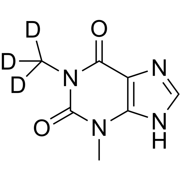
Theophylline-d3
Theophylline-d3 is deuterated labeled Theophylline (HY-B0809). Theophylline (1,3-Dimethylxanthine) is a potent phosphodiesterase (PDE) inhibitor, adenosine receptor antagonist, and histone deacetylase (HDAC) activator. Theophylline (1,3-Dimethylxanthine) inhibits PDE3 activity to relax airway smooth muscle. Theophylline (1,3-Dimethylxanthine) has anti-inflammatory activity by increase IL-10 and inhibit NF-κB into the nucleus. Theophylline (1,3-Dimethylxanthine) induces apoptosis. Theophylline (1,3-Dimethylxanthine) can be used for asthma and chronic obstructive pulmonary disease (COPD) research.
Nur für Forschungszwecke. Wir verkaufen nicht an Patienten.
- CAS. Nr.: 65566-68-9
- Formel: C7H5D3N4O2
- Molecular Weight:183.18
-
Speicherung:
Please store the product under the recommended conditions in the Certificate of Analysis.

To place orders, for customer services and technical support, please contact: MedChemExpress USA
Tel: 609-228-6898 E-mail: [email protected] [email protected]


-
 Biologische Aktivität
-
Chemical Information
- Protokoll
- Reinheit & Dokumentation
- Verweise
-
 Help & FAQs


Alle Endogenous Metabolite Isoform-spezifische Produkte anzeigen
MoreAlle TNF Receptor Isoform-spezifische Produkte anzeigen
MoreAlle Adenosine Receptor Isoform-spezifische Produkte anzeigen
More

Biologische Aktivität
Beschreibung
In Vitro
Stable heavy isotopes of hydrogen, carbon, and other elements have been incorporated into drug molecules, largely as tracers for quantitation during the drug development process. Deuteration has gained attention because of its potential to affect the pharmacokinetic and metabolic profiles of drugs[1].
Theophylline (1,3-Dimethylxanthine) (1-1000 μM) inhibits cAMP hydrolysis by PDE in homogenates of bronchial tissue to relax human bronchus and pulmonary arteries[2].
Theophylline (1,3-Dimethylxanthine) (10 μg/mL; 24 h; eosinophils) induces apoptosis through a reduction in the antiapoptotic protein Bcl-2[3].
Theophylline (1,3-Dimethylxanthine) (0-500 μM; 2 h; A549 cells) inhibits NF-κB activation, I kappa B alpha (I-κBα) degradation and decreases the level of IL-6 in a concentration-dependent manner[4].
Theophylline (1,3-Dimethylxanthine) (0-1000 μM; 30 min; A549 cells) induces histone deacetylase activity to decrease inflammatory gene expression[5].
MedChemExpress (MCE) has not independently confirmed the accuracy of these methods. They are for reference only.
In Vivo
MedChemExpress (MCE) has not independently confirmed the accuracy of these methods. They are for reference only.
Anwendung
1. This compound can be used as a tracer
2. This compound can be used as an internal standard for quantitative analysis by NMR, GC-MS, or LC-MS.


Chemical Information
-
CAS. Nr. 65566-68-9
-
Unlabeled CAS 58-55-9
-
Molecular Weight 183.18
-
Formel C7H5D3N4O2
-
SMILES
O=C(N1C([2H])([2H])[2H])N(C)C2=C(N=CN2)C1=O
-
Synonyms
1,3-Dimethylxanthine-d3; Theo-24-d3
-
Versand
Room temperature in continental US; may vary elsewhere.
-
Speicherung
Please store the product under the recommended conditions in the Certificate of Analysis.


Reinheit & Dokumentation
Verweise
[1]. Russak EM, et al. Impact of Deuterium Substitution on the Pharmacokinetics of Pharmaceuticals. Ann Pharmacother. 2019 Feb;53(2):211-216. [Content Brief]
[2]. Rabe KF, et, al. Theophylline and selective PDE inhibitors as bronchodilators and smooth muscle relaxants. Eur Respir J. 1995 Apr;8(4):637-42. [Content Brief]
[3]. Németh ZH, et, al. Amrinone and theophylline differentially regulate cytokine and nitric oxide production in endotoxemic mice. Shock. 1997 May;7(5):371-5. [Content Brief]
[4]. Ichiyama T, et, al. Theophylline inhibits NF-kappa B activation and I kappa B alpha degradation in human pulmonary epithelial cells. Naunyn Schmiedebergs Arch Pharmacol. 2001 Dec;364(6):558-61. [Content Brief]
[5]. Ito K, et, al, Adcock IM, Barnes PJ. A molecular mechanism of action of theophylline: Induction of histone deacetylase activity to decrease inflammatory gene expression. Proc Natl Acad Sci U S A. 2002 Jun 25;99(13):8921-6. [Content Brief]
[6]. Barnes PJ. Theophylline. Am J Respir Crit Care Med. 2013 Oct 15;188(8):901-6. [Content Brief]

Calculators
Konzentration (Stammlösung) × Volumen (Stammlösung) = Konzentration (Ziellösung) × Volumen (Ziellösung)


Theophylline-d3 Related Classifications
HY-B0809S1 Related Classifications
 Powered by Bioz
Keywords
- Theophylline-d3
- 65566-68-9
- 1,3-Dimethylxanthine-d3
- Theo-24-d3
- Endogenous Metabolite
- TNF Receptor
- Apoptosis
- HDAC
- Phosphodiesterase (PDE)
- Adenosine Receptor
- Interleukin Related
- Isotope-Labeled Compounds
- airway smooth muscle
- NF-κB
- I-κBα
- asthma
- chronic obstructive pulmonary disease
- A549 cells
- eosinophils
- Inhibitor
- inhibitor
- inhibit