



Kansuinine A

Based on 1 Customer Validation
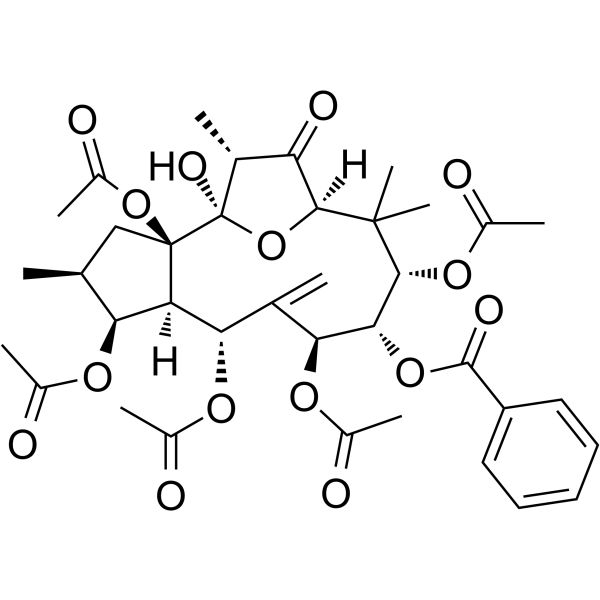
Kansuinine A is a diterpenoid compound. Kansuinine A is isolated from the roots of Euphorbia kansui. Kansuinine A inhibits H2O2-mediated upregulation of phosphorylated IKKβ, NF-κB and IκBα. Kansuinine A inhibits ROS production, reduces the Bax/Bcl-2 ratio and the expression of cleaved Caspase-3. Kansuinine A upregulates the expression of SOCS-3 and blocks IL-6-induced signal transduction. Kansuinine A decreases the expression of LOX-1 and inhibits Apoptosis. Kansuinine A reduces the atherosclerotic lesion area of the aortic arch, improves glucose/insulin tolerance, and enhances antioxidant capacity. Kansuinine A exhibits antiviral and antitumor activities. Kansuinine A can be used in research related to atherosclerosis, liver cancer and type 2 diabetes.
연구목적의 판매만을 진행합니다. 환자를 대상으로 한 판매는 하지 않습니다.
- Purity : 99.81%
- CAS No.: 57701-86-7
- 화학식: C37H46O15
- 분자량:730.75
-
보관:Powder -20°C, 3 years , 4°C, 2 years ; In solvent -80°C, 6 months , -20°C, 1 month

To place orders, for customer services and technical support, please contact: MedChemExpress USA
Tel: 609-228-6898 E-mail: [email protected] [email protected]


-
 Biological Activity
-
Chemical Information
-
용액&용해도
- Protocol
- 순도&문서
- References
-
 Help & FAQs


All Caspase Isoforms
More

Biological Activity
제품 설명
IC50 & Target
[2]|
IL-6 |
Caspase 3 |
Bcl-2 |
Bax |
IKKβ |
Cellular Effect
|
Cell Line

|
Type | Value | Description | References |
|---|---|---|---|---|
| NIH3T3 | ED50 |
3.28 μg/mL
Compound: 6
|
Survival effect on NGF-dependent mouse NIH/3T3 cells expressing TrkB assessed as cell viability after 2 days by MTT assay
Survival effect on NGF-dependent mouse NIH/3T3 cells expressing TrkB assessed as cell viability after 2 days by MTT assay
|
[PMID: 15387657] |
| NIH3T3 | ED50 |
7.92 μg/mL
Compound: 6
|
Survival effect on NGF-dependent mouse NIH/3T3 cells expressing TrkA assessed as cell viability after 2 days by MTT assay
Survival effect on NGF-dependent mouse NIH/3T3 cells expressing TrkA assessed as cell viability after 2 days by MTT assay
|
[PMID: 15387657] |
In Vitro
Kansuinine A (0.1-1.0 µM; 1 h) concentration-dependently reduces H2O2-induced intracellular reactive oxygen species production in aortic endothelial cells at concentrations of 0.1, 0.3, and 1.0 µM[1].
Kansuinine A (0.1-1 μM; 1 h) restores the activities of GPx and SOD antioxidant enzymes in AC3RL-treated RIN-m5F rat pancreatic β cells[3].
Kansuinine A (0.1-1.0 µM; 1 h) attenuates H2O2-induced activation of the pro-apoptotic signaling pathway in human aortic endothelial cells by reducing the Bax/Bcl-2 ratio at concentrations of 0.3 and 1.0 µM, and decreasing the expression of activated caspase-3 at a concentration of 1.0 µM[1].
Kansuinine A (0.1-1 μM) inhibits AC3RL-induced apoptosis of RIN-m5F rat pancreatic β cells[3].
Kansuinine A (6 μM; 15-120 min) increases the SOCS-3 mRNA expression level by approximately 17-fold in human hepatoma HepG2 cells at 60 min of treatment, and this effect depends on the PKC and MEK/ERK signaling pathways[2].
Kansuinine A (6 μM; 5-60 min) induces sustained phosphorylation of Stat3 at serine (Ser727) in human hepatoma HepG2 cells over a duration of 5 to 60 minutes, and this effect depends on the PKC and MEK/ERK signaling pathways[2].
MedChemExpress (MCE) has not independently confirmed the accuracy of these methods. They are for reference only.
-
Cell Line:human aortic endothelial cells (HAECs)
-
Concentration:0.1-3 µM (0.3, 1, 3 µM for toxicity testing; 0.1, 0.3, 1.0 µM for injury protection testing)
-
Incubation Time:24 h (toxicity testing); 60 min pre-incubation followed by 24 h H2O2 challenge (injury protection testing)
-
Result:Showed no significant cytotoxic effect on HAECs at concentrations up to 3 µM (p = 0.62, n = 3).
Protected HAECs from H2O2-induced cell damage, with significant effects observed at 0.1 µM (p < 0.01), 0.3 µM (p < 0.05), and 1.0 µM (p < 0.01, n = 3).
-
Cell Line:human aortic endothelial cells (HAECs)
-
Concentration:0.1-1.0 µM
-
Incubation Time:30 min pre-incubation followed by 24 h H2O2 challenge
-
Result:Reduced chromosomal breakage fragments and maintained cell membrane integrity in H2O2-treated HAECs.
Showed significant reduction in apoptosis rate at 0.3 µM and 1.0 µM (p < 0.001 vs.
H2O2 alone, n = 3).
-
Cell Line:human aortic endothelial cells (HAECs)
-
Concentration:0.1-1.0 µM
-
Incubation Time:1 h pre-incubation followed by 24 h H2O2 challenge
-
Result:Reduced the H2O2-induced Bax/Bcl-2 ratio at 0.3 and 1.0 µM (p < 0.05 vs.
H2O2 alone, n = 3).
Reversed H2O2-induced cleaved caspase-3 expression at 1.0 µM (p < 0.001 vs.
H2O2 alone, n = 3).\nReduced phosphorylated IKKβ levels at 1.0 µM (p < 0.01 vs.
H2O2 alone).
Reduced phosphorylated IκBα levels at 0.3 µM (p < 0.05) and 1.0 µM (p < 0.01 vs.
H2O2 alone).
Reduced phosphorylated NF-κB p65 levels at 0.3 and 1.0 µM (p < 0.05 vs.
H2O2 alone, n = 3).
In Vivo
MedChemExpress (MCE) has not independently confirmed the accuracy of these methods. They are for reference only.
-
Animal Model:Apolipoprotein E-deficient (ApoE-/-) mice (C57BL/6 background, n=5 per group, high-fat diet fed for 15 weeks)[1]
-
Dosage:20 µg/kg; 60 µg/kg
-
Administration:i.p.; three times a week; 15 weeks
-
Result:Significantly reduced body weight AUC values compared to HFD-only mice (p < 0.001).
Significantly lowered total cholesterol, LDL-cholesterol, and triglyceride levels compared to HFD-only mice (p < 0.05); the 60 µg/kg group significantly increased HDL-cholesterol levels (p < 0.05).
Reduced aortic arch lesion area by 23% in the 20 µg/kg group (p < 0.05) and by 61% in the 60 µg/kg group (p < 0.001) compared to HFD-only mice.
Significantly reduced mRNA expression of Bax (p < 0.05) and caspase-3 (p < 0.001), and protein expression of Bax and cleaved caspase-3 (p < 0.001) in aortic tissues compared to HFD-only mice.
Significantly increased aortic glutathione peroxidase (GPx) levels (p < 0.001) and significantly reduced aortic malondialdehyde (MDA) levels (p < 0.001) compared to HFD-only mice.
-
Animal Model:ApoE−/− C57BL/6 (10 weeks old, 20-25 g, mixed gender, fed high-fat diet for 15 weeks)[3]
-
Dosage:20 μg/kg; 60 μg/kg
-
Administration:i.p.; three times weekly; 15 weeks
-
Result:Significantly reduced body weight compared to untreated controls.
Significantly reduced serum triglycerides, total cholesterol, and LDL-C levels, and upregulated HDL-C levels at 60 μg/kg dose; reduced the ApoC3/ApoB-100 ratio in plasma at both doses.
Significantly reduced fasting blood glucose levels, decreased the area under the curve for intraperitoneal glucose tolerance tests and insulin tolerance tests, and lowered elevated plasma insulin levels compared to untreated controls at both doses.
Significantly increased pancreatic islet area, reduced TUNEL-positive apoptotic cell percentage in islets, and increased plasma glutathione peroxidase, superoxide dismutase, and catalase activity compared to untreated controls at both doses.
Significantly reduced plasma malondialdehyde levels and dose-dependently decreased pancreatic islet expression of phosphorylated NF-κB, total NF-κB, and cleaved caspase-3 compared to untreated controls at both doses.
- RNA extraction experimental
- Research Protocol for Infectious Diseases
- Detection of Apoptosis
- TUNEL staining for apoptotic DNA fragmentation
- Annexin V plus membrane-impermeant dye apoptosis staining
- ROS/oxidative-stress fluorescent staining
- Research Protocol for Endocrine Diseases
- Apoptosis Solutions
- Research Protocol for Cardiovascular Diseases
- Research Protocol for Inflammation-related Diseases
- Liver Cancer Modeling
- Research Protocol for Metabolic Diseases


Chemical Information
-
CAS No. 57701-86-7
-
Appearance Solid
-
분자량 730.75
-
화학식 C37H46O15
-
Color White to off-white
-
SMILES
O=C1[C@@]2([H])C(C)(C)[C@H](OC(C)=O)[C@H](OC(C3=CC=CC=C3)=O)[C@@H](OC(C)=O)C([C@H](OC(C)=O)[C@]([C@@H](OC(C)=O)[C@@H](C)C4)([H])[C@@]4(OC(C)=O)[C@@](O2)(O)[C@@H]1C)=C
-
Structure Classification
-
선적
Room temperature in continental US; may vary elsewhere.
-
보관
Powder -20°C 3 years 4°C 2 years In solvent -80°C 6 months -20°C 1 month


용액&용해도
In Vitro:
DMSO : ≥ 100 mg/mL (136.85 mM; Hygroscopic DMSO has a significant impact on the solubility of product, please use newly opened DMSO)
* "≥" means soluble, but saturation unknown.
Please refer to the solubility information to select the appropriate solvent. Once prepared, please aliquot and store the solution to prevent product inactivation from repeated freeze-thaw cycles.
Storage method and period of stock solution: -80°C, 6 months; -20°C, 1 month. When stored at -80°C, please use it within 6 months. When stored at -20°C, please use it within 1 month.
Please refer to the solubility information to select the appropriate solvent. Once prepared, please aliquot and store the solution to prevent product inactivation from repeated freeze-thaw cycles.
Storage method and period of stock solution: -80°C, 6 months; -20°C, 1 month. When stored at -80°C, please use it within 6 months. When stored at -20°C, please use it within 1 month.
Concentration (start) × Volume (start) = Concentration (final) × Volume (final)
In Vivo:
Select the appropriate dissolution method based on your experimental animal and administration route.
- For the following dissolution methods, please ensure to first prepare a clear stock solution using an In Vitro approach and then sequentially add co-solvents:
- To ensure reliable experimental results, the clarified stock solution can be appropriately stored based on storage conditions. As for the working solution for In Vivo experiments, it is recommended to prepare freshly and use it on the same day.
- The percentages shown for the solvents indicate their volumetric ratio in the final prepared solution. If precipitation or phase separation occurs during preparation, heat and/or sonication can be used to aid dissolution.
Add each solvent one by one: 10% DMSO 40% PEG300 5% Tween-80 45% Saline
Solubility: ≥ 2.5 mg/mL (3.42 mM); Clear solution
This protocol yields a clear solution of ≥ 2.5 mg/mL (saturation unknown).
Taking 1 mL working solution as an example, add 100 μL DMSO stock solution (25.0 mg/mL) to 400 μL PEG300, and mix evenly; then add 50 μL Tween-80 and mix evenly; then add 450 μL Saline to adjust the volume to 1 mL.
Preparation of Saline: Dissolve 0.9 g sodium chloride in ddH₂O and dilute to 100 mL to obtain a clear Saline solution.
In Vivo Dissolution Calculator
Please enter the basic information of animal experiments:
-
-
-
-
Recommended: Prepare an additional quantity of animals to account for potential losses during experiments.
Please enter your animal formula composition:
-
%DMSO +
Recommended: Keep the proportion of DMSO in working solution below 2% if your animal is weak.
-
%+
-
+%Tween-80 + +
-
%Saline +
The co-solvents required include: DMSO, . All of co-solvents are available by MedChemExpress (MCE). , Tween 80. All of co-solvents are available by MedChemExpress (MCE).
Working solution concentration: 0.22 mg/mL
Method for preparing stock solution: mg drug dissolved in μL DMSO. Stock solution concentration: mg/mL.
1. Take μL DMSO stock solution;
2. Add μL .
μL , mix evenly;
3. Then add μL Tween 80, mix evenly;
4. Then add μL
Please ensure that the stock solution in the first step is dissolved to a clear state, and add co-solvents in sequence. You can use ultrasonic heating (ultrasonic cleaner, recommended frequency 20-40 kHz), vortexing, etc. to assist dissolution.


순도&문서
-
Data Sheet (300 KB)
-
SDS (254 KB)
- English - EN (254 KB)
- Français - FR (254 KB)
- Deutsch - DE (254 KB)
- Norwegian - NO (254 KB)
- Español - ES (254 KB)
- Swedish - SV (254 KB)
- Italian - IT (254 KB)
- Korean - KR (254 KB)
- Portuguese - PT (254 KB)
-
Handling Instructions (2659 KB)
References
[1]. Chen CS, et al. Kansuinine A Ameliorates Atherosclerosis and Human Aortic Endothelial Cell Apoptosis by Inhibiting Reactive Oxygen Species Production and Suppressing IKKβ/IκBα/NF-κB Signaling. International journal of molecular sciences. 2021 Sep 24;22(19):10309. [Content Brief]
[2]. Chang JS, et al. Kansuinine A and Kansuinine B from Euphorbia kansui L. inhibit IL-6-induced Stat3 activation. Planta medica. 2010 Oct;76(14):1544-9. [Content Brief]
[3]. Lulji Taraqaz BP, et al. Pancreatic β-cell apoptosis caused by apolipoprotein C3-rich low-density lipoprotein is attenuated by kansuinine A through oxidative stress inhibition. Biomedicine & pharmacotherapy = Biomedecine & pharmacotherapie. 2025 Jun;187:118066. [Content Brief]
[4]. Shu X, et al. Bioassay-guided separation of the proinflammatory constituents from the roots of Euphorbia kansui. Journal of natural medicines. 2010 Jan;64(1):98-103. [Content Brief]


Complete Stock Solution Preparation Table
Please refer to the solubility information to select the appropriate solvent. Once prepared, please aliquot and store the solution to prevent product inactivation from repeated freeze-thaw cycles.
Storage method and period of stock solution: -80°C, 6 months; -20°C, 1 month. When stored at -80°C, please use it within 6 months. When stored at -20°C, please use it within 1 month.
| Optional Solvent | Concentration Solvent Mass | 1 mg | 5 mg | 10 mg | 25 mg |
|---|---|---|---|---|---|
| DMSO | 1 mM | 1.3685 mL | 6.8423 mL | 13.6846 mL | 34.2114 mL |
| 5 mM | 0.2737 mL | 1.3685 mL | 2.7369 mL | 6.8423 mL | |
| 10 mM | 0.1368 mL | 0.6842 mL | 1.3685 mL | 3.4211 mL | |
| 15 mM | 0.0912 mL | 0.4562 mL | 0.9123 mL | 2.2808 mL | |
| 20 mM | 0.0684 mL | 0.3421 mL | 0.6842 mL | 1.7106 mL | |
| 25 mM | 0.0547 mL | 0.2737 mL | 0.5474 mL | 1.3685 mL | |
| 30 mM | 0.0456 mL | 0.2281 mL | 0.4562 mL | 1.1404 mL | |
| 40 mM | 0.0342 mL | 0.1711 mL | 0.3421 mL | 0.8553 mL | |
| 50 mM | 0.0274 mL | 0.1368 mL | 0.2737 mL | 0.6842 mL | |
| 60 mM | 0.0228 mL | 0.1140 mL | 0.2281 mL | 0.5702 mL | |
| 80 mM | 0.0171 mL | 0.0855 mL | 0.1711 mL | 0.4276 mL | |
| 100 mM | 0.0137 mL | 0.0684 mL | 0.1368 mL | 0.3421 mL |


Kansuinine A Related Classifications
HY-126421 Related Classifications
 Powered by Bioz
Keywords
- Kansuinine A
- 57701-86-7
- Interleukin Related
- IKK
- NF-κB
- Reactive Oxygen Species (ROS)
- Bcl-2 Family
- Caspase
- LOX-1
- Apoptosis
- SOCS-3
- Euphorbia kansui roots
- IKKβ/NF-κB signaling pathway
- atherosclerosis
- ApoE-/- mice
- ERK1/2
- RIN-m5F rat pancreatic β-cells
- human aortic endothelial cells
- Stat3
- human hepatoma HepG2 cells
- Inhibitor
- inhibitor
- inhibit