



PRT543
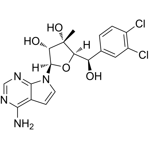
PRT543 is an orally active selective PRMT5 inhibitor. PRT543 reduces intracellular symmetric dimethylarginine (sDMA) levels, downregulates the expression of genes related to DNA damage repair and DNA replication pathways, and induces abnormal alternative splicing. PRT543 inhibits the MYB, NOTCH1 and PI3K/AKT signaling pathways, promotes nuclear translocation of FOXO1, upregulates the pro-apoptotic protein BAX, and enhances cellular sensitivity to BCL-2 inhibition. PRT543 disrupts the normal RNA splicing process and exerts a synthetic lethal effect on myeloid tumor cells carrying splicing factor mutations. PRT543 can be used in research related to various cancers including breast cancer, ovarian cancer and acute myeloid leukemia.
For research use only. We do not sell to patients.
- CAS No.: 2278356-90-2
- Formula: C18H18Cl2N4O4
- Molecular Weight:425.27
-
Storage:
Please store the product under the recommended conditions in the Certificate of Analysis.

To place orders, for customer services and technical support, please contact: MedChemExpress USA
Tel: 609-228-6898 E-mail: [email protected] [email protected]


-
 Biological Activity
-
Chemical Information
- Protocols
- Purity & Documentation
- References
-
 Help & FAQs




Biological Activity
Description
|
PRMT5 |
PI3K |
Akt |
Bax |
In Vitro
PRT543 (0.37-3.3 μM; 3 days) downregulates ACC-related gene signatures, MYB/selective MYB target genes, and RUNX1 target genes in CAL27, FaDu, and A253 head and neck squamous cell carcinoma (HNSCC) cells; and downregulates MYB target genes and alternatively spliced MYB transcripts in K562 and Jurkat leukemia cells[2].
PRT543 (0.37-10 μM; 4 days) can downregulate the expression of the NOTCH signaling pathway and target genes, reduce sDMA levels, and decrease NOTCH1 protein expression in NOTCH1-activated HPB.ALL T-ALL cells[2].
PRT543 exhibits anti-proliferative activity and dose-dependent SDMA inhibition in in vitro myeloid malignancy cell line models[3].
PRT543 (0.001-100 μmol/L; 10 days, or 4-day pretreatment followed by 6-day cotreatment) potently inhibits proliferation of UWB1.289, UWB1.289-BRCA1, and UWB1.289 Olaparib (HY-10162)-Res ovarian cancer cells, and overcomes Olaparib resistance driven by BRCA1 re-expression or acquired drug resistance[4].
PRT543 (10 days) acts synergistically with Olaparib, Cisplatin (HY-17394), and 5-Fluorouracil (HY-90006) to inhibit proliferation of HR-proficient A2780, ES-2, and OV-7 ovarian cancer cells[4].
PRT543 acts as a competitive, substrate-noncompetitive slow binding inhibitor of SAM and MTA, potently inhibiting PRMT5/MEP50 methyltransferase activity in vitro; its selectivity for PRMT5 is significantly higher than that of the enzyme group composed of 37 other methyltransferases; and it can dose-dependently reduce the level of PRMT5 substrate symmetric dimethylated SmD3 in cancer cells[5].
PRT543 (2-10 μM; 24 h) induces extensive global alternative splicing changes, including intron retention and exon skipping, in wild-type and splicing factor mutant (SRSF2P95H, SF3B1K700E, U2AF1Q157P) K562 leukemia cell lines, with the most events observed in SRSF2P95H knock-in cells[5].
MedChemExpress (MCE) has not independently confirmed the accuracy of these methods. They are for reference only.
-
Cell Line:CAL27, FaDu, A253 HNSCC cells
-
Concentration:0.04, 0.12, 0.37, 1.10, 3.30 μM
-
Incubation Time:3 days
-
Result:Downregulated ACC-associated gene signatures, MYB/alternative-MYB target genes, and RUNX1 target genes.
In Vivo
PRT543 (40 mg/kg; p.o.; 5 days on/2 days off; 36 days) monotherapy inhibits tumor growth in BRCA1-deleted ovarian cancer PDXs, and its combination with Olaparib provides additional tumor growth inhibition beyond Olaparib alone[4].
PRT543 (40 mg/kg; p.o.; 5 days on/2 days off; 21 days) monotherapy inhibites tumor growth in a breast cancer PDX model with normal heart rate function and a breast cancer PDX model resistant to Olaparib/BRCA1/2 hemizygosity[4].
PRT543 (40 mg/kg; p.o.; 5 days on/2 days off; 27 days) monotherapy induces tumor growth inhibition in Olaparib-resistant, BRCA1-deleted ovarian cancer PDXs, overcoming Olaparib resistance[4].
MedChemExpress (MCE) has not independently confirmed the accuracy of these methods. They are for reference only.
-
Animal Model:BALB/c nude (female, 6-9 weeks old, the A2780 cell line mixed 1:1 with Matrigel)[4]
-
Dosage:15 mg/kg; 40 mg/kg
-
Administration:p.o.; daily; 9 days
-
Result:Induced 56.2% tumor growth inhibition (TGI) compared with vehicle at 40 mg/kg.
Combined with olaparib at 15 mg/kg resulted in a 24.6% TGI improvement over PRT543 monotherapy at the same dose.
-
Animal Model:Nude-Foxn1nu (female, 6-8 weeks old)[4]
-
Dosage:40 mg/kg
-
Administration:p.o.; 5 days on/2 days off; 36 days
-
Result:Induced significant tumor growth inhibition as a single agent.
Combined with olaparib resulted in a 24% TGI improvement over olaparib monotherapy.
-
Animal Model:Nude-Foxn1nu (female, 6-8 weeks old)[4]
-
Dosage:40 mg/kg
-
Administration:p.o.; 5 days on/2 days off; 21 days
-
Result:Induced 53% TGI compared with vehicle as a single agent.
Combined with olaparib resulted in a 28% TGI improvement over olaparib monotherapy.\nInduced 52% TGI compared with vehicle as a single agent.
Olaparib showed no significant tumor growth inhibition.
-
Animal Model:Nude-Foxn1nu (female, 6-8 weeks old)[4]
-
Dosage:40 mg/kg
-
Administration:p.o.; 5 days on/2 days off; 27 days
-
Result:Induced 102% TGI compared with vehicle as a single agent.
Olaparib showed no significant tumor growth inhibition.
Clinical Trial
| NCT Number | Sponsor | Condition | Start Date |
Phase

|
|---|---|---|---|---|
| NCT01329991 | 2011-05 | PHASE1 |


Chemical Information
-
CAS No. 2278356-90-2
-
Molecular Weight 425.27
-
Formula C18H18Cl2N4O4
-
SMILES
O[C@@]1(C)[C@@H](O)[C@H](N2C3=NC=NC(N)=C3C=C2)O[C@@H]1[C@H](O)C4=CC=C(Cl)C(Cl)=C4
-
Shipping
Room temperature in continental US; may vary elsewhere.
-
Storage
Please store the product under the recommended conditions in the Certificate of Analysis.


Purity & Documentation
References
[4]. Carter J, et al. PRMT5 Inhibitors Regulate DNA Damage Repair Pathways in Cancer Cells and Improve Response to PARP Inhibition and Chemotherapies. Cancer Res Commun. 2023;3(11):2233-2243. [Content Brief]
[5]. Bewersdorf JP, et al. Phase Ib study of PRT543, an oral protein arginine methyltransferase 5 (PRMT5) inhibitor, in patients with advanced splicing factor-mutant myeloid malignancies. Leukemia. 2025;39(3):765-769. [Content Brief]

Calculators
Concentration (start) × Volume (start) = Concentration (final) × Volume (final)


PRT543 Related Classifications
HY-141876 Related Classifications
 Powered by Bioz