



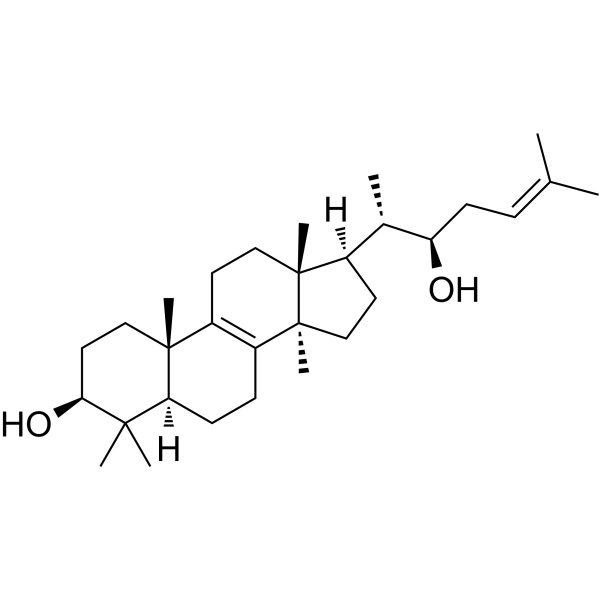
Inotodiol

Based on 1 Customer Validation
Inotodiol activates p53 signaling pathway, inhibits MMP-2/9, and exhibits antitumor activity in cancer cell HeLa. Inotodiol inhibits the generation of ROS and exhibits antioxidant and neuroprotective effects. Inotodiol inhibits the activation of MAPK and NF-κB signaling pathway, and exhibits anti-inflammatory activity. Inotodiol inhibits TLR-4 mediated TNF-α production (IC50s in BMMC and BMDM is 0.7 μM and 3.0 μM), inhibits the degranulation in mast cell, exhibits anti-allergic activity. Inotodiol is orally active.
Nur für Forschungszwecke. Wir verkaufen nicht an Patienten.
- Reinheit : 98.29%
- CAS. Nr.: 35963-37-2
- Formel: C30H50O2
- Molecular Weight:442.72
-
Speicherung:
4°C, sealed storage, away from moisture and light
* In solvent : -80°C, 6 months; -20°C, 1 month (sealed storage, away from moisture and light)

To place orders, for customer services and technical support, please contact: MedChemExpress USA
Tel: 609-228-6898 E-mail: [email protected] [email protected]


-
 Biologische Aktivität
-
Chemical Information
- Protokoll
- Reinheit & Dokumentation
- Verweise
-
 Help & FAQs


Alle Caspase Isoform-spezifische Produkte anzeigen
More

Biologische Aktivität
Beschreibung
Cellular Effect
|
Cell Line

|
Type | Value | Description | References |
|---|---|---|---|---|
| COLO 205 | IC50 |
171 μM
Compound: 147
|
Cytotoxicity against human COLO205 cells
Cytotoxicity against human COLO205 cells
|
[PMID: 23092389] |
| HL-60 | IC50 |
25 μM
Compound: 147
|
Cytotoxicity against human HL60 cells
Cytotoxicity against human HL60 cells
|
[PMID: 23092389] |
| L1210 | IC50 |
110 μM
Compound: 147
|
Cytotoxicity against mouse L1210 cells
Cytotoxicity against mouse L1210 cells
|
[PMID: 23092389] |
| P388 | IC50 |
13.9 μM
Compound: 147
|
Antiproliferative activity against mouse P388 cells
Antiproliferative activity against mouse P388 cells
|
[PMID: 23092389] |
In Vitro
Inotodiol (0-100 μM, 72 h) inhibits the proliferation of cell HeLa, inhibits the cell migration and invasion, induces apoptosis i HeLa[2].
Inotodiol (0.1-10 μM, 28 h) inhibits the ROS generation, activates SOD, improves oxygen and glucose deprivation-induced mitochondrial dysfunction, reduces the leakage of lactate dehydrogenase and exhibits neuroprotective effect in PC12 cell[3].
Inotodiol (2 μg/mL, 24 h) inhibits H2O2-induced expression of IL-1β, IL-6, TNF-α and COX-2, and blocks the senescence in human dermal fibroblasts (HDF)[4].
MedChemExpress (MCE) has not independently confirmed the accuracy of these methods. They are for reference only.
-
Cell Line:PC12
-
Concentration:0.1-10 μM
-
Incubation Time:28 h
-
Result:Inproved the cell viability.
-
Cell Line:HeLa
-
Concentration:0-100 μM
-
Incubation Time:72 h
-
Result:Induced apoptosis.
-
Cell Line:HeLa
-
Concentration:30-50 μM
-
Incubation Time:48 h
-
Result:Upregulated the expression of p53, p21 and Bax, downregulated the expression of MMP-2/9, Bcl-2.
-
Cell Line:HeLa
-
Concentration:30-50 μM
-
Incubation Time:48 h
-
Result:Inhibited cell migration.
In Vivo
MedChemExpress (MCE) has not independently confirmed the accuracy of these methods. They are for reference only.
-
Animal Model:Mouse passive anaphylaxis models[5]
-
Dosage:2-20 mpk
-
Administration:po, once daily for 3-5 days
-
Result:Reduced the decrease in body temperature and ear swelling.
- Mitochondrial membrane-potential fluorescent assay
- ROS/oxidative-stress fluorescent staining
- Cotton Pellet Granuloma
- Carrageenan-Induced Paw Edema
- Mitochondrial membrane-potential and mitochondrial mass staining
- Research Protocol for Inflammation-related Diseases
- Subcutaneous Cell-Line-Derived Xenograft


Chemical Information
-
CAS. Nr. 35963-37-2
-
Appearance Solid
-
Molecular Weight 442.72
-
Formel C30H50O2
-
Color White to off-white
-
SMILES
C[C@@]1(CC[C@@H]2O)C(CC[C@@]34C)=C(CC[C@@]1([H])C2(C)C)[C@@]3(CC[C@]4([H])[C@H](C)[C@H](O)C/C=C(C)\C)C
-
Structure Classification
-
Initial Source
Inonotus obliquus Chaga
-
Versand
Room temperature in continental US; may vary elsewhere.
-
Speicherung
4°C, sealed storage, away from moisture and light
* In solvent : -80°C, 6 months; -20°C, 1 month (sealed storage, away from moisture and light)


Reinheit & Dokumentation
-
Data Sheet (274 KB)
-
SDS (252 KB)
- English - EN (252 KB)
- Français - FR (252 KB)
- Deutsch - DE (252 KB)
- Norwegian - NO (252 KB)
- Español - ES (252 KB)
- Swedish - SV (252 KB)
- Italian - IT (252 KB)
- Korean - KR (252 KB)
- Portuguese - PT (252 KB)
-
Handling Instructions (2659 KB)
Verweise
[1]. Nakata T, et al. Structure determination of inonotsuoxides A and B and in vivo anti-tumor promoting activity of inotodiol from the sclerotia of Inonotus obliquus. Bioorg Med Chem. 2007;15(1):257-264. [Content Brief]
[2]. Zhang SD, et al., Inotodiol inhibits cells migration and invasion and induces apoptosis via p53-dependent pathway in HeLa cells. Phytomedicine. 2019 Jul;60:152957. [Content Brief]
[4]. Lee SH, et al., Antiaging effect of inotodiol on oxidative stress in human dermal fibroblasts. Biomed Pharmacother. 2022 Sep;153:113311. [Content Brief]
[5]. Liu Y, et al., Inotodiol, an antiasthmatic agent with efficacy and safety, preferentially impairs membrane-proximal signaling for mast cell activation. Int Immunopharmacol. 2023 Apr;117:109854. [Content Brief]

Calculators
Konzentration (Stammlösung) × Volumen (Stammlösung) = Konzentration (Ziellösung) × Volumen (Ziellösung)


Inotodiol Related Classifications
HY-120149 Related Classifications
 Powered by Bioz