



2-Methoxyestradiol
Based on 34 publication(s) in Google Scholar
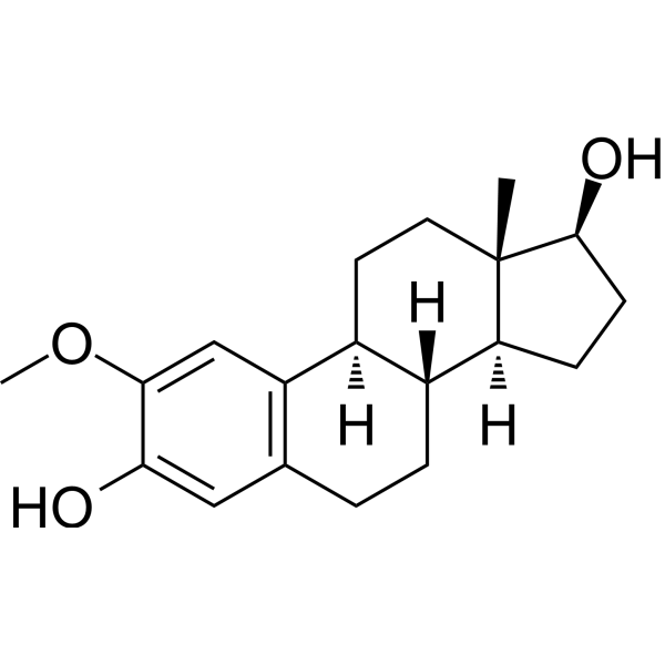
2-Methoxyestradiol (2-ME2; NSC-659853) is an endogenous steroid metabolite generated from 17β-estradiol, possessing various activities such as antitumor and antiangiogenic effects. 2-Methoxyestradiol competitively binds to the colchicine-binding site of tubulin (tubulin), interfering with microtubule assembly dynamics. 2-Methoxyestradiol inhibits CYP1A1/CYP1B1, HIF-1α/2α, and superoxide dismutase, activates the JNK signaling pathway, elevates ROS levels, and induces cell cycle arrest and apoptosis (apoptosis). 2-Methoxyestradiol is used in cancer-related research, such as breast cancer, prostate cancer, and multiple myeloma.
For research use only. We do not sell to patients.
- Purity : 99.76%
- CAS No.: 362-07-2
- Formula: C19H26O3
- Molecular Weight:302.41
-
Storage:Powder -20°C, 3 years , 4°C, 2 years ; In solvent -80°C, 2 years , -20°C, 1 year

To place orders, for customer services and technical support, please contact: MedChemExpress USA
Tel: 609-228-6898 E-mail: [email protected] [email protected]


-
 Biological Activity
-
Chemical Information
-
Solvent & Solubility
- Protocols
- Purity & Documentation
- References
-
 Help & FAQs


-
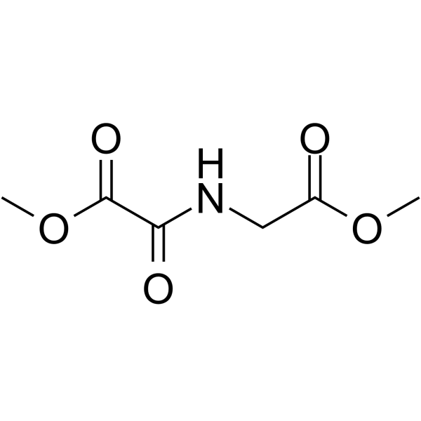
HY-15893
DMOGHIF-PH Inhibitor -
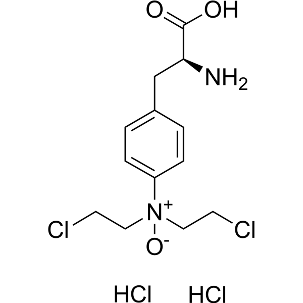
HY-10231
PX-478HIF-1α Inhibitor -

HY-100579
Ferrostatin-1Ferroptosis Inhibitor -
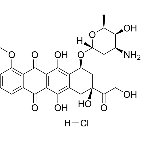
HY-15142
Doxorubicin hydrochlorideTopoisomerase Inhibitor -
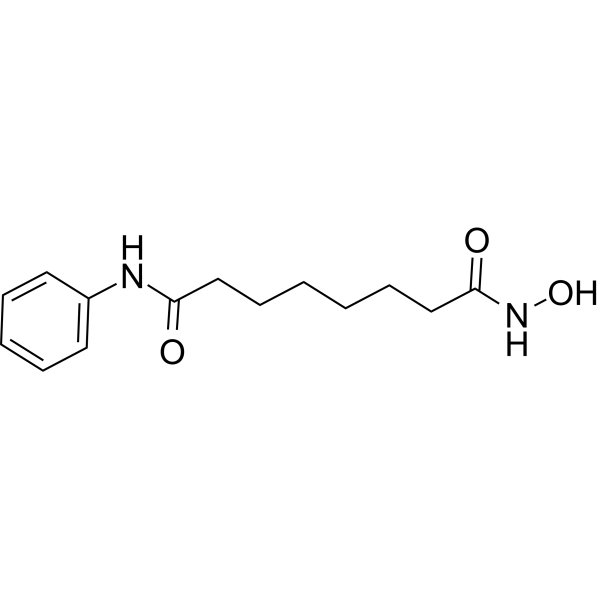
HY-10221
VorinostatHDAC Inhibitor -
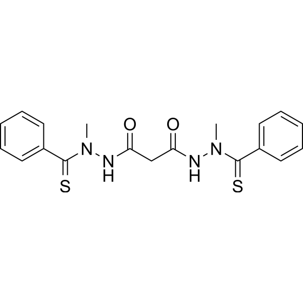
HY-12040
ElesclomolCuproptosis Inducer -
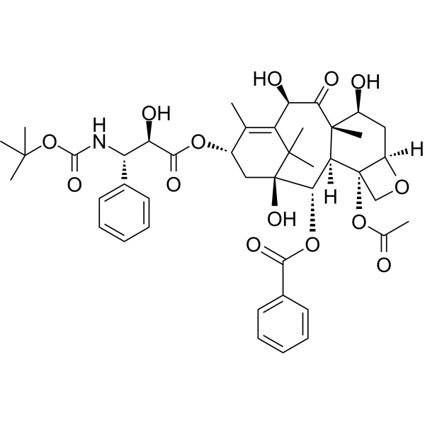
HY-B0011
DocetaxelTubulin Stabilizer -
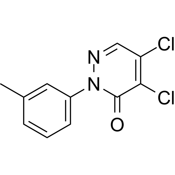
HY-115445
LCS-1SOD1 Inhibitor -
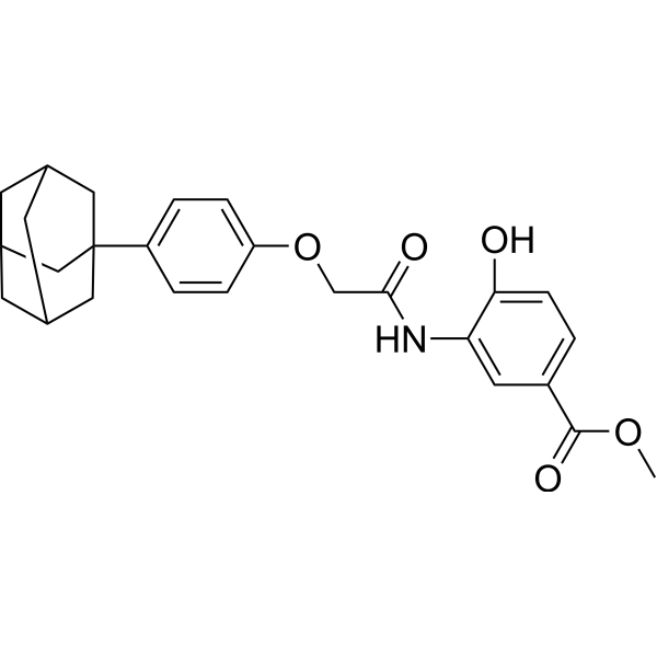
HY-13671
LW6Apoptosis Inducer -

HY-10219
RapamycinmTOR Inhibitor -
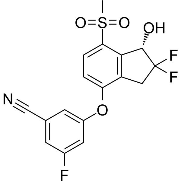
HY-12867
PT-2385HIF-2α Inhibitor -
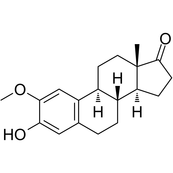
HY-113252
2-MethoxyestroneEstrogen Metabolite
Publications Citing Use of MedChemExpress (MCE) 2-Methoxyestradiol
More- Cell Res. 2026 Mar;36(3):219-232. [Abstract]
- J Nanobiotechnology. 2023 Jul 22;21(1):233. [Abstract]
- Theranostics. 2024 May 27;14(8):3339-3357. [Abstract]
- J Exp Clin Cancer Res. 2018 Jun 28;37(1):128. [Abstract]
- Carbohydr Polym. 2025 Aug 15:362:123685. [Abstract]
- Cell Death Dis. 2025 Jun 20;16(1):462. [Abstract]
- Cancer Lett. 2016 Nov 1;382(1):44-52. [Abstract]
- Mol Ther. 2021 Dec 1;29(12):3449-3464. [Abstract]
- Acta Biomater. 2025 Aug:202:248-261. [Abstract]
- J Transl Med. 2023 Oct 26;21(1):754. [Abstract]
- Free Radic Biol Med. 2021 Jun:169:271-282. [Abstract]
- Stem Cell Res Ther. 2022 Feb 5;13(1):59. [Abstract]
- Front Immunol. 2021 May 12:12:639049. [Abstract]
- Ann Hepatol. 2023 Nov-Dec;28(6):101135. [Abstract]
- Eur J Pharmacol. 2022 Oct 15:933:175276. [Abstract]
- Cancer Biol Ther. 2016 Jun 2;17(6):625-34. [Abstract]
- Int J Mol Sci. 2025 Jul 8;26(14):6554. [Abstract]
- Front Cell Dev Biol. 2021 Feb 23:9:607142. [Abstract]
- Chem Biol Interact. 2023 Jan 5:369:110277. [Abstract]
- Pestic Biochem Physiol. 2023 Dec:197:105647. [Abstract]
- Rheumatology (Oxford). 2019 Nov 1;58(11):1966-1975. [Abstract]
- Aquaculture. 2020, 735393.
- Front Cell Neurosci. 2018 Dec 21:12:504. [Abstract]
- Am J Physiol Lung Cell Mol Physiol. 2025 Aug 1;329(2):L282-L295. [Abstract]
- Peptides. 2024 Nov:181:171299. [Abstract]
- J Immunol Res. 2022 Apr 28;2022:6324326. [Abstract]
- Biomed Res Int. 2016:2016:7948345. [Abstract]
- Thorac Cancer. 2019 Apr;10(4):676-685. [Abstract]
- Exp Ther Med. 2017 Aug;14(2):1825-1831. [Abstract]
- Res Sq. 2026 Aug 14.
- SSRN. 2025 Mar 17.
- Research Square Print. 2022 Aug.
- Research Square Print. 2022 May.
- Patent. US20180263995A1.
 Customer Validation & Images

-
WB
-
WB
-
WB
All Endogenous Metabolite Isoforms
MoreAll Caspase Isoforms
More

Biological Activity
Description
|
Aromatase |
CYP1A1 |
CYP1B1 |
Caspase-8 |
Caspase-9 |
HIF-1α |
HIF-2α |
Caspase 3 |
ERα 21 nM (Ki) |
ERβ 417 nM (Ki) |
Cellular Effect
|
Cell Line

|
Type | Value | Description | References |
|---|---|---|---|---|
| MCF7 | IC50 |
1.5 μM
|
Antiproliferative activity against human MCF7 breast carcinoma cells in charcoal-dextran-stripped serum assessed by cell counting after 8 or 10 days.
Antiproliferative activity against human MCF7 breast carcinoma cells in charcoal-dextran-stripped serum assessed by cell counting after 8 or 10 days.
|
12097276 |
| MDA-MB-435 | IC50 |
1.3 μM
|
Antiproliferative activity against human MDA-MB-435 breast carcinoma cells assessed by cell counting after 72 hrs.
Antiproliferative activity against human MDA-MB-435 breast carcinoma cells assessed by cell counting after 72 hrs.
|
12097276 |
| MDA-MB-231 | IC50 |
1.1 μM
|
Antiproliferative activity against human MDA-MB-231 breast carcinoma cells assessed by cell counting after 72 hrs.
Antiproliferative activity against human MDA-MB-231 breast carcinoma cells assessed by cell counting after 72 hrs.
|
12097276 |
| HOP-62 | IC50 |
0.7 μM
|
Antiproliferative activity against human HOP-62 cells assessed by metabolic labeling or cell count.
Antiproliferative activity against human HOP-62 cells assessed by metabolic labeling or cell count.
|
11191057 |
| NCI-H460 | IC50 |
5.0 μM
|
Antiproliferative activity against human H460 cells assessed by metabolic labeling or cell count.
Antiproliferative activity against human H460 cells assessed by metabolic labeling or cell count.
|
11191057 |
| A549 | IC50 |
5.0 μM
|
Antiproliferative activity against human A549 cells assessed by metabolic labeling or cell count.
Antiproliferative activity against human A549 cells assessed by metabolic labeling or cell count.
|
11191057 |
| HCT-116 | IC50 |
0.47 μM
|
Antiproliferative activity against human HCT-116 cells assessed by metabolic labeling or cell count.
Antiproliferative activity against human HCT-116 cells assessed by metabolic labeling or cell count.
|
11191057 |
| SF-539 | IC50 |
0.32 μM
|
Antiproliferative activity against human SF-539 cells assessed by metabolic labeling or cell count.
Antiproliferative activity against human SF-539 cells assessed by metabolic labeling or cell count.
|
11191057 |
| SH-SY5Y | IC50 |
1.3 μM
|
Antiproliferative activity against human SH-SY5Y cells assessed by metabolic labeling or cell count.
Antiproliferative activity against human SH-SY5Y cells assessed by metabolic labeling or cell count.
|
11191057 |
| UACC-62 | IC50 |
0.36 μM
|
Antiproliferative activity against human UACC-62 cells assessed by metabolic labeling or cell count.
Antiproliferative activity against human UACC-62 cells assessed by metabolic labeling or cell count.
|
11191057 |
| OVCAR-3 | IC50 |
0.21 μM
|
Antiproliferative activity against human OVCAR-3 cells assessed by metabolic labeling or cell count.
Antiproliferative activity against human OVCAR-3 cells assessed by metabolic labeling or cell count.
|
11191057 |
| SN12C | IC50 |
0.95 μM
|
Antiproliferative activity against human SN12-C cells assessed by metabolic labeling or cell count.
Antiproliferative activity against human SN12-C cells assessed by metabolic labeling or cell count.
|
11191057 |
| DU-145 | IC50 |
1.8 μM
|
Antiproliferative activity against human DU-145 cells assessed by metabolic labeling or cell count.
Antiproliferative activity against human DU-145 cells assessed by metabolic labeling or cell count.
|
11191057 |
| MDA-MB-435 | IC50 |
0.08-0.61 μM
|
Antiproliferative activity against human MDA-MB-435 cells assessed by metabolic labeling or cell count.
Antiproliferative activity against human MDA-MB-435 cells assessed by metabolic labeling or cell count.
|
11191057 |
| MDA-MB-231 | IC50 |
1.03 μM
|
Antiproliferative activity against human MDA231 cells assessed by metabolic labeling or cell count.
Antiproliferative activity against human MDA231 cells assessed by metabolic labeling or cell count.
|
11191057 |
| MCF7 | IC50 |
0.45 μM
|
Antiproliferative activity against human MCF-7 cells assessed by metabolic labeling or cell count.
Antiproliferative activity against human MCF-7 cells assessed by metabolic labeling or cell count.
|
11191057 |
| Jurkat | IC50 |
0.3 μM
|
Antiproliferative activity against human Jurkat cells assessed by metabolic labeling or cell count.
Antiproliferative activity against human Jurkat cells assessed by metabolic labeling or cell count.
|
11191057 |
| TK6 | IC50 |
1-2 μM
|
Antiproliferative activity against human TK6 cells assessed by metabolic labeling or cell count.
Antiproliferative activity against human TK6 cells assessed by metabolic labeling or cell count.
|
11191057 |
| HUVEC | IC50 |
0.45 μM
|
Antiproliferative activity against human HUVEC cells assessed by metabolic labeling or cell count.
Antiproliferative activity against human HUVEC cells assessed by metabolic labeling or cell count.
|
11191057 |
| Lewis lung carcinoma cell line | IC50 |
1.68 μM
|
Antiproliferative activity against murine Lewis lung cells assessed by metabolic labeling or cell count.
Antiproliferative activity against murine Lewis lung cells assessed by metabolic labeling or cell count.
|
11191057 |
| B16-BL6 | IC50 |
0.40 μM
|
Antiproliferative activity against murine B16BL6 cells assessed by metabolic labeling or cell count.
Antiproliferative activity against murine B16BL6 cells assessed by metabolic labeling or cell count.
|
11191057 |
| B16-F10 | IC50 |
0.30 μM
|
Antiproliferative activity against murine B16F10 cells assessed by metabolic labeling or cell count.
Antiproliferative activity against murine B16F10 cells assessed by metabolic labeling or cell count.
|
11191057 |
| V79 | IC50 |
3.0 μM
|
Antiproliferative activity against hamster V79 cells assessed by metabolic labeling or cell count.
Antiproliferative activity against hamster V79 cells assessed by metabolic labeling or cell count.
|
11191057 |
| Adipocyte | IC50 |
1.7 μM
|
Antiproliferative activity against murine adipocytes assessed by metabolic labeling or cell count.
Antiproliferative activity against murine adipocytes assessed by metabolic labeling or cell count.
|
11191057 |
In Vitro
2-Methoxyestradiol has low ERα/ERβ binding affinity, and its antiproliferative/pro-apoptotic effects are independent of ER; its binding affinities for estrogen receptor α and β are 500-fold and 3200-fold lower than those of Estradiol (HY-B0141), respectively[1][2][4].
2-Methoxyestradiol competitively inhibits the binding of colchicine to tubulin and inhibits the polymerization of purified tubulin through the colchicine binding site[1][2][4].
2-Methoxyestradiol is demethylated to 2-hydroxyestradiol (HY-124489) by purified CYP1A1 and 1B1, and inhibits CYP1A1/1B1 in a concentration-dependent manner[2].
2-Methoxyestradiol (2ME2) binds to recombinant human ERα with a Ki of 21 nM and to ERβ with a Ki of 417 nM, showing low affinity for both estrogen receptor subtypes[3].
2-Methoxyestradiol is rapidly and extensively converted by liver microsomes from multiple species into catechol estrogens with estrogenic activity[4].
2-Methoxyestradiol exhibits antiproliferative activity against multiple human and murine tumor cell lines (IC50 = 0.08‑5 μM); moreover, it inhibits the growth of pancreatic cancer cell lines PaTu 8902, 8988t, and 8988s by 50-90% in a dose- and time-dependent manner[1][4].
2-Methoxyestradiol does not sustain the proliferation of estrogen-dependent MCF-7 cells[1].
2-Methoxyestradiol stimulates proliferation in ER-positive cells through CYP450-dependent metabolism and inhibits growth at higher concentrations[2].
2-Methoxyestradiol (HUVEC/MDA-MB-435 treated for 72 h; MCF7 treated for 8-10 days) exhibits antiproliferative activity in MDA-MB-435, HUVEC, and MCF7 cells, and this activity is not altered by the ER antagonist ICI 182,780 (HY-13636) and agonist E2, indicating that the antiproliferative effect is independent of ERα and ERβ[3].
2-Methoxyestradiol acts synergistically with radiotherapy, produces radiosensitizing effects in human lung cancer and breast cancer cell lines, and exhibits p53-dependent enhancement in human lung cancer cell lines[4].
2-Methoxyestradiol induces caspase-dependent apoptosis and G2-M phase arrest in SC-M1 and NUGC-3 gastric cancer cells[1].
2-Methoxyestradiol reduces aconitase activity by 53% in HL-60 human leukemia cells and supports increased superoxide production[1].
2-Methoxyestradiol disrupts microtubules and increases mitotic figures in V79, MCF-7, and endothelial cells, while inhibiting aromatase activity[1].
2-Methoxyestradiol (2 μM; 3 days) completely blocks bFGF-induced angiogenesis in the chick chorioallantoic membrane model[1].
2-Methoxyestradiol inhibits neovascularization in the rat aortic ring assay[1].
2-Methoxyestradiol (2.5 μM; 5 days) inhibits microvessel growth from rat aortic rings in collagen gels[4].
2-Methoxyestradiol inhibits the expression of HIF-1α/HIF-2α in cancer cell lines and the expression of HIF-1α in HUVECs, reducing VEGF transcription and secretion[2].
2-Methoxyestradiol upregulates DR5 and sensitizes tumor/endothelial cells to TRAIL, activating caspase 8/9 within 24 hours; in Ewing sarcoma cells, it induces H2O2-dependent JNK signaling and mitochondrial apoptosis[2].
2-Methoxyestradiol inhibits the proliferation of estrogen-independent MCF7, MDA-MB-435, and MDA-MB-231 cells with IC50 values of 1.5, 1.3, and 1.1 μM, respectively, and induces apoptosis independently of ER expression[3].
2-Methoxyestradiol upregulates DR5 and activates caspase 8/3 in endothelial cells and tumor cell lines, sensitizing them to TRAIL-induced apoptosis through a caspase-dependent mechanism[4].
2-Methoxyestradiol rapidly activates JNK and induces Bcl-2/Bcl-xL phosphorylation in multiple cancer cell lines; in wild-type p53 cells, it induces p53/p21 accumulation and regulates NF-κB in a p53 status-dependent manner in medulloblastoma and LNCaP cells[2].
2-Methoxyestradiol (24 h) increases superoxide in CLL cells, and higher basal superoxide levels correlate with greater sensitivity and loss of viability[2].
2-Methoxyestradiol inhibits the proliferation, adhesion, motility, and migration of Bcr-Abl-transformed mouse pre-B cells in a manner independent of superoxide generation[2].
MedChemExpress (MCE) has not independently confirmed the accuracy of these methods. They are for reference only. Further protocols information, click here.
Clinical Trial
| NCT Number | Sponsor | Condition | Start Date |
Phase
|
|---|---|---|---|---|
| NCT01329991 | Plexxikon| | 2011-05 | PHASE1 |


Chemical Information
-
CAS No. 362-07-2
-
Appearance Solid
-
Molecular Weight 302.41
-
Formula C19H26O3
-
Color White to off-white
-
SMILES
OC1=CC2=C([C@]3(CC[C@@]4([C@H](CC[C@]4([C@@]3(CC2)[H])[H])O)C)[H])C=C1OC
-
Synonyms
2-ME2; NSC-659853
-
Structure Classification
-
Shipping
Room temperature in continental US; may vary elsewhere.
-
Storage
Powder -20°C 3 years 4°C 2 years In solvent -80°C 2 years -20°C 1 year


Publications (34)
-
Journal Impact Factor
-
Most Recent
-
Cell Res
2026 Mar;36(3):219-232. PMID: 41634384 -
J Nanobiotechnology
Mesenchymal stem cells, as glioma exosomal immunosuppressive signal multipliers, enhance MDSCs immunosuppressive activity through the miR-21/SP1/DNMT1 positive feedback loop. [Abstract]2023 Jul 22;21(1):233. PMID: 37481646 -
Theranostics
Energy competition remodels the metabolic glucose landscape of psoriatic epidermal cells. [Abstract]2024 May 27;14(8):3339-3357. PMID: 38855186 -
J Exp Clin Cancer Res
Supervillin promotes epithelial-mesenchymal transition and metastasis of hepatocellular carcinoma in hypoxia via activation of the RhoA/ROCK-ERK/p38 pathway. [Abstract]2018 Jun 28;37(1):128. PMID: 29954442 -
Carbohydr Polym
Bioactive glycyrrhizic acid-astragalus polysaccharide hydrogel facilitates gastric ulcer healing via ROS scavenging and anti-apoptotic effects. [Abstract]2025 Aug 15:362:123685. PMID: 40409809 -
Cell Death Dis
ISCU-p53 axis orchestrates macrophage polarization to dictate immunotherapy response in esophageal squamous cell carcinoma. [Abstract]2025 Jun 20;16(1):462. PMID: 40541964 -
Cancer Lett
Sulfated fucoidan FP08S2 inhibits lung cancer cell growth in vivo by disrupting angiogenesis via targeting VEGFR2/VEGF and blocking VEGFR2/Erk/VEGF signaling. [Abstract]2016 Nov 1;382(1):44-52. PMID: 27569654
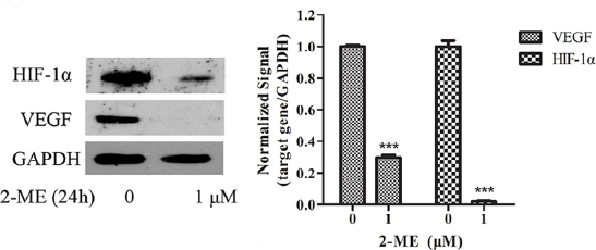
2-Methoxyestradiol purchased from MedChemExpress. Usage Cited in: Cancer Lett. 2016 Nov 1;382(1):44-52. [Abstract]
Downregulation of VEGF and HIF-1α by FP08S2 is dependent on Erk inactivation. VEGF and HIF-1α expression at protein level is detected after 2-ME treatment by Western blotting. 18S rRNA is used as internal control in qPCR while GAPDH is used as loading control in Western blotting.
-
Mol Ther
Exosomal miR-1246 from glioma patient body fluids drives the differentiation and activation of myeloid-derived suppressor cells. [Abstract]2021 Dec 1;29(12):3449-3464. PMID: 34217892 -
Acta Biomater
Redox-responsive dendritic copolymer-drug conjugates enhance therapeutic mitophagy through coordinated microtubule destabilization for synergistic triple-negative breast cancer therapy. [Abstract]2025 Aug:202:248-261. PMID: 40555336 -
J Transl Med
Fasting mimicking diet inhibits tumor-associated macrophage survival and pro-tumor function in hypoxia: implications for combination therapy with anti-angiogenic agent. [Abstract]2023 Oct 26;21(1):754. PMID: 37884960 -
Free Radic Biol Med
2021 Jun:169:271-282. PMID: 33895289 -
Stem Cell Res Ther
2022 Feb 5;13(1):59. PMID: 35123567 -
Front Immunol
Rapamycin Modulates the Proinflammatory Memory-Like Response of Microglia Induced by BAFF. [Abstract]2021 May 12:12:639049. PMID: 34054807 -
Ann Hepatol
Metformin ameliorates liver fibrosis induced by congestive hepatopathy via the mTOR/HIF-1α signaling pathway. [Abstract]2023 Nov-Dec;28(6):101135. PMID: 37451514 -
Eur J Pharmacol
Inhibition of JAK1/STAT3 pathway by 2-methoxyestradiol ameliorates psoriatic features in vitro and in an imiquimod-induced psoriasis-like mouse model. [Abstract]2022 Oct 15:933:175276. PMID: 36130639 -
Cancer Biol Ther
HIF-1α inhibition by 2-methoxyestradiol induces cell death via activation of the mitochondrial apoptotic pathway in acute myeloid leukemia. [Abstract]2016 Jun 2;17(6):625-34. PMID: 27082496
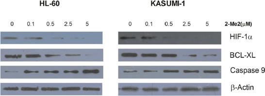
2-Methoxyestradiol purchased from MedChemExpress. Usage Cited in: Cancer Biol Ther. 2016 Jun 2;17(6):625-34. [Abstract]
HL-60 and Kasumi-1 cells are treated with different concentration of 2ME2 (0.1, 0.5, 2.5, or 5μM) for 24 hours or diluent control, and intracelluar protein levels are examined by Western blot.
-
Int J Mol Sci
A High-Calorie Diet Aggravates Lipopolysaccharide-Induced Pulmonary Inflammation in Juvenile Rats via Hypothalamic-Pituitary-Adrenal Axis-Related Pathways. [Abstract]2025 Jul 8;26(14):6554. PMID: 40724811 -
Front Cell Dev Biol
Hypoxia-Mediated Complement 1q Binding Protein Regulates Metastasis and Chemoresistance in Triple-Negative Breast Cancer and Modulates the PKC-NF-κB-VCAM-1 Signaling Pathway. [Abstract]2021 Feb 23:9:607142. PMID: 33708767 -
Chem Biol Interact
2023 Jan 5:369:110277. PMID: 36414027 -
Pestic Biochem Physiol
2-Methoxyestradiol ameliorates paraquat-induced pulmonary fibrosis by inhibiting the TGF-β1/Smad2/3 signaling pathway. [Abstract]2023 Dec:197:105647. PMID: 38072522 -
Rheumatology (Oxford)
Autophagy mediates 2-methoxyestradiol-inhibited scleroderma collagen synthesis and endothelial-to-mesenchymal transition induced by hypoxia. [Abstract]2019 Nov 1;58(11):1966-1975. PMID: 31049569 -
-
Front Cell Neurosci
IRAK-M Deficiency Exacerbates Ischemic Neurovascular Injuries in Experimental Stroke Mice. [Abstract]2018 Dec 21:12:504. PMID: 30622459 -
Am J Physiol Lung Cell Mol Physiol
Intrauterine inflammation-induced neonatal lung injury via succinic acid-mediated alveolar epithelial E-cadherin downregulation. [Abstract]2025 Aug 1;329(2):L282-L295. PMID: 40643013 -
Peptides
Cholecystokinin regulates atrial natriuretic peptide secretion through activation of NOX4-Sirt1-LEF1 signaling in beating rat hypoxic atria. [Abstract]2024 Nov:181:171299. PMID: 39326462 -
J Immunol Res
Combined Effects of 2-Methoxyestradiol (Hypoxia-Inducible Factor 1 α Inhibitor) and Dasatinib (A Second-Generation Tyrosine Kinase Inhibitor) on Chronic Myelocytic Leukemia Cells. [Abstract]2022 Apr 28;2022:6324326. PMID: 35528614 -
Biomed Res Int
2-Methoxyestradiol Alleviates Experimental Autoimmune Uveitis by Inhibiting Lymphocytes Proliferation and T Cell Differentiation. [Abstract]2016:2016:7948345. PMID: 27243036 -
Thorac Cancer
Mitochondrial NDUFA4L2 protein promotes the vitality of lung cancer cells by repressing oxidative stress. [Abstract]2019 Apr;10(4):676-685. PMID: 30710412 -
Exp Ther Med
Hypoxia-inducible factor 1α participates in hypoxia-induced epithelial-mesenchymal transition via response gene to complement 32. [Abstract]2017 Aug;14(2):1825-1831. PMID: 28810656
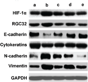
2-Methoxyestradiol purchased from MedChemExpress. Usage Cited in: Exp Ther Med. 2017 Aug;14(2):1825-1831. [Abstract]
HIF 1α regulates the protein levels of RGC 32 and epithelial mesenchymal transition associated genes induced by hypoxia. Proteins are extracted from cells after different treatments. Protein levels of HIF 1α, RGC 32, E cadherin, cytokeratins, N cadherin and vimentin are detected by western blotting. a, no treatment cells under normoxia; b, cells under hypoxia; c, cells are pretreated with HIF 1α inhibitor for 30 min, and then incubated under hypoxia; d, cells are transfected with negative contro
-
-
-
-
-


Solvent & Solubility
In Vitro:
DMSO : 250 mg/mL (826.69 mM; Need ultrasonic; Hygroscopic DMSO has a significant impact on the solubility of product, please use newly opened DMSO)
H2O : < 0.1 mg/mL (insoluble)
Please refer to the solubility information to select the appropriate solvent. Once prepared, please aliquot and store the solution to prevent product inactivation from repeated freeze-thaw cycles.
Storage method and period of stock solution: -80°C, 2 years; -20°C, 1 year. When stored at -80°C, please use it within 2 years. When stored at -20°C, please use it within 1 year.
Please refer to the solubility information to select the appropriate solvent. Once prepared, please aliquot and store the solution to prevent product inactivation from repeated freeze-thaw cycles.
Storage method and period of stock solution: -80°C, 2 years; -20°C, 1 year. When stored at -80°C, please use it within 2 years. When stored at -20°C, please use it within 1 year.
Concentration (start) × Volume (start) = Concentration (final) × Volume (final)
In Vivo:
Select the appropriate dissolution method based on your experimental animal and administration route.
- For the following dissolution methods, please ensure to first prepare a clear stock solution using an In Vitro approach and then sequentially add co-solvents:
- To ensure reliable experimental results, the clarified stock solution can be appropriately stored based on storage conditions. As for the working solution for In Vivo experiments, it is recommended to prepare freshly and use it on the same day.
- The percentages shown for the solvents indicate their volumetric ratio in the final prepared solution. If precipitation or phase separation occurs during preparation, heat and/or sonication can be used to aid dissolution.
Add each solvent one by one: 10% DMSO 40% PEG300 5% Tween-80 45% Saline
Solubility: ≥ 2.08 mg/mL (6.88 mM); Clear solution
This protocol yields a clear solution of ≥ 2.08 mg/mL (saturation unknown).
Taking 1 mL working solution as an example, add 100 μL DMSO stock solution (20.8 mg/mL) to 400 μL PEG300, and mix evenly; then add 50 μL Tween-80 and mix evenly; then add 450 μL Saline to adjust the volume to 1 mL.
Preparation of Saline: Dissolve 0.9 g sodium chloride in ddH₂O and dilute to 100 mL to obtain a clear Saline solution.
Add each solvent one by one: 10% DMSO 90% (20% SBE-β-CD in Saline)
Solubility: ≥ 2.08 mg/mL (6.88 mM); Clear solution
This protocol yields a clear solution of ≥ 2.08 mg/mL (saturation unknown).
Taking 1 mL working solution as an example, add 100 μL DMSO stock solution (20.8 mg/mL) to 900 μL 20% SBE-β-CD in Saline, and mix evenly.
Preparation of 20% SBE-β-CD in Saline (4°C, storage for one week): 2 g SBE-β-CD powder is dissolved in 10 mL Saline, completely dissolve until clear.
In Vivo Dissolution Calculator
Please enter the basic information of animal experiments:
-
-
-
-
Recommended: Prepare an additional quantity of animals to account for potential losses during experiments.
Please enter your animal formula composition:
-
%DMSO +
Recommended: Keep the proportion of DMSO in working solution below 2% if your animal is weak.
-
%+
-
+%Tween-80 + +
-
%Saline +
The co-solvents required include: DMSO, . All of co-solvents are available by MedChemExpress (MCE). , Tween 80. All of co-solvents are available by MedChemExpress (MCE).
Working solution concentration: 0.22 mg/mL
Method for preparing stock solution: mg drug dissolved in μL DMSO. Stock solution concentration: mg/mL.
1. Take μL DMSO stock solution;
2. Add μL .
μL , mix evenly;
3. Then add μL Tween 80, mix evenly;
4. Then add μL
Please ensure that the stock solution in the first step is dissolved to a clear state, and add co-solvents in sequence. You can use ultrasonic heating (ultrasonic cleaner, recommended frequency 20-40 kHz), vortexing, etc. to assist dissolution.

Protocols
-
Detection of Apoptosis
Apoptosis, also called programmed cell death, is generally characterized by distinct morphological characteristics.
-
TUNEL staining for apoptotic DNA fragmentation
TUNEL staining detects DNA strand breaks by using terminal deoxynucleotidyl transferase to add labeled nucleotides to exposed 3′-OH DNA termini, generating either microscopic staining in fixed cells or tissue sections, or fluorescence/cytometric signal in cell suspensions. TUNEL positivity reflects DNA fragmentation but should not be interpreted alone as definitive apoptosis, because TUNEL can also label necrotic, autolytic, mechanically damaged, or DNA-repair-associated DNA breaks.
-
Flow cytometric DNA-content cell-cycle staining
Flow cytometric DNA-content cell-cycle staining measures the fluorescence intensity of DNA-bound fluorochromes in single cells or nuclei to estimate DNA content distributions, allowing assignment of populations to G0/G1, S, and G2/M phases by DNA histogram deconvolution. Propidium iodide (PI) intercalates into DNA, and PI fluorescence is proportional to cellular DNA content when staining is performed under conditions that make DNA accessible and minimize non-DNA signal. Cells with G2/M DNA content are expected to show approximately twice the fluorescence intensity of G0/G1 cells, while S-phase cells occupy intermediate fluorescence values. PI-based DNA-content analysis can also detect cells with fractional DNA content, often reported as sub-G1, when DNA fragmentation and extraction during staining reduce retained DNA signal in apoptotic cells. DAPI is an alternative DNA fluorochrome for univariate DNA-content analysis, while bivariate approaches combining DNA content with proliferation
-
Annexin V plus membrane-impermeant dye apoptosis staining
Annexin V-based apoptosis assays rely on the detection of phosphatidylserine (PS) externalization from the inner leaflet of the plasma membrane to the outer leaflet, an early biochemical hallmark of apoptosis. Fluorescently labeled Annexin V binds PS in a calcium-dependent manner, enabling identification of early apoptotic cells by flow cytometry or fluorescence microscopy. When combined with a membrane-impermeant DNA-binding dye (e. g. , propidium iodide), this approach allows discrimination between viable (Annexin V−/dye−), early apoptotic (Annexin V+/dye−), and late apoptotic or necrotic (Annexin V+/dye+) cell populations by assessing membrane integrity and PS exposure.
-
ROS/oxidative-stress fluorescent staining
ROS/oxidative-stress fluorescent staining uses cell-permeant fluorogenic probes that become fluorescent after oxidation inside cells or tissues; commonly used examples include DCFH-DA/DCFDA for broad cellular oxidant detection, DHE for superoxide-related signal detection, MitoSOX for mitochondrial superoxide-related signal detection, and CellROX probes for oxidative-stress-associated fluorescence readouts. The assay detects probe oxidation rather than a single ROS species unless the probe and analysis method have been chemically validated for that species. DCFH-DA enters cells, is deacetylated by intracellular esterases to DCFH, and produces fluorescent DCF after oxidation, so the readout is used as an operational measure of total cellular oxidative stress rather than a species-specific ROS measurement. DHE and MitoSOX can report superoxide-related oxidation, but red fluorescence alone can include non-specific ethidium-like oxidation products; HPLC or optimized spectral approaches are
-
BrdU Incorporation Assay
Bromodeoxyuridine (BrdU) incorporation assay is based on the principle that BrdU, a thymidine analog, is incorporated into newly synthesized DNA during the S phase of the cell cycle, thereby serving as a marker of DNA replication and cellular proliferation. Incorporated BrdU can be detected using anti-BrdU antibodies following DNA denaturation, enabling visualization or quantification of proliferating cells through immunochemical detection methods such as immunofluorescence or immunohistochemistry.
-
Apoptosis Solutions
Apoptosis is a regulated, generally non-lytic cell-death pathway that removes unwanted, damaged, infected, or abnormal cells through coordinated morphological changes, caspase activation, DNA fragmentation, and membrane remodeling. The intrinsic apoptosis pathway is controlled mainly by mitochondrial outer membrane permeabilization, BCL-2 family proteins, cytochrome c release, apoptosome formation, caspase-9 activation, and downstream executioner caspase-3/7 activation. The extrinsic apoptosis pathway is initiated by death receptors such as Fas, TNFR, and TRAIL receptors, which recruit adaptor proteins and activate caspase-8 before engaging executioner caspases or mitochondrial amplification through BID cleavage. Apoptosis is linked to many phenotypes, including cancer cell killing, tissue homeostasis, immune regulation, neurodegeneration, infection response, and treatment-induced cytotoxicity; unresolved questions include how apoptosis interacts with necroptosis, pyroptosis, ferroptos
-
Protocol for Cell Cycle
Cell-cycle analysis by flow cytometry measures DNA content in single cells to estimate the fraction of cells in G0/G1, S, and G2/M phases. Propidium iodide intercalates into DNA, and after RNA removal with RNase, fluorescence intensity reflects cellular DNA content: 2N cells are assigned to G0/G1, cells between 2N and 4N to S phase, and 4N cells to G2/M. DNA-content analysis alone cannot reliably separate G0 from G1 or G2 from M. Ki-67 can distinguish quiescent G0 cells from cycling cells, EdU or BrdU incorporation marks active DNA synthesis in S phase, and phospho-histone H3 staining identifies mitotic cells within the 4N population.
-
Breast Cancer Modeling
Breast cancer is a heterogeneous cancer, and it has been distinguished into four subtypes: luminal A, luminal B, HER2-positive and basal-like. Molecular mutations, epigenetic alterations, hormone exposure and immune microenvironment are related to the progression of breast cancer.


Purity & Documentation
-
Data Sheet (299 KB)
-
SDS (644 KB)
- English - EN (644 KB)
- Français - FR (644 KB)
- Deutsch - DE (644 KB)
- Norwegian - NO (644 KB)
- Español - ES (644 KB)
- Swedish - SV (644 KB)
- Italian - IT (644 KB)
- Korean - KR (644 KB)
- Portuguese - PT (644 KB)
-
Handling Instructions (2659 KB)
References
[2]. Mooberry SL, et al. Mechanism of action of 2-methoxyestradiol: new developments. Drug resistance updates : reviews and commentaries in antimicrobial and anticancer chemotherapy. 2003 Dec;6(6):355-61. [Content Brief]
[3]. LaVallee TM, et al. 2-Methoxyestradiol inhibits proliferation and induces apoptosis independently of estrogen receptors alpha and beta. Cancer research. 2002 Jul 01;62(13):3691-7. [Content Brief]


Complete Stock Solution Preparation Table
Please refer to the solubility information to select the appropriate solvent. Once prepared, please aliquot and store the solution to prevent product inactivation from repeated freeze-thaw cycles.
Storage method and period of stock solution: -80°C, 2 years; -20°C, 1 year. When stored at -80°C, please use it within 2 years. When stored at -20°C, please use it within 1 year.
| Optional Solvent | Concentration Solvent Mass | 1 mg | 5 mg | 10 mg | 25 mg |
|---|---|---|---|---|---|
| DMSO | 1 mM | 3.3068 mL | 16.5338 mL | 33.0677 mL | 82.6692 mL |
| 5 mM | 0.6614 mL | 3.3068 mL | 6.6135 mL | 16.5338 mL | |
| 10 mM | 0.3307 mL | 1.6534 mL | 3.3068 mL | 8.2669 mL | |
| 15 mM | 0.2205 mL | 1.1023 mL | 2.2045 mL | 5.5113 mL | |
| 20 mM | 0.1653 mL | 0.8267 mL | 1.6534 mL | 4.1335 mL | |
| 25 mM | 0.1323 mL | 0.6614 mL | 1.3227 mL | 3.3068 mL | |
| 30 mM | 0.1102 mL | 0.5511 mL | 1.1023 mL | 2.7556 mL | |
| 40 mM | 0.0827 mL | 0.4133 mL | 0.8267 mL | 2.0667 mL | |
| 50 mM | 0.0661 mL | 0.3307 mL | 0.6614 mL | 1.6534 mL | |
| 60 mM | 0.0551 mL | 0.2756 mL | 0.5511 mL | 1.3778 mL | |
| 80 mM | 0.0413 mL | 0.2067 mL | 0.4133 mL | 1.0334 mL | |
| 100 mM | 0.0331 mL | 0.1653 mL | 0.3307 mL | 0.8267 mL |


2-Methoxyestradiol Related Classifications
HY-12033 Related Classifications
 Powered by Bioz
Keywords
- 2-Methoxyestradiol
- 362-07-2
- 2-ME2
- NSC-659853
- NSC659853
- NSC 659853
- NSC-659853
- Endogenous Metabolite
- Microtubule/Tubulin
- HIF/HIF Prolyl-Hydroxylase
- Cytochrome P450
- JNK
- Caspase
- Estrogen Receptor/ERR
- Reactive Oxygen Species (ROS)
- Apoptosis
- Autophagy
- CYP1A1
- CYP1B1
- G2-M arrest
- HIF-1α
- antiangiogenesis
- apoptosis
- aromatase inhibitor
- estrogen receptors
- superoxide dismutase
- tubulin inhibitor
- Inhibitor
- inhibitor
- inhibit