



Amitriptyline
Based on 9 publication(s) in Google Scholar
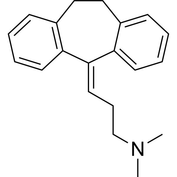
Amitriptyline is an orally active tricyclic antidepressant (TCA). Amitriptyline mainly exerts its antidepressant effect by blocking SERT (Ki = 3.45 nM) and NET (Ki = 13.3 nM), thereby increasing the concentrations of 5-hydroxytryptamine (5-HT) and norepinephrine (NE) in the synaptic cleft. Amitriptyline is also an agonist at α2A and TrkA/TrkB receptors, thereby exerting analgesic and neurotrophic activities (inhibiting cell apoptosis). Amitriptyline can reduce inflammation, angiogenesis and fibrosis. Amitriptyline binds to DAT (with Ki = 2.58 μM). Amitriptyline has high affinity for a series of receptors and can antagonize muscarinic cholinergic receptors (M1/M2/M3/M4/M5 receptors) (Ki = 11-24 nM), H1 receptors (Ki = 0.5-1.1 nM), adrenergic α1 receptors (Ki = 4.4 nM), etc., resulting in a series of side effects. Amitriptyline can block sodium channels and hERG potassium channel (IC50 = 4.78 μM) and it has cardiotoxicity.
For research use only. We do not sell to patients.
- Purity : 99.93%
- CAS No.: 50-48-6
- Formula: C20H23N
- Molecular Weight:277.40
-
Storage:
4°C, protect from light
* In solvent : -80°C, 6 months; -20°C, 1 month (protect from light)

To place orders, for customer services and technical support, please contact: MedChemExpress USA
Tel: 609-228-6898 E-mail: [email protected] [email protected]


-
 Biological Activity
-
Chemical Information
-
Solvent & Solubility
- Protocols
- Purity & Documentation
- References
-
 Help & FAQs


-
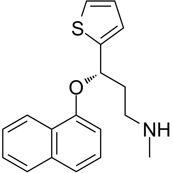
HY-B0161
DuloxetineSerotonin-norepinephrine Reuptake Inhibitor -
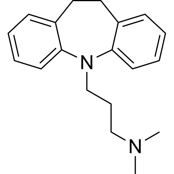
HY-B1490A
ImipramineAntidepressant -
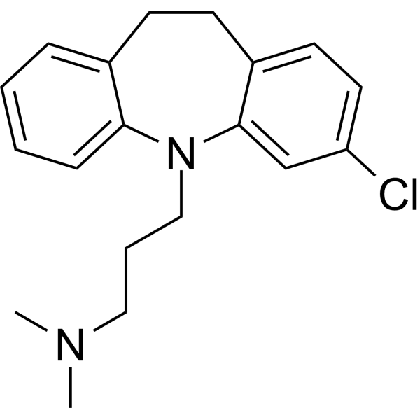
HY-B0457A
Clomipramine5-HT Reuptake Inhibitor -
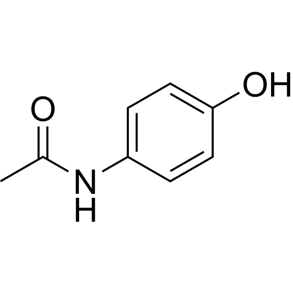
HY-66005
AcetaminophenCOX-2 Inhibitor -
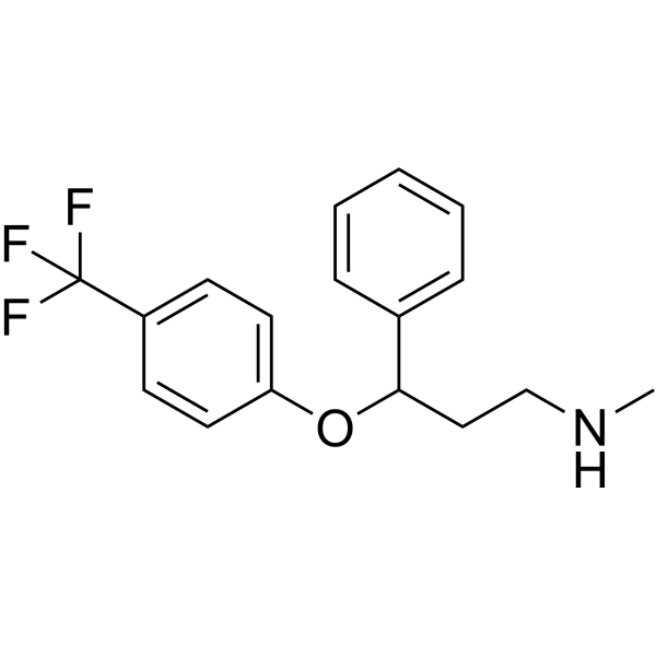
HY-B0102
Fluoxetine -
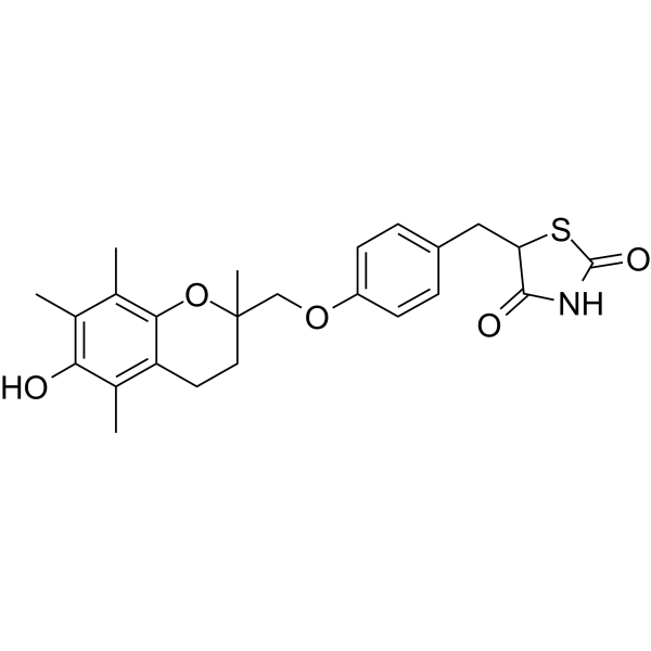
HY-50935
TroglitazonePPARγ Agonist -
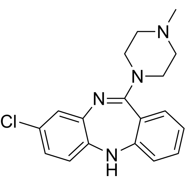
HY-14539
ClozapineAntipsychotic Agent -
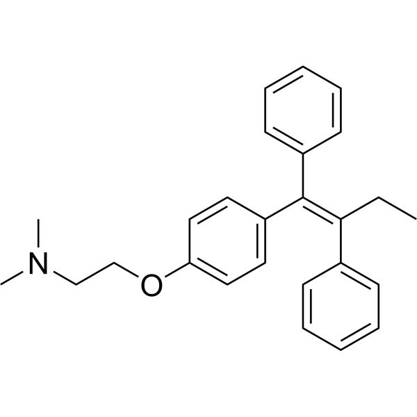
HY-13757A
TamoxifenEstrogen Receptor Modulator -
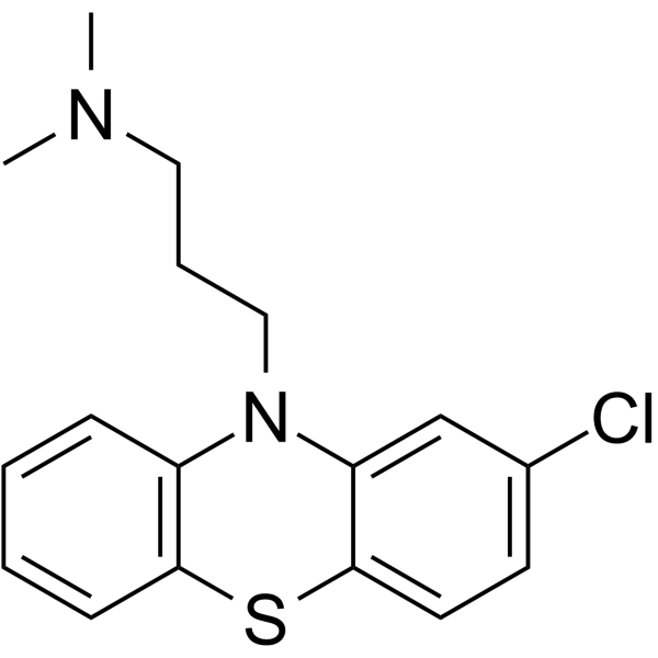
HY-12708
ChlorpromazineAntipsychotic Agent -
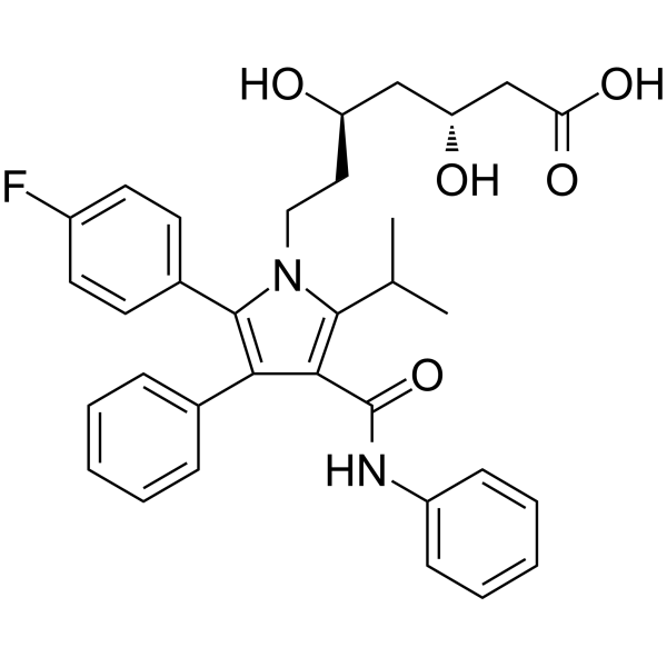
HY-B0589
AtorvastatinHMG-CoA Reductase Inhibitor -
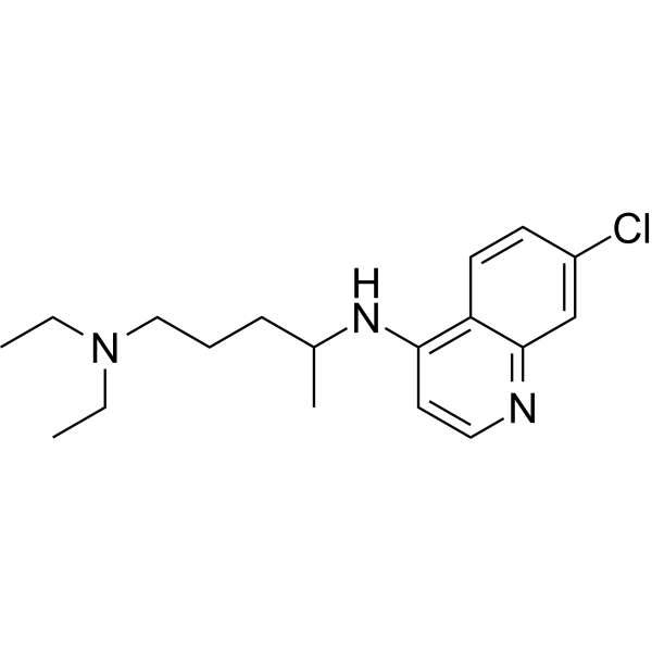
HY-17589A
ChloroquineAntimalarial Agent -
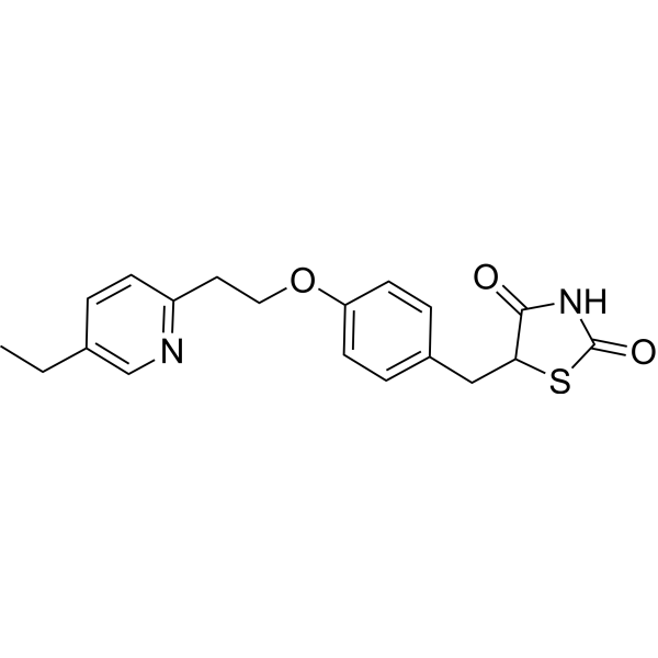
HY-13956
PioglitazonePPARγ Agonist
Publications Citing Use of MedChemExpress (MCE) Amitriptyline
More- NPJ Digit Med. 2025 Nov 17;8(1):663. [Abstract]
- J Am Chem Soc. 2025 Mar 26. [Abstract]
- Cell Death Dis. 2026 Jul 10.
- Cell Commun Signal. 2023 May 25;21(1):123. [Abstract]
- Neurosci Bull. 2025 Mar 17. [Abstract]
- Int Immunopharmacol. 2025 Feb 6:147:113969. [Abstract]
- Skelet Muscle. 2024 Jul 18;14(1):16. [Abstract]
- J Med Virol. 2023 Jan;95(1):e28266. [Abstract]
- PLoS Negl Trop Dis. 2019 Aug 20;13(8):e0007681. [Abstract]
All 5-HT Receptor Isoforms
MoreAll Adrenergic Receptor Isoforms
MoreAll Histamine Receptor Isoforms
More

Biological Activity
Description
|
TrkA |
TrkB |
5-HT Receptor |
Histamine Receptor |
Cellular Effect
|
Cell Line

|
Type | Value | Description | References |
|---|---|---|---|---|
| CHO | IC50 |
3 μM
Compound: Amitryptline
|
Inhibition of human ERG expressed in CHO cells at -80 mV holding potential by whole-cell patch clamp assay
Inhibition of human ERG expressed in CHO cells at -80 mV holding potential by whole-cell patch clamp assay
|
[PMID: 30653317] |
| CHO-K1 | IC50 |
1.6 μM
Compound: amitriptyline
|
Inhibition of human NaV1.5 alpha subunit expressed in CHOK1 cells at -90 mV holding potential by patch clamp electrophysiological assay
Inhibition of human NaV1.5 alpha subunit expressed in CHOK1 cells at -90 mV holding potential by patch clamp electrophysiological assay
|
[PMID: 22770500] |
| CHO-K1 | IC50 |
3.1 μM
Compound: amitriptyline
|
Inhibition of human NaV1.2 alpha subunit expressed in CHOK1 cells at -65 mV holding potential by patch clamp electrophysiological assay
Inhibition of human NaV1.2 alpha subunit expressed in CHOK1 cells at -65 mV holding potential by patch clamp electrophysiological assay
|
[PMID: 22770500] |
| HEK293 | IC50 |
16.9 μM
Compound: amitriptyline
|
Inhibition of 4-(4-(dimethylamino)styryl)-N-methylpyridinium uptake at human OCT1 expressed in HEK293 cells by confocal microscopy
Inhibition of 4-(4-(dimethylamino)styryl)-N-methylpyridinium uptake at human OCT1 expressed in HEK293 cells by confocal microscopy
|
[PMID: 18788725] |
| HEK293 | IC50 |
1 μM
Compound: amitriptyline
|
Inhibition of recombinant human NaV1.7 alpha subunit expressed in HEK293 cells at -65 mV holding potential by patch clamp electrophysiological assay
Inhibition of recombinant human NaV1.7 alpha subunit expressed in HEK293 cells at -65 mV holding potential by patch clamp electrophysiological assay
|
[PMID: 22770500] |
| HEK293 | IC50 |
12 μM
Compound: amitriptyline
|
Inhibition of human NaV1.7 F1737A mutant expressed in HEK293 cells at -65 mV holding potential by patch clamp electrophysiological assay
Inhibition of human NaV1.7 F1737A mutant expressed in HEK293 cells at -65 mV holding potential by patch clamp electrophysiological assay
|
[PMID: 22770500] |
| HEK293 | IC50 |
7 μM
Compound: amitriptyline
|
Inhibition of recombinant human NaV1.7 alpha subunit expressed in HEK293 cells at -90 mV holding potential by patch clamp electrophysiological assay
Inhibition of recombinant human NaV1.7 alpha subunit expressed in HEK293 cells at -90 mV holding potential by patch clamp electrophysiological assay
|
[PMID: 22770500] |
| HEK293 | IC50 |
9.6 μM
Compound: Amitriptyline
|
Inhibition of [3H]-dofetilide binding to human ERG expressed in HEK293 cell membranes measured after 1 hr by liquid scintillation counting method
Inhibition of [3H]-dofetilide binding to human ERG expressed in HEK293 cell membranes measured after 1 hr by liquid scintillation counting method
|
[PMID: 30597328] |
| HepG2 | IC50 |
29.6 μM
Compound: Amitriptyline
|
Cytotoxicity against human HepG2 cells assessed as reduction in cell viability by measuring ATP content incubated for 24 hrs by luminescent analysis
Cytotoxicity against human HepG2 cells assessed as reduction in cell viability by measuring ATP content incubated for 24 hrs by luminescent analysis
|
[PMID: 32442850] |
| NCI-H1650 | GI50 |
>40 μM
Compound: Amitriptyline
|
Cytotoxicity against human NCI-H1650 cells assessed as growth inhibition after 48 hrs by MTT assay
Cytotoxicity against human NCI-H1650 cells assessed as growth inhibition after 48 hrs by MTT assay
|
[PMID: 26372073] |
| SH-SY5Y | IC50 |
48.1 μM
Compound: Amitriptyline
|
Cytotoxicity against human SH-SY5Y cells assessed as reduction in cell viability by measuring ATP content incubated for 24 hrs by luminescent analysis
Cytotoxicity against human SH-SY5Y cells assessed as reduction in cell viability by measuring ATP content incubated for 24 hrs by luminescent analysis
|
[PMID: 32442850] |
In Vitro
Amitriptyline (0.5-10 μM, 3.5-16.5 h) effectively resists cell apotosis (EC50 = 50 nM) in T17 cells and shows no protective effect on the SN56 cells, and in primary rat hippocampal neurons stimulated by glutamate and subjected to oxygen-glucose deprivation (OGD), the neuronal apoptosis is significantly reduced[3].
Amitriptyline (0.5 μM, 30 min) induces TrkA and TrkB receptor phosphorylation and activation in hippocampal neurons[3].
Amitriptyline (0.5 μM, 5 days) induces neurite outgrowth in PC12 cells[3].
MedChemExpress (MCE) has not independently confirmed the accuracy of these methods. They are for reference only.
-
Cell Line:Primary rat hippocampal neurons
-
Concentration:0.5 μM
-
Incubation Time:30 min
-
Result:Strongly induced phosphorylation of TrkA, TrkB, Akt, and ERK.
Induced TrkA-TrkA homodimerization, TrkB-TrkB homodimerization, and TrkA-TrkB heterodimerization.
In Vivo
Amitriptyline (15 mg/kg, i.p., single dose) produces antinociception in a mouse model by activating α2A-adrenoceptor receptors in the central nervous system[4].
Amitriptyline (5 mg/kg, p.o., once daily for 7 days) is able to down-regulate angiogenesis and foreign body reaction (FBR) in 14-day-old implants[5].
MedChemExpress (MCE) has not independently confirmed the accuracy of these methods. They are for reference only.
-
Animal Model:Kainic acid induced excitatory epilepsy model established in male C57BL/6 mice[3]
-
Dosage:15 mg/kg
-
Administration:Intraperitoneal injection (i.p.), once daily for 5 days
-
Result:Effectively activated TrkA, TrkB and their downstream signaling pathways in the brain. Significantly reduced KA-induced hippocampal (70%).
-
Animal Model:Hot-plate test and abdominal constriction test established in male Swiss albino mice (23-30 g)[4]
-
Dosage:15 mg/kg
-
Administration:Intraperitoneal injection (i.p.), single dose
-
Result:Significantly increased pain threshold in both models (increased hot plate latency and reduced writhing times).
Required the presence of endogenous monoamine neurotransmitters (such as NE).
Exhibited the analgesic effect be completely blocked by BRL 44408 (HY-12716).
-
Animal Model:Polyether-polyurethane sponge disks induced FBR model established in male C57BL/6 mice[5]
-
Dosage:5 mg/kg
-
Administration:Oral administration (p.o.), once daily for 7 days
-
Result:Had no effect on 7-day implants.
Significantly reduced all key parameters of the 14-day implants, reducing angiogenesis, fibrosis markers, and FBR markers.
- Fibrosis/Collagen Morphometry
- Collagen: Sirius Red Staining
- Detection of Apoptosis
- TUNEL staining for apoptotic DNA fragmentation
- Human pluripotent stem cell endothelial-cell differentiation
- Vascular/Branching Fractal Analysis
- Annexin V plus membrane-impermeant dye apoptosis staining
- Connective Tissue: Masson's Trichrome/Collagen Trichrome Staining
- Endothelial Tube Formation Assay
- Apoptosis Solutions
- Research Protocol for Inflammation-related Diseases
- Endothelial Cell Migration/Angiogenic Sprouting Assay


Chemical Information
-
CAS No. 50-48-6
-
Appearance Liquid (Density: 1.076±0.06 g/cm3)
-
Molecular Weight 277.40
-
Formula C20H23N
-
Color Colorless to light yellow
-
SMILES
CN(CC/C=C1C2=CC=CC=C2CCC3=C\1C=CC=C3)C
-
Shipping
Room temperature in continental US; may vary elsewhere.
-
Storage
4°C, protect from light
* In solvent : -80°C, 6 months; -20°C, 1 month (protect from light)


Publications (9)
-
Journal Impact Factor
-
Most Recent
-
NPJ Digit Med
Tandospirone augments cisplatin treatment by lowering cholesterol and managing distress in NSCLC patients. [Abstract]2025 Nov 17;8(1):663. PMID: 41249389 -
J Am Chem Soc
Fluorinated Ribonucleocarbohydrate Nanoparticles Allow Ultraefficient mRNA Delivery and Protein Expression in Tumor-Associated Myeloid Cells. [Abstract]2025 Mar 26. PMID: 40135499 -
-
Cell Commun Signal
Tricyclic antidepressants induce liver inflammation by targeting NLRP3 inflammasome activation. [Abstract]2023 May 25;21(1):123. PMID: 37231437 -
Neurosci Bull
Electrophysiological Abnormalities and Pharmacological Corrections of Pathogenic Missense Variants in KCNQ3. [Abstract]2025 Mar 17. PMID: 40095209 -
Int Immunopharmacol
Innovative role of the antidepressant imipramine in esophageal squamous cell carcinoma treatment: Promoting apoptosis and protective autophagy. [Abstract]2025 Feb 6:147:113969. PMID: 39764996 -
Skelet Muscle
ASM is a therapeutic target in dermatomyositis by regulating the differentiation of naive CD4 + T cells into Th17 and Treg subsets. [Abstract]2024 Jul 18;14(1):16. PMID: 39026344 -
J Med Virol
2023 Jan;95(1):e28266. PMID: 36319186 -
PLoS Negl Trop Dis
Identification of anti-flaviviral drugs with mosquitocidal and anti-Zika virus activity in Aedes aegypti. [Abstract]2019 Aug 20;13(8):e0007681. PMID: 31430351


Solvent & Solubility
In Vitro:
DMSO : 100 mg/mL (360.49 mM; Need ultrasonic; Hygroscopic DMSO has a significant impact on the solubility of product, please use newly opened DMSO)
Please refer to the solubility information to select the appropriate solvent. Once prepared, please aliquot and store the solution to prevent product inactivation from repeated freeze-thaw cycles.
Storage method and period of stock solution: -80°C, 6 months; -20°C, 1 month (protect from light). When stored at -80°C, please use it within 6 months. When stored at -20°C, please use it within 1 month.
Please refer to the solubility information to select the appropriate solvent. Once prepared, please aliquot and store the solution to prevent product inactivation from repeated freeze-thaw cycles.
Storage method and period of stock solution: -80°C, 6 months; -20°C, 1 month (protect from light). When stored at -80°C, please use it within 6 months. When stored at -20°C, please use it within 1 month.
Concentration (start) × Volume (start) = Concentration (final) × Volume (final)
In Vivo:
Select the appropriate dissolution method based on your experimental animal and administration route.
- For the following dissolution methods, please ensure to first prepare a clear stock solution using an In Vitro approach and then sequentially add co-solvents:
- To ensure reliable experimental results, the clarified stock solution can be appropriately stored based on storage conditions. As for the working solution for In Vivo experiments, it is recommended to prepare freshly and use it on the same day.
- The percentages shown for the solvents indicate their volumetric ratio in the final prepared solution. If precipitation or phase separation occurs during preparation, heat and/or sonication can be used to aid dissolution.
Add each solvent one by one: 10% DMSO 40% PEG300 5% Tween-80 45% Saline
Solubility: ≥ 2.5 mg/mL (9.01 mM); Clear solution
This protocol yields a clear solution of ≥ 2.5 mg/mL (saturation unknown).
Taking 1 mL working solution as an example, add 100 μL DMSO stock solution (25.0 mg/mL) to 400 μL PEG300, and mix evenly; then add 50 μL Tween-80 and mix evenly; then add 450 μL Saline to adjust the volume to 1 mL.
Preparation of Saline: Dissolve 0.9 g sodium chloride in ddH₂O and dilute to 100 mL to obtain a clear Saline solution.
Add each solvent one by one: 10% DMSO 90% (20% SBE-β-CD in Saline)
Solubility: ≥ 2.5 mg/mL (9.01 mM); Clear solution
This protocol yields a clear solution of ≥ 2.5 mg/mL (saturation unknown).
Taking 1 mL working solution as an example, add 100 μL DMSO stock solution (25.0 mg/mL) to 900 μL 20% SBE-β-CD in Saline, and mix evenly.
Preparation of 20% SBE-β-CD in Saline (4°C, storage for one week): 2 g SBE-β-CD powder is dissolved in 10 mL Saline, completely dissolve until clear.
In Vivo Dissolution Calculator
Please enter the basic information of animal experiments:
-
-
-
-
Recommended: Prepare an additional quantity of animals to account for potential losses during experiments.
Please enter your animal formula composition:
-
%DMSO +
Recommended: Keep the proportion of DMSO in working solution below 2% if your animal is weak.
-
%+
-
+%Tween-80 + +
-
%Saline +
The co-solvents required include: DMSO, . All of co-solvents are available by MedChemExpress (MCE). , Tween 80. All of co-solvents are available by MedChemExpress (MCE).
Working solution concentration: 0.22 mg/mL
Method for preparing stock solution: mg drug dissolved in μL DMSO. Stock solution concentration: mg/mL. * In solvent : -80°C, 6 months; -20°C, 1 month (protect from light)
1. Take μL DMSO stock solution;
2. Add μL .
μL , mix evenly;
3. Then add μL Tween 80, mix evenly;
4. Then add μL
Please ensure that the stock solution in the first step is dissolved to a clear state, and add co-solvents in sequence. You can use ultrasonic heating (ultrasonic cleaner, recommended frequency 20-40 kHz), vortexing, etc. to assist dissolution.


Purity & Documentation
-
Data Sheet (286 KB)
-
SDS (952 KB)
- English - EN (952 KB)
- Français - FR (952 KB)
- Deutsch - DE (952 KB)
- Norwegian - NO (952 KB)
- Español - ES (952 KB)
- Swedish - SV (952 KB)
- Italian - IT (952 KB)
- Korean - KR (952 KB)
- Portuguese - PT (952 KB)
-
Handling Instructions (2659 KB)
References
[1]. Kim Lawson. A Brief Review of the Pharmacology of Amitriptyline and Clinical Outcomes in Treating Fibromyalgia. Biomedicines. 2017 Jun; 5(2): 24. [Content Brief]
[2]. S Neil Vaishnavi , et al. Milnacipran: a comparative analysis of human monoamine uptake and transporter binding affinity. Biol Psychiatry. 2004 Feb 1;55(3):320-2. [Content Brief]
[3]. Jang, S.W., et al., Amitriptyline is a TrkA and TrkB receptor agonist that promotes TrkA/TrkB heterodimerization and has potent neurotrophic activity. Chem Biol, 2009. 16(6): p. 644-56. [Content Brief]
[4]. Ghelardini C, et al. Antinociception induced by amitriptyline and imipramine is mediated by alpha2A-adrenoceptors. Jpn J Pharmacol. 2000 Feb;82(2):130-7. [Content Brief]
[5]. Scheuermann K, et al. Amitriptyline efficacy in decreasing implant-induced foreign body reaction. IUBMB Life. 2023 Sep;75(9):732-742. [Content Brief]


Complete Stock Solution Preparation Table
Please refer to the solubility information to select the appropriate solvent. Once prepared, please aliquot and store the solution to prevent product inactivation from repeated freeze-thaw cycles.
Storage method and period of stock solution: -80°C, 6 months; -20°C, 1 month (protect from light). When stored at -80°C, please use it within 6 months. When stored at -20°C, please use it within 1 month.
| Optional Solvent | Concentration Solvent Mass | 1 mg | 5 mg | 10 mg | 25 mg |
|---|---|---|---|---|---|
| DMSO | 1 mM | 3.6049 mL | 18.0245 mL | 36.0490 mL | 90.1226 mL |
| 5 mM | 0.7210 mL | 3.6049 mL | 7.2098 mL | 18.0245 mL | |
| 10 mM | 0.3605 mL | 1.8025 mL | 3.6049 mL | 9.0123 mL | |
| 15 mM | 0.2403 mL | 1.2016 mL | 2.4033 mL | 6.0082 mL | |
| 20 mM | 0.1802 mL | 0.9012 mL | 1.8025 mL | 4.5061 mL | |
| 25 mM | 0.1442 mL | 0.7210 mL | 1.4420 mL | 3.6049 mL | |
| 30 mM | 0.1202 mL | 0.6008 mL | 1.2016 mL | 3.0041 mL | |
| 40 mM | 0.0901 mL | 0.4506 mL | 0.9012 mL | 2.2531 mL | |
| 50 mM | 0.0721 mL | 0.3605 mL | 0.7210 mL | 1.8025 mL | |
| 60 mM | 0.0601 mL | 0.3004 mL | 0.6008 mL | 1.5020 mL | |
| 80 mM | 0.0451 mL | 0.2253 mL | 0.4506 mL | 1.1265 mL | |
| 100 mM | 0.0360 mL | 0.1802 mL | 0.3605 mL | 0.9012 mL |


Amitriptyline Related Classifications
HY-B0527 Related Classifications
 Powered by Bioz