



XZ739
Based on 1 publication(s) in Google Scholar
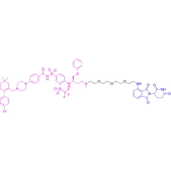
XZ739 is a potent and selective BCL-XL PROTAC degrader with a DC50 of 2.5 nM. XZ739 recruits CRBN to ubiquitinate and degrade BCL-XL via the ubiquitin-proteasome system. XZ739 also induces cell cycle arrest and caspase-mediated apoptosis. XZ739 can be used in research related to T-cell acute lymphoblastic leukemia and cholangiocarcinoma.
(Pink: Bcl-xL ligand (HY-131232); Blue: Cereblon ligand (HY-10984); Black: linker).
For research use only. We do not sell to patients.
- Purity : 99.08%
- CAS No.: 2365172-19-4
- Formula: C65H76ClF3N8O12S3
- Molecular Weight:1349.99
-
Storage:
-20°C, stored under nitrogen
* In solvent : -80°C, 6 months; -20°C, 1 month (stored under nitrogen)

To place orders, for customer services and technical support, please contact: MedChemExpress USA
Tel: 609-228-6898 E-mail: [email protected] [email protected]


-
 Biological Activity
-
Chemical Information
-
Solvent & Solubility
- Protocols
- Purity & Documentation
- References
-
 Help & FAQs


Publications Citing Use of MedChemExpress (MCE) XZ739
MoreAll PROTACs Isoforms
MoreAll Caspase Isoforms
More

Biological Activity
Description
IC50 & Target
[1]|
Bcl-xL 2.5 nM (DC50) |
Caspase-3 |
Cereblon |
Cellular Effect
|
Cell Line

|
Type | Value | Description | References |
|---|---|---|---|---|
| MOLT-4 | DC50 |
2.5 nM
|
BCL-XL degradation in human MOLT-4 T-cell acute lymphoblastic leukemia cells after 16 h treatment measured via Western blot.
BCL-XL degradation in human MOLT-4 T-cell acute lymphoblastic leukemia cells after 16 h treatment measured via Western blot.
|
32145645 |
| MOLT-4 | IC50 |
10.1 nM
|
Inhibition of viability of human MOLT-4 cancer cells.
Inhibition of viability of human MOLT-4 cancer cells.
|
32145645 |
| RS4-11 | IC50 |
41.8 nM
|
Inhibition of viability of human RS4;11 cancer cells.
Inhibition of viability of human RS4;11 cancer cells.
|
32145645 |
| NCI-H146 | IC50 |
25.3 nM
|
Inhibition of viability of human H146 cancer cells.
Inhibition of viability of human H146 cancer cells.
|
32145645 |
In Vitro
XZ739 (1.2 nM-1.0 μM; 1-48 h) rapidly induces BCL-XL degradation in a dose- and time-dependent manner in MOLT-4 cells, with a DC50 of 2.5 nM, and triggers PARP and caspase-3 cleavage as well as apoptosis, while it does not significantly degrade BCL-XL in human platelets[1].
XZ739 (0.001-100 μM; 24-72 h) inhibits cell growth, induces caspase-dependent apoptosis, suppresses colony formation, inhibits cell migration, degrades BCL-XL, and upregulates cPARP expression in SNU1079 cells[2].
XZ739 (30 nM-0.3 μM; 24-48 h) in combination with Gemcitabine (HY-17026) (10 nM-0.1 μM) synergistically induces apoptosis and increases cPARP cleavage in SNU1079 cells[2].
MedChemExpress (MCE) has not independently confirmed the accuracy of these methods. They are for reference only.
-
Cell Line:MOLT-4 cells
-
Concentration:1.2, 3.7, 11, 33, 100, 300 nM
-
Incubation Time:1, 2, 4, 8, 12, 16 h
-
Result:Induced BCL-XL degradation in a dose- and time-dependent manner in MOLT-4 cells, with a DC50 of 2.5 nM.
Significantly promoted the cleavage of the apoptosis-related proteins PARP and caspase-3.
-
Cell Line:MOLT-4 cells
-
Concentration:10 nM, 100 nM
-
Incubation Time:48 h
-
Result:Significantly increased the percentage of Annexin-V positive cells, inducing marked cell apoptosis.
-
Cell Line:SNU1079 cells
-
Concentration:0.3 μM
-
Incubation Time:48 h
-
Result:Significantly increased the apoptosis rate, and this apoptotic process was completely blocked by a pan-caspase inhibitor.
-
Cell Line:SNU1079 cells
-
Concentration:0.1 μM, 0.3 μM, 1 μM
-
Incubation Time:72 h
-
Result:Decreased the number of formed cell colonies in a dose-dependent manner.
-
Cell Line:SNU1079 cells
-
Concentration:0.1 μM
-
Incubation Time:48 h
-
Result:Significantly inhibited cell migration ability and reduced the proportion of wound healing.
In Vivo
XZ739 (2.5 mg/kg; i.p.; every 3 days) synergizes with gemcitabine to suppress cholangiocarcinoma xenograft growth while improving gemcitabine's safety profile[2].
XZ739 (0.5-6 mg/kg; i.p.; single dose) induces transient platelet reduction at 2 mg/kg and sustained thrombocytopenia at doses ≥3 mg/kg in healthy C57BL/6J mice[2].
MedChemExpress (MCE) has not independently confirmed the accuracy of these methods. They are for reference only.
-
Animal Model:C57BL/6J mice (7 weeks old, ~25 g, equal numbers of males and females)[3]
-
Dosage:0.5, 1, 2, 3, 6 mg/kg
-
Administration:i.p.; single dose; 24, 72 h
-
Result:Caused transient platelet reduction recoverable by 72 h at 2 mg/kg.
Induced sustained thrombocytopenia with ≥50% reduction at doses ≥3 mg/kg.
Left monocyte counts unchanged across all doses.
-
Animal Model:BALB/c nude mice (male, ~5 weeks old) were subcutaneously inoculated into the right flank with 1.5 x 10⁶ to 1.8 x 10⁶ human cholangiocarcinoma SNU1079 cells[3]
-
Dosage:1.5, 2.5, 5 mg/kg (monotherapy experiment); 2.5 mg/kg (combination therapy experiment)
-
Administration:i.p.; once every 3 days or once every 2 days; monotherapy treatment time was about 25 days, and combination therapy treatment lasted up to approximately 31 days
-
Result:Failed to effectively inhibit the growth of cholangiocarcinoma xenograft tumors as a single agent, but successfully reduced BCL-XL protein levels in tumor tissues.
Greatly and synergistically suppressed the growth of the xenograft tumors when was used in combination with the chemotherapeutic drug Gemcitabine.
The combination regimen not only improved anti-tumor efficacy but also effectively restored the body weight of mice by appropriately adjusting the chemotherapy dose, thereby widening the therapeutic safety window.


Chemical Information
-
CAS No. 2365172-19-4
-
Appearance Solid
-
Molecular Weight 1349.99
-
Formula C65H76ClF3N8O12S3
-
Color Light yellow to yellow
-
SMILES
O=C(C1=CC=C(N2CCN(CC3=C(C4=CC=C(Cl)C=C4)CCC(C)(C)C3)CC2)C=C1)NS(C5=CC(S(=O)(C(F)(F)F)=O)=C(N[C@@H](CSC6=CC=CC=C6)CCN(C)CCOCCOCCOCCNC7=C8C(C(N(C9CCC(NC9=O)=O)C8=O)=O)=CC=C7)C=C5)(=O)=O
-
Shipping
Room temperature in continental US; may vary elsewhere.
-
Storage
-20°C, stored under nitrogen
* In solvent : -80°C, 6 months; -20°C, 1 month (stored under nitrogen)


Publications (1)
-
Journal Impact Factor
-
Most Recent
-
Cancer Res Commun
Direct Co-Targeting of Bcl-xL and Mcl-1 Exhibits Synergistic Effects in AR-V7-Expressing CRPC Models. [Abstract]2025 Aug 1;5(8):1396-1408. PMID: 40704654


Solvent & Solubility
In Vitro:
DMSO : 20 mg/mL (14.81 mM; Need ultrasonic; Hygroscopic DMSO has a significant impact on the solubility of product, please use newly opened DMSO)
Please refer to the solubility information to select the appropriate solvent. Once prepared, please aliquot and store the solution to prevent product inactivation from repeated freeze-thaw cycles.
Storage method and period of stock solution: -80°C, 6 months; -20°C, 1 month (stored under nitrogen). When stored at -80°C, please use it within 6 months. When stored at -20°C, please use it within 1 month.
Please refer to the solubility information to select the appropriate solvent. Once prepared, please aliquot and store the solution to prevent product inactivation from repeated freeze-thaw cycles.
Storage method and period of stock solution: -80°C, 6 months; -20°C, 1 month (stored under nitrogen). When stored at -80°C, please use it within 6 months. When stored at -20°C, please use it within 1 month.
Concentration (start) × Volume (start) = Concentration (final) × Volume (final)
In Vivo:
Select the appropriate dissolution method based on your experimental animal and administration route.
- For the following dissolution methods, please ensure to first prepare a clear stock solution using an In Vitro approach and then sequentially add co-solvents:
- To ensure reliable experimental results, the clarified stock solution can be appropriately stored based on storage conditions. As for the working solution for In Vivo experiments, it is recommended to prepare freshly and use it on the same day.
- The percentages shown for the solvents indicate their volumetric ratio in the final prepared solution. If precipitation or phase separation occurs during preparation, heat and/or sonication can be used to aid dissolution.
Add each solvent one by one: 10% DMSO 40% PEG300 5% Tween-80 45% Saline
Solubility: 2 mg/mL (1.48 mM); Suspended solution; Need ultrasonic
This protocol yields a suspended solution of 2 mg/mL. Suspended solution can be used for oral and intraperitoneal injection.
Taking 1 mL working solution as an example, add 100 μL DMSO stock solution (20.0 mg/mL) to 400 μL PEG300, and mix evenly; then add 50 μL Tween-80 and mix evenly; then add 450 μL Saline to adjust the volume to 1 mL.
Preparation of Saline: Dissolve 0.9 g sodium chloride in ddH₂O and dilute to 100 mL to obtain a clear Saline solution.
In Vivo Dissolution Calculator
Please enter the basic information of animal experiments:
-
-
-
-
Recommended: Prepare an additional quantity of animals to account for potential losses during experiments.
Please enter your animal formula composition:
-
%DMSO +
Recommended: Keep the proportion of DMSO in working solution below 2% if your animal is weak.
-
%+
-
+%Tween-80 + +
-
%Saline +
The co-solvents required include: DMSO, . All of co-solvents are available by MedChemExpress (MCE). , Tween 80. All of co-solvents are available by MedChemExpress (MCE).
Working solution concentration: 0.22 mg/mL
Method for preparing stock solution: mg drug dissolved in μL DMSO. Stock solution concentration: mg/mL. * In solvent : -80°C, 6 months; -20°C, 1 month (stored under nitrogen)
1. Take μL DMSO stock solution;
2. Add μL .
μL , mix evenly;
3. Then add μL Tween 80, mix evenly;
4. Then add μL
Please ensure that the stock solution in the first step is dissolved to a clear state, and add co-solvents in sequence. You can use ultrasonic heating (ultrasonic cleaner, recommended frequency 20-40 kHz), vortexing, etc. to assist dissolution.


Purity & Documentation
-
Data Sheet (283 KB)
-
SDS (393 KB)
- English - EN (393 KB)
- Français - FR (393 KB)
- Deutsch - DE (393 KB)
- Norwegian - NO (393 KB)
- Español - ES (393 KB)
- Swedish - SV (393 KB)
- Italian - IT (393 KB)
- Korean - KR (393 KB)
- Portuguese - PT (393 KB)
-
Handling Instructions (2659 KB)
References
[1]. Zhang X, et al. Discovery of PROTAC BCL-X degraders as potent anticancer agents with low on-target platelet toxicity. European journal of medicinal chemistry. 2020 Apr 15;192:112186. [Content Brief]
[2]. Zeng Q, et al. Targeting BCL-XL for degradation synergizes with gemcitabine against cholangiocarcinoma. BMC medicine. 2026 Jan 30;24(1):126. [Content Brief]
[3]. Zhang Z, et al. Trends in targeting Bcl-2 anti-apoptotic proteins for cancer treatment. European journal of medicinal chemistry. 2022 Mar 15;232:114184. [Content Brief]


Complete Stock Solution Preparation Table
Please refer to the solubility information to select the appropriate solvent. Once prepared, please aliquot and store the solution to prevent product inactivation from repeated freeze-thaw cycles.
Storage method and period of stock solution: -80°C, 6 months; -20°C, 1 month (stored under nitrogen). When stored at -80°C, please use it within 6 months. When stored at -20°C, please use it within 1 month.
| Optional Solvent | Concentration Solvent Mass | 1 mg | 5 mg | 10 mg | 25 mg |
|---|---|---|---|---|---|
| DMSO | 1 mM | 0.7407 mL | 3.7037 mL | 7.4075 mL | 18.5187 mL |
| 5 mM | 0.1481 mL | 0.7407 mL | 1.4815 mL | 3.7037 mL | |
| 10 mM | 0.0741 mL | 0.3704 mL | 0.7407 mL | 1.8519 mL |


XZ739 Related Classifications
HY-133557 Related Classifications
 Powered by Bioz