



12(S)-HEPE

Based on 1 Customer Validation
12 (S)-HEPE is an oxidation product of Eicosapentaenoic acid (HY-B0660). 12 (S)-HEPE activates p38α, GSKβ, CREB, and ERK1/2. 12 (S)-HEPE induces DNA synthesis. 12 (S)-HEPE leads to activation of the PI3K-mTORC2-Akt signaling pathway and translocation of Glut4 to the plasma membrane. 12 (S)-HEPE increases glucose uptake. 12 (S)-HEPE reduces body weight gain in HFD mice. 12 (S)-HEPE can be used in research on colorectal cancer, zymosan-induced peritonitis, diabetes, and obesity.
For research use only. We do not sell to patients.
- Purity : 99.9%
- CAS No.: 116180-17-7
- Formula: C₂₀H₃₀O₃
- Molecular Weight:318.45
-
Storage:
Solution, -20°C, 2 years


To place orders, for customer services and technical support, please contact: MedChemExpress USA
Tel: 609-228-6898 E-mail: [email protected] [email protected]


-
 Biological Activity
-
Chemical Information
- Protocols
- Purity & Documentation
- References
-
 Help & FAQs


-

HY-113490
15-KETE -
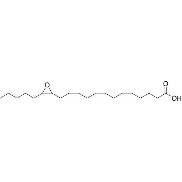
HY-150084
(±)14,15-Epoxyeicosatrienoic acidProliferation Inducer -

HY-130675A
15(S)-HEPEAsthma Marker -
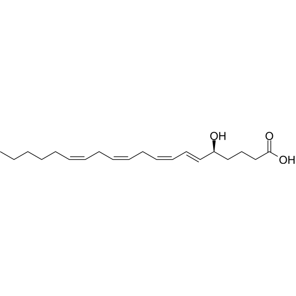
HY-113434
5(S)-HETEEndogenous Metabolite -
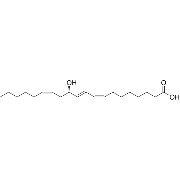
HY-130060
12(S)-HETrEPlatelet Aggregation Inhibitor -

HY-130309
(±)8(9)-EpETEEpoxide -
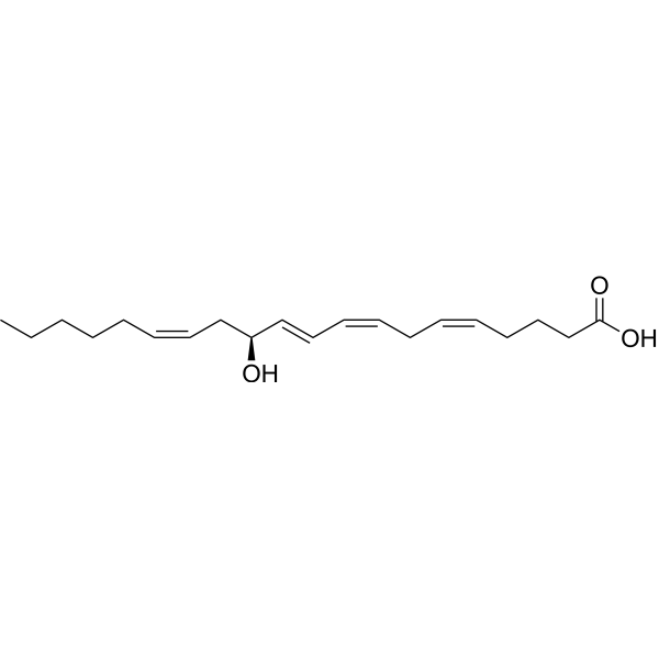
HY-124404A
12(S)-HETEP38 MAPK/ERK Activator -
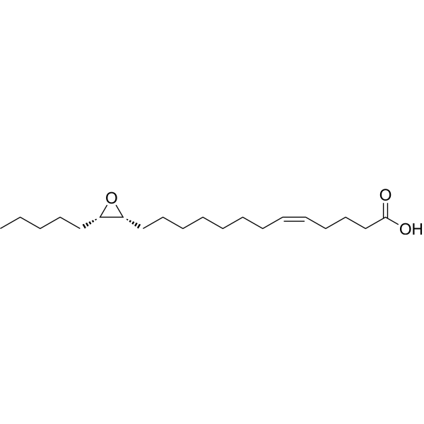
HY-120954
14,15-EE-5(Z)-E14,15-EET structural analogue -

HY-130140
8,9-DiHETEEster Product -
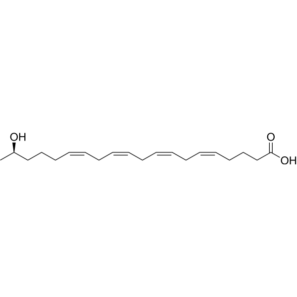
HY-118332
19(R)-HETEVasodilator -
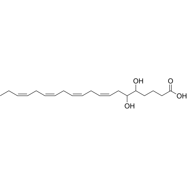
HY-130138C
5,6-DiHETEAnti-inflammatory Lipid Mediators -

HY-113711
15-OxoEDEEster Product
All DNA/RNA Synthesis Isoforms
More

Biological Activity
Description
IC50 & Target
[1]|
ERK1 |
ERK2 |
p38α |
mTORC2 |
GLUT4 |
In Vitro
12 (S)-HEPE (100-1000 nM; 48 h) induces Caco-2 cell growth and DNA synthesis, and this effect is blocked by COX and BLT antagonists[1].
12 (S)-HEPE (100 nM; 5-15 min) activates phosphorylation of p38α, GSKβ, CREB, and ERK1/2 in Caco-2 cells, but does not activate AKT1 or AKT2[1].
12 (S)-HEPE (0.01-1 μM; 30 min) increases glucose uptake in WT-1 mouse brown adipocytes through a Gs/PI3K/mTORC2-dependent pathway[3].
12 (S)-HEPE (0.01-1 μM; 30 min) increases glucose uptake in differentiated C2C12 myotubes[3].
12 (S)-HEPE (5-45 min) activates the Akt/mTORC2/AS160 signaling pathway in WT-1 mouse brown adipocytes[3].
12 (S)-HEPE supports the formation of receptor-G protein complexes in primary hepatocyte plasma membrane fractions, thereby enabling the identification of 8 candidate proteins[4].
MedChemExpress (MCE) has not independently confirmed the accuracy of these methods. They are for reference only. Further protocols information, click here.
-
Cell Line:human intestinal Caco-2 cells
-
Concentration:10, 100, 1000 nM; 100 nM plus U75302 5 μM, LY255283 25 μM, or ketoprofen 5 μM
-
Incubation Time:48 h
-
Result:Induced significant cell growth and DNA synthesis at 100 and 1000 nM.
This mitogenic action was blocked by ketoprofen (COX inhibitor), U75302 (BLT1 antagonist), and LY255283 (BLT1/BLT2 antagonist).
-
Cell Line:human intestinal Caco-2 cells
-
Concentration:100 nM
-
Incubation Time:5 or 15 min
-
Result:O.D. 450 nm values for 12(S)-HEPE were AKT1 0.19; AKT2 0.21; p38α 0.85; GSKβ 0.48; CREB 0.76; ERK1/2 0.95.
Increased phosphorylation of p38α, GSKβ, CREB, and ERK1/2, with no effect on AKT1 or AKT2.
In Vivo
12(S)-HEPE (200 µg/kg; intravenous injection; single dose) acutely increases glucose uptake in BAT and skeletal muscle of lean mice[3].
12(S)-HEPE (8 weeks) reduces body weight gain and improves glucose and insulin tolerance in HFD-fed wild-type mice, but has no such effect in Olfr110-/- mice[4].
MedChemExpress (MCE) has not independently confirmed the accuracy of these methods. They are for reference only.
-
Animal Model:C57BL/6J (male, 6 weeks old at start of high-fat diet; diet-induced obese after 16 weeks of high-fat diet)[3]
-
Dosage:200 μg/kg body weight
-
Administration:i.p.; once daily; for 2 weeks
-
Result:Circulating 12-HEPE levels increased 2.5-fold 30 min after i.p. administration.
No change in body weight gain or fasting glucose.
12(S)-HEPE-treated DIO mice showed improved glucose tolerance and improved insulin sensitivity compared with vehicle-treated mice.
No difference in serum insulin before or after glucose injection.
No change in food intake, physical activity, VCO2, or VO2.
BAT weight was increased.
BAT mRNA expression of Glut-1, Chrebp-b, and Fasn was increased.
IL-10 expression was increased in pgWAT; no differences in M1/M2 macrophage populations in pgWAT.
-
Animal Model:C57BL/6J (male, 12 weeks old)[3]
-
Dosage:200 µg/kg
-
Administration:retro-orbital (i.v.); single dose
-
Result:Significantly increased [3H]2-deoxyglucose uptake into BAT and skeletal muscle but not into pgWAT, ingWAT, or liver.


Chemical Information
-
CAS No. 116180-17-7
-
Appearance Liquid
-
Molecular Weight 318.45
-
Formula C₂₀H₃₀O₃
-
Color Colorless to light yellow
-
SMILES
CC/C=C\C/C=C\C[C@H](O)/C=C/C=C\C/C=C\CCCC(O)=O
-
Shipping
Room temperature in continental US; may vary elsewhere.
-
Storage
Solution, -20°C, 2 years

Protocols
-
EdU Incorporation Assay (Click Chemistry-Based DNA Synthesis Measurement)
The EdU incorporation assay measures DNA synthesis by adding the thymidine analog 5-ethynyl-2′-deoxyuridine to cells or tissues, where it is incorporated into newly synthesized DNA during S phase. Incorporated EdU is detected by copper-catalyzed azide-alkyne cycloaddition, in which a fluorescent azide covalently reacts with the ethynyl group on EdU, allowing S-phase cells to be detected by fluorescence microscopy, flow cytometry, or high-content imaging. EdU detection does not require DNA denaturation or anti-BrdU antibody access, which preserves sample structure and improves compatibility with immunostaining and multiparameter cytometry compared with BrdU-based detection. EdU can be cytotoxic in a cell-type- and exposure-dependent manner, so pulse duration, concentration, and continuous-labeling designs should be validated for each cell type.
-
BrdU Incorporation Assay
Bromodeoxyuridine (BrdU) incorporation assay is based on the principle that BrdU, a thymidine analog, is incorporated into newly synthesized DNA during the S phase of the cell cycle, thereby serving as a marker of DNA replication and cellular proliferation. Incorporated BrdU can be detected using anti-BrdU antibodies following DNA denaturation, enabling visualization or quantification of proliferating cells through immunochemical detection methods such as immunofluorescence or immunohistochemistry.
-
Zymosan-Induced Peritonitis
Zymosan-induced peritonitis is a sterile acute-inflammation model produced by intraperitoneal injection of zymosan, a yeast cell-wall particle preparation, followed by quantification of leukocyte recruitment and soluble inflammatory mediators in peritoneal lavage fluid. Low-dose zymosan peritonitis is commonly used as a self-resolving acute inflammation model in which neutrophil recruitment occurs early and monocyte/macrophage accumulation follows later. The assay readouts include total peritoneal leukocyte number, differential neutrophil and monocyte/macrophage counts, peritoneal cytokines and chemokines, plasma or peritoneal exudation, and optional lipidomic or metabolomic changes during inflammation and resolution. Early neutrophil recruitment after zymosan depends strongly on complement and mast-cell C5a receptor signaling, whereas later monocyte recruitment is linked to MCP-1/CCL2 production.
-
Research Protocol for Metabolic Diseases
AMP-activated protein kinase, AMPK, is a conserved cellular energy sensor that responds to reduced cellular energy status and coordinates metabolism by increasing ATP-generating catabolic pathways while suppressing ATP-consuming anabolic processes. In metabolic disease research, the AMPK pathway is experimentally relevant because it regulates hepatic lipid synthesis, fatty acid oxidation, glucose production, skeletal-muscle glucose disposal, mTORC1-linked biosynthesis, autophagy, mitochondrial homeostasis, and whole-body energy balance. The central pathway logic is that energy stress, metformin, exercise-like stimulation, or direct AMPK activators increase AMPKα Thr172 phosphorylation and downstream substrate phosphorylation, including ACC and RAPTOR. Phosphorylation of ACC suppresses lipogenesis and supports fatty acid oxidation, whereas phosphorylation of RAPTOR suppresses mTORC1 signaling and links cellular energy status to growth and protein synthesis control. The pathway is linked


Purity & Documentation
-
Data Sheet (275 KB)
-
SDS (393 KB)
- English - EN (393 KB)
- Français - FR (393 KB)
- Deutsch - DE (393 KB)
- Norwegian - NO (393 KB)
- Español - ES (393 KB)
- Swedish - SV (393 KB)
- Italian - IT (393 KB)
- Korean - KR (393 KB)
- Portuguese - PT (393 KB)
-
Handling Instructions (2659 KB)
References

Calculators
Concentration (start) × Volume (start) = Concentration (final) × Volume (final)


12(S)-HEPE Related Classifications
HY-W747548 Related Classifications
 Powered by Bioz