



Croconic acid disodium

Based on 1 Customer Validation
Croconic acid disodium (Nacr) is a lysine crotonylation (Kcr) activator and electroactive material. Croconic acid disodium reduces the expression of HDAC2, HDAC3, SIRT1, and SIRT3, and increases the expression of EP300, CITED1, ACSS2, DPF2, CDYL, MLLT3, and YEATS2. Croconic acid disodium elevates intracellular crotonyl-CoA content and global histone lysine crotonylation levels. Croconic acid disodium promotes the growth of bovine fibroblasts, regulates cell cycle progression, and inhibits bovine fibroblast apoptosis (apoptosis). Croconic acid disodium improves the blastocyst development efficiency of bovine somatic cell nuclear transfer embryos. Croconic acid disodium undergoes reversible lithium intercalation/deintercalation reactions via sodium-lithium ion exchange. Croconic acid disodium is applicable to research related to cell growth promotion.
For research use only. We do not sell to patients.
- Purity : 99.98%
- CAS No.: 14379-00-1
- Formula: C5Na2O5
- Molecular Weight:186.03
-
Storage:
Store at room temperature, keep dry and cool.
In solvent -80°C, 1 year , -20°C, 6 months


To place orders, for customer services and technical support, please contact: MedChemExpress USA
Tel: 609-228-6898 E-mail: [email protected] [email protected]


-
 Biological Activity
-
Chemical Information
-
Solvent & Solubility
- Protocols
- Purity & Documentation
- References
-
 Help & FAQs


-
HY-W110551
2-Methacryloyloxyethyl phosphorylcholinePolymer Monomer -
HY-W110584
Carboxybetaine methacrylateAmphoteric Ionic Compound -
HY-W013538
1,2-Bis(maleimido)ethaneProtein Cross-Linking Agent -

HY-W089777
3-GlycidyloxypropyltrimethoxysilaneBiochemical Assay Reagent -
HY-W012723
2-Hydroxyethyl methacrylateHydroxyester Compound -

HY-W105423
Methacrylic acid N-hydroxysuccinimide esterPolymer -

HY-W011957
1,4-Bis-maleimidobutaneBiochemical Reagent -

HY-W095198
N-(2-Hydroxyethyl)acrylamideAmphiphilic Polymer -
HY-W020027
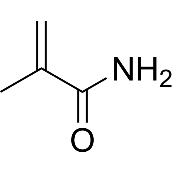
MethacrylamideGelatin Derivatization Reagent -
HY-W011103
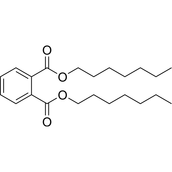
Diheptyl phthalateBiochemical Assay Reagent -
![[2-(Methacryloyloxy)ethyl]trimethylammonium chloride](https://file.medchemexpress.com/product_pic/hy-w014539.gif)
HY-W014539
[2-(Methacryloyloxy)ethyl]trimethylammonium chlorideBiochemical Assay Reagent -
HY-128833
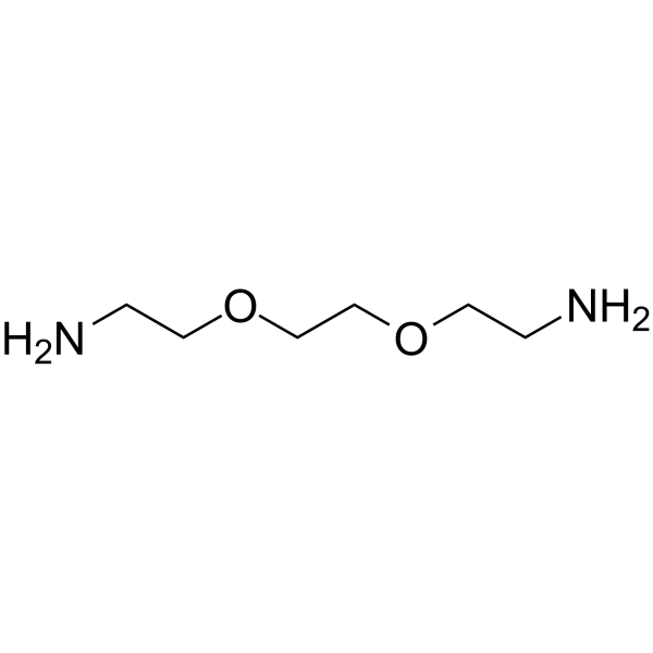
Bis-NH2-PEG2PROTAC Linkers


Biological Activity
Description
In Vitro
Croconic acid disodium (2.5-10 mM; 4-8 h) increases the cell density and maintains the viability of bovine fibroblasts[1].
Disodium croconate (2.5-10 mM; 4-8 h) significantly increases the proliferation rate of bovine fibroblasts[1].
Croconic acid disodium (2.5-10 mM; 8 h) extremely significantly upregulates the messenger ribonucleic acid expression levels of PCNA, CDK1, MK167 and CDK2, and downregulates the expression of CDKN1A and CDKN2A in bovine fibroblasts[1].
Croconic acid disodium (2.5-10 mM; 8 h) upregulates the mRNA expression levels of CDK4, CDK6, CCNB1, CCNA2, CCNE2, and CCND1 in bovine fibroblasts[1].
Croconic acid disodium (2.5-10 mM; 8 h) downregulates the expression of BAD, CYCS, CASP3 and CASP9 genes and upregulates the expression of BCL2 and BCL-XL genes in bovine fibroblasts[1].
Croconic acid disodium (2.5-10 mM; 8 h) exerts the most significant up-regulatory effects on Kcr writer genes (EP300, CITED1, ACSS2) and reader genes (DPF2, CDYL, MLLT3, YEATS2), while down-regulating Kcr eraser genes (HDAC2, HDAC3, SIRT1, SIRT3) in bovine fibroblasts[1].
Croconic acid disodium (2.5-10 mM; 8 h) reduces the proportion of bovine fibroblasts in the G0/G1 phase, while increasing the proportion of cells in the S and G2/M phases (compared with the control group level), thereby promoting cell cycle progression[1].
Disodium croconate (2.5-10 mM; 8 h) significantly reduces the proportion of total apoptotic cells in bovine fibroblasts[1].
Disodium croconate (5 mM; 8 h) significantly increases the level of crotonyl-CoA in bovine fibroblasts[1].
Croconic acid disodium (5 mM; 8 h) significantly increases global histone crotonylation and H3K9 crotonylation levels in bovine fibroblasts[1].
Treatment of bovine ear tip fibroblasts with croconic acid disodium (5 mM; added to cell culture medium; single dose; 8 hours) significantly increases the blastocyst development rate of the resulting somatic cell nuclear transfer embryos to 38.1%[1].
Croconic acid disodium (110 charge-discharge cycles; 0.1-6 C current density) exhibits better lithium-ion battery performance than larger-sized CADS micro-columns and microwires[2].
MedChemExpress (MCE) has not independently confirmed the accuracy of these methods. They are for reference only. Further protocols information, click here.
-
Cell Line:bovine fibroblasts
-
Concentration:2.5, 5, 7.5, 10 mM
-
Incubation Time:4 h, 8 h, 12 h
-
Result:Significantly reduced cell density at 5 mM, 7.5 mM, and 10 mM after 4 h.
Significantly reduced viable cell proportion only at 10 mM after 4 h.
Significantly increased cell density at 5 mM, reduced cell density at 7.5 mM and 10 mM, maintained viable cell proportion at 5 mM, reduced viable cell proportion at 7.5 mM and 10 mM, and showed no significant effect at 2.5 mM on either measure after 8 h.
Significantly reduced cell density and viable cell proportion at all concentrations after 12 h.
-
Cell Line:bovine fibroblasts
-
Concentration:2.5, 5, 7.5, 10 mM
-
Incubation Time:4 h, 8 h, 12 h
-
Result:Showed no significant effect on percentage of cells in DNA replication at 2.5 mM and 7.5 mM, significantly increased this percentage at 5 mM, and significantly decreased this percentage at 10 mM after 4 h.
Significantly decreased percentage of cells in DNA replication at 2.5 mM, 7.5 mM, and 10 mM, significantly increased this percentage at 5 mM after 8 h.
Significantly decreased percentage of cells in DNA replication only at 10 mM, showed no effect at 2.5 mM, 5 mM, and 7.5 mM after 12 h.
-
Cell Line:bovine fibroblasts
-
Concentration:2.5, 5, 7.5, 10 mM
-
Incubation Time:8 h
-
Result:Significantly upregulated mRNA expression of PCNA, CDK1, MK167, and CDK2, and significantly downregulated mRNA expression of CDKN1A and CDKN2A at 2.5 mM, 5 mM, and 7.5 mM, with most significant effects at 5 mM.
Downregulated mRNA expression of PCNA, CDK1, MK167, and CDK2, and upregulated mRNA expression of CDKN1A and CDKN2A at 10 mM.\nSignificantly upregulated mRNA expression of CDK4, CDK6, CCNB1, CCNA2, CCNE2, and CCND1 at 2.5 mM, 5 mM, and 7.5 mM.
Showed no effect on the expression of these genes at 10 mM.\nDownregulated mRNA expression of proapoptotic genes BAD, CYCS, CASP3, and CASP9, and upregulated mRNA expression of antiapoptotic genes BCL2 and BCL-XL at 2.5 mM and 5 mM.
Upregulated mRNA expression of proapoptotic genes and downregulated mRNA expression of antiapoptotic genes at 7.5 mM and 10 mM.\nUpregulated mRNA expression of Kcr writer genes EP300, CITED1, and ACSS2, and downregulated mRNA expression of Kcr eraser genes HDAC2, HDAC3, SIRT1, and SIRT3 at all concentrations.
Upregulated mRNA expression of Kcr reader genes DPF2, CDYL, MLLT3, and YEATS2 at 2.5 mM and 5 mM, with most significant effects at 5 mM.
-
Cell Line:bovine fibroblasts
-
Concentration:2.5, 5, 7.5, 10 mM
-
Incubation Time:4 h, 8 h, 12 h
-
Result:Significantly reduced G0/G1 phase cell proportion (65.05% vs. control 72.84%) and increased S phase cell proportion (10.58% vs. control 7.11%) at 5 mM, decreased S phase cell proportion at 10 mM, and showed no notable effect at other concentrations after 4 h.
Increased S phase cell proportion to 8.84% and 8.37% respectively (vs. control 7.98% and 7.89% respectively), increased G2/M phase cell proportion to 15.59% and 15.16% respectively (vs. control 12.93% for both), and significantly reduced G0/G1 phase cell proportion at 5 mM and 7.5 mM after 8 h.
Increased G0/G1 phase cell proportion and decreased S and G2/M phase cell proportions at all concentrations after 12 h.
-
Cell Line:bovine fibroblasts
-
Concentration:2.5, 5, 7.5, 10 mM
-
Incubation Time:4 h, 8 h, 12 h
-
Result:Significantly reduced total apoptotic cell proportion from control 18.6% to 16% at 2.5 mM, to 12.61% at 5 mM, to 13.18% at 7.5 mM, and to 13.6% at 10 mM after 4 h.
Significantly reduced total apoptotic cell proportion from control 21.44% to 10.42% (early apoptosis reduced from 3.67% to 2.53%, late apoptosis reduced from 17.77% to 7.89%) at 5 mM, reduced total apoptotic cell proportion to 13.18% at 7.5 mM after 8 h.
Increased total apoptotic cell proportion at all concentrations after 12 h.
-
Cell Line:bovine fibroblasts
-
Concentration:5 mM
-
Incubation Time:8 h
-
Result:Significantly increased intracellular crotonyl-CoA content compared to control.
-
Cell Line:bovine fibroblasts
-
Concentration:5 mM
-
Incubation Time:8 h
-
Result:Significantly increased overall pan-Kcr fluorescence intensity and specifically increased H3K9cr fluorescence intensity compared to control.


Chemical Information
-
CAS No. 14379-00-1
-
Appearance Solid
-
Molecular Weight 186.03
-
Formula C5Na2O5
-
Color Light yellow to yellow
-
SMILES
O=C1C(C(O[Na])=C(C1=O)O[Na])=O
-
Synonyms
Nacr
-
Shipping
Room temperature in continental US; may vary elsewhere.
-
Storage
Store at room temperature, keep dry and cool
In solvent -80°C 1 year -20°C 6 months


Solvent & Solubility
In Vitro:
H2O : 50 mg/mL (268.77 mM; ultrasonic and warming and heat to 60°C)
Please refer to the solubility information to select the appropriate solvent. Once prepared, please aliquot and store the solution to prevent product inactivation from repeated freeze-thaw cycles.
Storage method and period of stock solution: -80°C, 1 year; -20°C, 6 months. When stored at -80°C, please use it within 1 year. When stored at -20°C, please use it within 6 months.
* Note: If you choose water as the stock solution, please dilute it to the working solution, then filter and sterilize it with a 0.22 μm filter before use.
Please refer to the solubility information to select the appropriate solvent. Once prepared, please aliquot and store the solution to prevent product inactivation from repeated freeze-thaw cycles.
Storage method and period of stock solution: -80°C, 1 year; -20°C, 6 months. When stored at -80°C, please use it within 1 year. When stored at -20°C, please use it within 6 months.
* Note: If you choose water as the stock solution, please dilute it to the working solution, then filter and sterilize it with a 0.22 μm filter before use.
Concentration (start) × Volume (start) = Concentration (final) × Volume (final)

Protocols
-
RNA extraction experimental
By lysing cells, releasing RNA, and removing impurities such as proteins and DNA, high-purity RNA products are finally obtained. The commonly used traditional method is the guanidine isothiocyanate/phenol/chloroform method (Trizol), which is suitable for a variety of animal materials including animal tissues, microorganisms, cultured cells, etc., and most plant materials.
-
Annexin V plus membrane-impermeant dye apoptosis staining
Annexin V-based apoptosis assays rely on the detection of phosphatidylserine (PS) externalization from the inner leaflet of the plasma membrane to the outer leaflet, an early biochemical hallmark of apoptosis. Fluorescently labeled Annexin V binds PS in a calcium-dependent manner, enabling identification of early apoptotic cells by flow cytometry or fluorescence microscopy. When combined with a membrane-impermeant DNA-binding dye (e. g. , propidium iodide), this approach allows discrimination between viable (Annexin V−/dye−), early apoptotic (Annexin V+/dye−), and late apoptotic or necrotic (Annexin V+/dye+) cell populations by assessing membrane integrity and PS exposure.
-
BrdU Incorporation Assay
Bromodeoxyuridine (BrdU) incorporation assay is based on the principle that BrdU, a thymidine analog, is incorporated into newly synthesized DNA during the S phase of the cell cycle, thereby serving as a marker of DNA replication and cellular proliferation. Incorporated BrdU can be detected using anti-BrdU antibodies following DNA denaturation, enabling visualization or quantification of proliferating cells through immunochemical detection methods such as immunofluorescence or immunohistochemistry.
-
Somatic Cell Culture
A method of simulating the in vivo environment in vitro to maintain the cell growth, differentation and main functions.
-
Protocol for Cell Cycle
Cell-cycle analysis by flow cytometry measures DNA content in single cells to estimate the fraction of cells in G0/G1, S, and G2/M phases. Propidium iodide intercalates into DNA, and after RNA removal with RNase, fluorescence intensity reflects cellular DNA content: 2N cells are assigned to G0/G1, cells between 2N and 4N to S phase, and 4N cells to G2/M. DNA-content analysis alone cannot reliably separate G0 from G1 or G2 from M. Ki-67 can distinguish quiescent G0 cells from cycling cells, EdU or BrdU incorporation marks active DNA synthesis in S phase, and phospho-histone H3 staining identifies mitotic cells within the 4N population.
-
CCK-8/WST-8 Cell Proliferation Assay
The CCK-8/WST-8 assay is based on the reduction of the water-soluble tetrazolium salt WST-8 to a water-soluble formazan product by cellular dehydrogenases in metabolically active cells, where the generated formazan amount is proportional to the number of living cells and is quantified by measuring absorbance in the visible range, providing a colorimetric readout for cell viability and proliferation assessment. This class of tetrazolium-based assays improves upon earlier MTT-based systems by producing a water-soluble formazan, eliminating the need for organic solubilization steps and enabling direct spectrophotometric measurement in culture medium.
-
Cell Counting-Based Growth Curve Assay
Cell counting-based growth curve assays quantify cell proliferation by directly measuring changes in viable cell number over time using manual or automated counting methods such as hemocytometer-based counting or instrument-assisted cell enumeration, enabling construction of growth curves that reflect population expansion dynamics in response to culture conditions. A widely used approach is trypan blue exclusion with hemocytometer counting, where membrane-compromised (non-viable) cells take up the dye, allowing discrimination between viable and non-viable cells while simultaneously enabling total cell number quantification. Repeated sampling across time points allows estimation of proliferation rate, growth phases, and comparative growth kinetics between experimental conditions.
-
Cell Viability Determination by MTT Colorimetric Assay
The following protocol uses the MTT colorimetric assay as a classic literature-established method for assessing cell viability/metabolic activity in cultured mammalian cells. MTT[3-(4,5-dimethylthiazol-2-yl)-2,5-diphenyltetrazolium bromide] is reduced by metabolically active cells to a colored formazan product; the amount of formazan is quantified spectrophotometrically and provides an indirect measure of metabolically active viable cells. Importantly, MTT reduction reflects cellular oxidoreductase/metabolic activity rather than an absolute direct count of living cells, so changes in cellular metabolism can alter the signal independently of cell number.
-
Apoptosis
Apoptosis, also called programmed cell death, is generally characterized by distinct morphological characteristics.
-
TUNEL staining for apoptotic DNA fragmentation
TUNEL staining detects DNA strand breaks by using terminal deoxynucleotidyl transferase to add labeled nucleotides to exposed 3′-OH DNA termini, generating either microscopic staining in fixed cells or tissue sections, or fluorescence/cytometric signal in cell suspensions. TUNEL positivity reflects DNA fragmentation but should not be interpreted alone as definitive apoptosis, because TUNEL can also label necrotic, autolytic, mechanically damaged, or DNA-repair-associated DNA breaks.
-
Flow cytometric DNA-content cell-cycle staining
Flow cytometric DNA-content cell-cycle staining measures the fluorescence intensity of DNA-bound fluorochromes in single cells or nuclei to estimate DNA content distributions, allowing assignment of populations to G0/G1, S, and G2/M phases by DNA histogram deconvolution. Propidium iodide (PI) intercalates into DNA, and PI fluorescence is proportional to cellular DNA content when staining is performed under conditions that make DNA accessible and minimize non-DNA signal. Cells with G2/M DNA content are expected to show approximately twice the fluorescence intensity of G0/G1 cells, while S-phase cells occupy intermediate fluorescence values. PI-based DNA-content analysis can also detect cells with fractional DNA content, often reported as sub-G1, when DNA fragmentation and extraction during staining reduce retained DNA signal in apoptotic cells. DAPI is an alternative DNA fluorochrome for univariate DNA-content analysis, while bivariate approaches combining DNA content with proliferation
-
Protocol for Cell Counting and Cell Density Analysis
Cell counting and cell-density analysis estimate the number of cells in a known volume or field area. Manual hemocytometer counting uses a chamber of defined geometry to convert counted cells into cells/mL, while automated counters and image-analysis workflows detect cell objects from optical, brightfield, fluorescence, impedance, or digital-image features. Trypan blue viability counting is based on dye exclusion: viable cells with intact membranes exclude dye, while non-viable cells with compromised membranes stain blue. The readout is total cell density, viable-cell density, dead-cell density, and percent viability. Cell density can also be estimated from microscopy images by counting objects per image area, from flow cytometry using calibrated volume or reference particles, or from in situ microscopy in bioreactors after calibration against reference methods such as hemocytometer or flow cytometry.
-
Apoptosis Solutions
Apoptosis is a regulated, generally non-lytic cell-death pathway that removes unwanted, damaged, infected, or abnormal cells through coordinated morphological changes, caspase activation, DNA fragmentation, and membrane remodeling. The intrinsic apoptosis pathway is controlled mainly by mitochondrial outer membrane permeabilization, BCL-2 family proteins, cytochrome c release, apoptosome formation, caspase-9 activation, and downstream executioner caspase-3/7 activation. The extrinsic apoptosis pathway is initiated by death receptors such as Fas, TNFR, and TRAIL receptors, which recruit adaptor proteins and activate caspase-8 before engaging executioner caspases or mitochondrial amplification through BID cleavage. Apoptosis is linked to many phenotypes, including cancer cell killing, tissue homeostasis, immune regulation, neurodegeneration, infection response, and treatment-induced cytotoxicity; unresolved questions include how apoptosis interacts with necroptosis, pyroptosis, ferroptos
-
MTT Cell Proliferation Assay
The MTT assay is a colorimetric endpoint assay for estimating viable cell number, cell growth, cytotoxicity, or cell activation in cultured mammalian cells. Living cells reduce the yellow tetrazolium salt MTT into purple/blue formazan, while dead cells do not generate the same signal; the resulting color can be quantified with a multiwell spectrophotometer. MTT reduction is commonly interpreted as a readout of metabolic activity that often correlates with viable cell number, but it should not be treated as a direct cell-counting method unless the assay is optimized for the cell type and experimental condition. Studies show that MTT reduction can involve mitochondrial and non-mitochondrial reducing systems, and formazan may accumulate in intracellular lipid droplets rather than simply marking mitochondria.


Purity & Documentation
-
Data Sheet (288 KB)
-
SDS (396 KB)
- English - EN (396 KB)
- Français - FR (396 KB)
- Deutsch - DE (396 KB)
- Norwegian - NO (396 KB)
- Español - ES (396 KB)
- Swedish - SV (396 KB)
- Italian - IT (396 KB)
- Korean - KR (396 KB)
- Portuguese - PT (396 KB)
-
Handling Instructions (2659 KB)
References
[1]. Zhao X, et al. High histone crotonylation modification in bovine fibroblasts promotes cell proliferation and the developmental efficiency of preimplantation nuclear transfer embryos. Sci Rep. 2024;14(1):10295. Published 2024 May 4. [Content Brief]
[2]. Luo C, et al. Self-assembled organic nanowires for high power density lithium ion batteries. Nano Lett. 2014;14(3):1596-1602. [Content Brief]


Complete Stock Solution Preparation Table
Please refer to the solubility information to select the appropriate solvent. Once prepared, please aliquot and store the solution to prevent product inactivation from repeated freeze-thaw cycles.
Storage method and period of stock solution: -80°C, 1 year; -20°C, 6 months. When stored at -80°C, please use it within 1 year. When stored at -20°C, please use it within 6 months.
| Optional Solvent | Concentration Solvent Mass | 1 mg | 5 mg | 10 mg | 25 mg |
|---|---|---|---|---|---|
| H2O | 1 mM | 5.3755 mL | 26.8774 mL | 53.7548 mL | 134.3869 mL |
| 5 mM | 1.0751 mL | 5.3755 mL | 10.7510 mL | 26.8774 mL | |
| 10 mM | 0.5375 mL | 2.6877 mL | 5.3755 mL | 13.4387 mL | |
| 15 mM | 0.3584 mL | 1.7918 mL | 3.5837 mL | 8.9591 mL | |
| 20 mM | 0.2688 mL | 1.3439 mL | 2.6877 mL | 6.7193 mL | |
| 25 mM | 0.2150 mL | 1.0751 mL | 2.1502 mL | 5.3755 mL | |
| 30 mM | 0.1792 mL | 0.8959 mL | 1.7918 mL | 4.4796 mL | |
| 40 mM | 0.1344 mL | 0.6719 mL | 1.3439 mL | 3.3597 mL | |
| 50 mM | 0.1075 mL | 0.5375 mL | 1.0751 mL | 2.6877 mL | |
| 60 mM | 0.0896 mL | 0.4480 mL | 0.8959 mL | 2.2398 mL | |
| 80 mM | 0.0672 mL | 0.3360 mL | 0.6719 mL | 1.6798 mL | |
| 100 mM | 0.0538 mL | 0.2688 mL | 0.5375 mL | 1.3439 mL |
* Note: If you choose water as the stock solution, please dilute it to the working solution, then filter and sterilize it with a 0.22 μm filter before use.


Croconic acid disodium Related Classifications
HY-W105310A Related Classifications
 Powered by Bioz