



Gossypin

Based on 1 Customer Validation
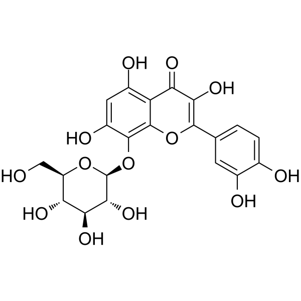
Gossypin is an orally active flavone isolated from Hibiscus vitifolius. Gossypin has antioxidant, antiinflammatory, anticancer, anticataract, antidiabetic, and hepatoprotective activities. Gossypin inhibits NF-κB and NF-κB-regulated gene expression. Gossypin inhibits AURKA and RSK2. Gossypin inhibits invasion and induces apoptosis. Gossypin can be used for gastric cancer study.
Nur für Forschungszwecke. Wir verkaufen nicht an Patienten.
- Reinheit : 98.29%
- CAS. Nr.: 652-78-8
- Formel: C21H20O13
- Molecular Weight:480.38
-
Speicherung:Powder -20°C, 3 years , 4°C, 2 years ; In solvent -80°C, 6 months , -20°C, 1 month

To place orders, for customer services and technical support, please contact: MedChemExpress USA
Tel: 609-228-6898 E-mail: [email protected] [email protected]


-
 Biologische Aktivität
-
Chemical Information
-
Lösungsmittel & Löslichkeit
- Protokoll
- Reinheit & Dokumentation
- Verweise
-
 Help & FAQs



Alle Aurora Kinase Isoform-spezifische Produkte anzeigen
MoreAlle Caspase Isoform-spezifische Produkte anzeigen
More

Biologische Aktivität
Beschreibung
IC50 & Target
IC50: NF-κB[1]
In Vitro
Gossypin (10-100 μM, 0.5-8 h and then treats with TNF for 30 minutes) inhibits NF-κB activation, leading to potentiation of apoptosis, suppression of invasion in KBM-5 cells[1].
Gossypin (50 μM, 3-5 days) inhibits RANKL-induced osteoclastogenesis in RAW 264.7 and mouse (C57BL/6J) primary bone marrow cells[1].
Gossypin (10-60 μM, 72h or 2 weeks) inhibits gastric cancer cell growth[3].
Gossypin (20-40 μM, 48 h) significantly suppresses migration in gastric cancer cell [3].
Gossypin (5-60 μM, 1-24 h) inhibits AURKA and RSK2 in JB6 cells and HGC27 cells[3].
Gossypin (40 μM, 48 h) induces G2/M phase cell cycle arrest[3].
Gossypin (40-60 μM, 72 h) increases apoptosis of gastric cancer cells[3].
MedChemExpress (MCE) has not independently confirmed the accuracy of these methods. They are for reference only.
-
Cell Line:HGC27 human gastric cancer cells, AGS human gastric cancer cells
-
Concentration:20, 40, 60 μM (72 h), or 10, 20, 40 μM (2 weeks)
-
Incubation Time:(40 μM) 48 h or 72 h or 2 weeks
-
Result:Reduced S phase and induced G2 phase cell cycle arrest.
Reduced the expression of cyclin A2, cyclin B1, and phosphorylated CDC2 proteins.
Growth of gastric cancer cells was significantly inhibited in a dose dependent manner.
-
Cell Line:HGC27 human gastric cancer cells
-
Concentration:40, 60 μM
-
Incubation Time:72 h
-
Result:Increased the number of suspended cells (dead), decreased the number of adherent cells (live).
Early and late apoptosis cells were strongly increased compared to control cells.
Increased expression of pro-apoptotic marker proteins (cleaved caspase 3, 7 and 9, cleaved PARP and cytochrome c), decreased expression of anti-apoptotic marker protein (BcL-xL).
-
Cell Line:HGC27 human gastric cancer cells, AGS human gastric cancer cells
-
Concentration:20, 40 μM
-
Incubation Time:48 h
-
Result:Significantly suppressed migration.
-
Cell Line:KBM-5 (chronic myeloid leukemia)
-
Concentration:0.5, 1, 2, 4, 8 h, and then treated with TNF for 30 minutes; or 2 h then treated with TNF for 16 h; or Preincubated for 30 minutes before add TNF, H2O2, PMA, LPS, IL-1β, OA, CSC
-
Incubation Time:2.5 h (2 hours alone and then treated with TNF for 30 minutes)
-
Result:Blocked NF-κB activation induced by tumor necrosis factor (TNF), H2O2, Phorbol myristate acetate (PMA), Lipopolysaccharides (LPS) (HY-D1056), interleukin-1β (IL-1β), okadaic acid (OA), cigarette smoke condensate (CSC).
Suppressed TNF-induced NF-κB in a dose-dependent manner.
Suppressed TNF-induced NF-κB in a time-dependent manner.
Enhanced TNF-induced apoptosis.
-
Cell Line:RAW 264.7 cells, Mouse (C57BL/6J) primary bone marrow cells
-
Concentration:50 μM
-
Incubation Time:5 days (RAW 264.7 cells) or 3 days (primary bone marrow cells)
-
Result:Decreased RANKL-induced osteoclast differentiation in RAW 264.7 cells.
50 μM concentration was sufficient to reduce osteoclastogenesis by more than 65% in RAW 264.7 cells.
Under these conditions, RAW 264.7cells remained fully viable.
Substantially decreased RANKL-induced osteoclast differentiation in primary bone marrow cells.
In Vivo
MedChemExpress (MCE) has not independently confirmed the accuracy of these methods. They are for reference only.
-
Animal Model:Sprague Dawley (SD) rats (200-225 g; male; 8-10 weeks)[2]
-
Dosage:10, 20 mg/kg
-
Administration:Oral gavage (p.o.); 30 days
-
Result:Reduced the infarct size induced by Isoprenaline (ISO) (HY-108353).
Showed the boosted level of LVESP, SW, ESPVR, DP, EDPVR and suppressed level of LVEDP.
Increased the bodyweight and suppressed the heart weight and heart/body weight ratio.
Suppressed the level of triglyceride (TG), total cholesterol (TC), low-density lipoprotein (LDL), very low-density lipoprotein (VLDL) and increased the level of high-density lipoprotein (HDL).
Suppressed the level of nitric oxide synthase (iNOS) and nitric oxide (NO).
Decreased the level of MMP-2 and MMP-9.
Suppressed the level of inflammatory cytokines (TNF-α, IL-1β, IL-6) and decreased the level of NF-κB in the serum and heart tissue.
Suppressed the apoptosis of muscle fibres with edema and necrosis.


Chemical Information
-
CAS. Nr. 652-78-8
-
Appearance Solid
-
Molecular Weight 480.38
-
Formel C21H20O13
-
Color Light yellow to yellow
-
SMILES
O=C1C(O)=C(C2=CC=C(O)C(O)=C2)OC3=C(O[C@H]4[C@@H]([C@H]([C@@H]([C@@H](CO)O4)O)O)O)C(O)=CC(O)=C13
-
Structure Classification
-
Initial Source
-
Versand
Room temperature in continental US; may vary elsewhere.
-
Speicherung
Powder -20°C 3 years 4°C 2 years In solvent -80°C 6 months -20°C 1 month


Lösungsmittel & Löslichkeit
In Vitro:
DMSO : 200 mg/mL (416.34 mM; Need ultrasonic; Hygroscopic DMSO has a significant impact on the solubility of product, please use newly opened DMSO)
Please refer to the solubility information to select the appropriate solvent. Once prepared, please aliquot and store the solution to prevent product inactivation from repeated freeze-thaw cycles.
Storage method and period of stock solution: -80°C, 6 months; -20°C, 1 month. When stored at -80°C, please use it within 6 months. When stored at -20°C, please use it within 1 month.
Please refer to the solubility information to select the appropriate solvent. Once prepared, please aliquot and store the solution to prevent product inactivation from repeated freeze-thaw cycles.
Storage method and period of stock solution: -80°C, 6 months; -20°C, 1 month. When stored at -80°C, please use it within 6 months. When stored at -20°C, please use it within 1 month.
Konzentration (Stammlösung) × Volumen (Stammlösung) = Konzentration (Ziellösung) × Volumen (Ziellösung)
In Vivo:
Select the appropriate dissolution method based on your experimental animal and administration route.
- For the following dissolution methods, please ensure to first prepare a clear stock solution using an In Vitro approach and then sequentially add co-solvents:
- To ensure reliable experimental results, the clarified stock solution can be appropriately stored based on storage conditions. As for the working solution for In Vivo experiments, it is recommended to prepare freshly and use it on the same day.
- The percentages shown for the solvents indicate their volumetric ratio in the final prepared solution. If precipitation or phase separation occurs during preparation, heat and/or sonication can be used to aid dissolution.
Add each solvent one by one: 10% DMSO 40% PEG300 5% Tween-80 45% Saline
Solubility: ≥ 2.08 mg/mL (4.33 mM); Clear solution
This protocol yields a clear solution of ≥ 2.08 mg/mL (saturation unknown).
Taking 1 mL working solution as an example, add 100 μL DMSO stock solution (20.8 mg/mL) to 400 μL PEG300, and mix evenly; then add 50 μL Tween-80 and mix evenly; then add 450 μL Saline to adjust the volume to 1 mL.
Preparation of Saline: Dissolve 0.9 g sodium chloride in ddH₂O and dilute to 100 mL to obtain a clear Saline solution.
Add each solvent one by one: 10% DMSO 90% (20% SBE-β-CD in Saline)
Solubility: ≥ 2.08 mg/mL (4.33 mM); Clear solution
This protocol yields a clear solution of ≥ 2.08 mg/mL (saturation unknown).
Taking 1 mL working solution as an example, add 100 μL DMSO stock solution (20.8 mg/mL) to 900 μL 20% SBE-β-CD in Saline, and mix evenly.
Preparation of 20% SBE-β-CD in Saline (4°C, storage for one week): 2 g SBE-β-CD powder is dissolved in 10 mL Saline, completely dissolve until clear.
In Vivo Dissolution Calculator
Please enter the basic information of animal experiments:
-
-
-
-
Recommended: Prepare an additional quantity of animals to account for potential losses during experiments.
Please enter your animal formula composition:
-
%DMSO +
Recommended: Keep the proportion of DMSO in working solution below 2% if your animal is weak.
-
%+
-
+%Tween-80 + +
-
%Saline +
The co-solvents required include: DMSO, . All of co-solvents are available by MedChemExpress (MCE). , Tween 80. All of co-solvents are available by MedChemExpress (MCE).
Working solution concentration: 0.22 mg/mL
Method for preparing stock solution: mg drug dissolved in μL DMSO. Stock solution concentration: mg/mL.
1. Take μL DMSO stock solution;
2. Add μL .
μL , mix evenly;
3. Then add μL Tween 80, mix evenly;
4. Then add μL
Please ensure that the stock solution in the first step is dissolved to a clear state, and add co-solvents in sequence. You can use ultrasonic heating (ultrasonic cleaner, recommended frequency 20-40 kHz), vortexing, etc. to assist dissolution.


Reinheit & Dokumentation
-
Data Sheet (286 KB)
-
SDS (393 KB)
- English - EN (393 KB)
- Français - FR (393 KB)
- Deutsch - DE (393 KB)
- Norwegian - NO (393 KB)
- Español - ES (393 KB)
- Swedish - SV (393 KB)
- Italian - IT (393 KB)
- Korean - KR (393 KB)
- Portuguese - PT (393 KB)
-
Handling Instructions (2659 KB)
Verweise
[1]. Ajaikumar B Kunnumakkara, et al. Gossypin, a pentahydroxy glucosyl flavone, inhibits the transforming growth factor beta-activated kinase-1-mediated NF-kappaB activation pathway, leading to potentiation of apoptosis, suppression of invasion, and abrogation of osteoclastogenesis. Blood. 2007 Jun 15;109(12):5112-21. [Content Brief]
[2]. Cheng G, et al. Cardioprotective Effect of Gossypin Against Myocardial Ischemic/Reperfusion in Rats via Alteration of Oxidative Stress, Inflammation and Gut Microbiota. J Inflamm Res. 2022 Mar 5;15:1637-1651. [Content Brief]
[3]. Wang L, et al. Gossypin inhibits gastric cancer growth by direct targeting of AURKA and RSK2. Phytother Res. 2019 Mar;33(3):640-650. [Content Brief]


Complete Stock Solution Preparation Table
Please refer to the solubility information to select the appropriate solvent. Once prepared, please aliquot and store the solution to prevent product inactivation from repeated freeze-thaw cycles.
Storage method and period of stock solution: -80°C, 6 months; -20°C, 1 month. When stored at -80°C, please use it within 6 months. When stored at -20°C, please use it within 1 month.
| Optional Solvent | Concentration Solvent Mass | 1 mg | 5 mg | 10 mg | 25 mg |
|---|---|---|---|---|---|
| DMSO | 1 mM | 2.0817 mL | 10.4084 mL | 20.8169 mL | 52.0421 mL |
| 5 mM | 0.4163 mL | 2.0817 mL | 4.1634 mL | 10.4084 mL | |
| 10 mM | 0.2082 mL | 1.0408 mL | 2.0817 mL | 5.2042 mL | |
| 15 mM | 0.1388 mL | 0.6939 mL | 1.3878 mL | 3.4695 mL | |
| 20 mM | 0.1041 mL | 0.5204 mL | 1.0408 mL | 2.6021 mL | |
| 25 mM | 0.0833 mL | 0.4163 mL | 0.8327 mL | 2.0817 mL | |
| 30 mM | 0.0694 mL | 0.3469 mL | 0.6939 mL | 1.7347 mL | |
| 40 mM | 0.0520 mL | 0.2602 mL | 0.5204 mL | 1.3011 mL | |
| 50 mM | 0.0416 mL | 0.2082 mL | 0.4163 mL | 1.0408 mL | |
| 60 mM | 0.0347 mL | 0.1735 mL | 0.3469 mL | 0.8674 mL | |
| 80 mM | 0.0260 mL | 0.1301 mL | 0.2602 mL | 0.6505 mL | |
| 100 mM | 0.0208 mL | 0.1041 mL | 0.2082 mL | 0.5204 mL |


Gossypin Related Classifications
HY-125911 Related Classifications
 Powered by Bioz