



Quinalizarin

Based on 1 Customer Validation
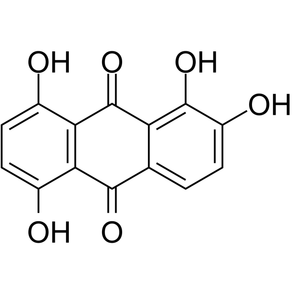
Quinalizarin is a protein kinase CK2 inhibitor with a Ki of 0.052 μM. Quinalizarin exhibits antifungal and anticancer activities. Quinalizarin induces ROS production, apoptotic signaling, mitochondrial pathway activation, cell cycle arrest, and cytotoxicity in cancer cells. Quinalizarin inhibits hyphal growth, biofilm formation, and mature biofilm integrity of Candida albicans. Quinalizarin can be used in research related to cancer and fungal infections.
For research use only. We do not sell to patients.
- Purity : 97.23%
- CAS No.: 81-61-8
- Formula: C14H8O6
- Molecular Weight:272.21
-
Storage:
-20°C, sealed storage, away from moisture and light
* In solvent : -80°C, 6 months; -20°C, 1 month (sealed storage, away from moisture and light)

To place orders, for customer services and technical support, please contact: MedChemExpress USA
Tel: 609-228-6898 E-mail: [email protected] [email protected]


-
 Biological Activity
-
Chemical Information
-
Solvent & Solubility
- Protocols
- Purity & Documentation
- References
-
 Help & FAQs


-
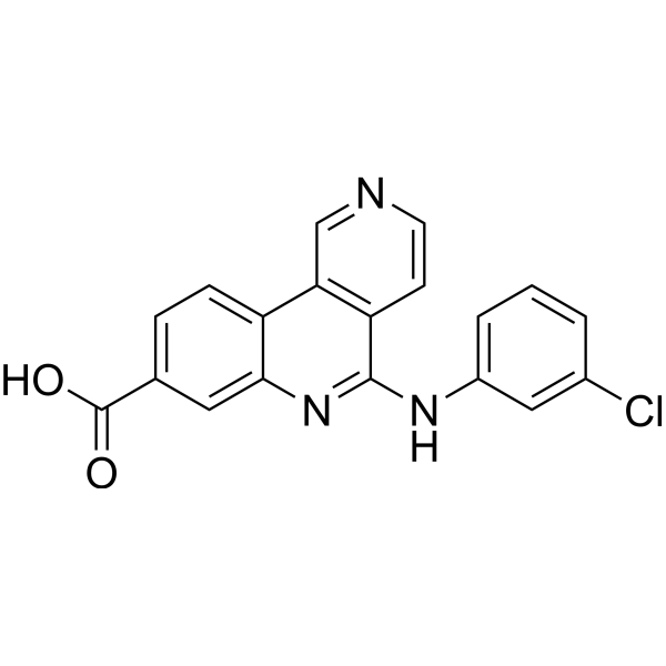
HY-50855
SilmitasertibCasein Kinase 2 Inhibitor -
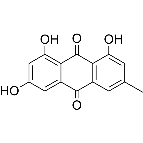
HY-14393
EmodinNatural Anthraquinone -
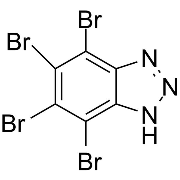
HY-14394
TBBCK2 Inhibitor -
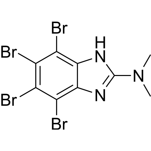
HY-15535
DMATCasein Kinase Inhibitor -
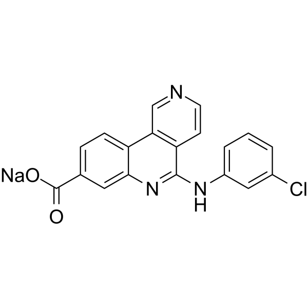
HY-50855B
Silmitasertib sodium saltCasein Kinase 2 Inhibitor -
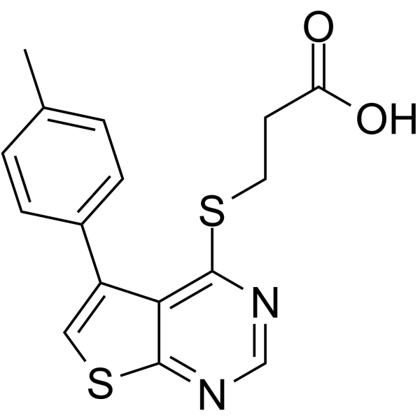
HY-15479
TTP 22Casein Kinase Inhibitor -
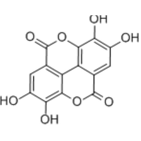
HY-B0183
Ellagic acidSHP2 Inhibitor -
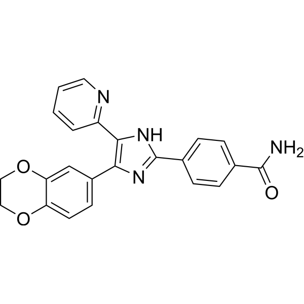
HY-10324
D4476CK1 Inhibitor -
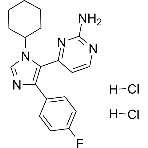
HY-15490
PF-670462 dihydrochlorideCasein Kinase Inhibitor -
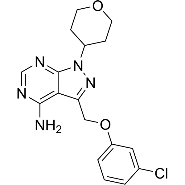
HY-12470
PF-4800567CK1 Inhibitor -
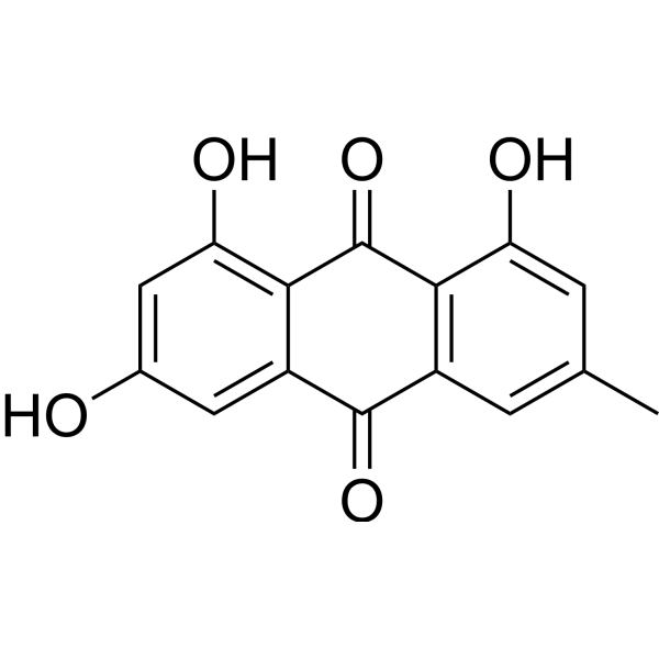
HY-14393R
Emodin (Standard) -
HY-119751
HemateinCasein Kinase Inhibitor
All Caspase Isoforms
More

Biological Activity
Description
IC50 & Target
[2]|
CK2 ~50 nM (Ki) |
In Vitro
Quinalizarin (1-100 μM; 24 h) potently inhibits the viability of SW480 and HCT-116 colorectal cancer cells in a dose-dependent manner[1].
Quinalizarin (10 μM; 3-24 h) reduces the protein expression levels of cyclin B1 and CDK1/2 in a time-dependent manner, and induces G2/M phase cell cycle arrest in SW480 colon cancer cells[1].
Quinalizarin (10 μM; 3-24 h) induces caspase-3-dependent apoptosis in SW480 colorectal cancer cells, which is characterized by upregulated expression of p-p53, Bad, activated caspase-3 and activated PARP, as well as downregulated expression of Bcl-2[1].
Quinalizarin (10 μmol/L; 3-24 h) reduces the phosphorylation levels of Akt, ERK and STAT3, and increases the phosphorylation levels of JNK and p38 in SW480 colon cancer cells[1].
Quinalizarin (5, 20 μM; 4-24 h) is cell-permeable and inhibits endogenous CK2 activity in HEK-293T and Jurkat cells[2].
Quinalizarin (1-100 µM; 24 h) potently inhibits the viability of human lung cancer A549, NCI-H23 and NCI-H460 cells, with IC50 values of 12.1, 20.24 and 27.94 µM, respectively, and exerts no significant cytotoxicity against normal liver QSG-7701 cells[3].
Quinalizarin (12.1 µM; 0-24 h) induces time-dependent G0/G1 cell cycle arrest and apoptosis in human lung cancer A549 cells[3].
Quinalizarin (12.1 µM; 0-24 h) regulates the Akt, MAPK, STAT3 and p53 signaling pathways in human lung cancer A549 cells, and promotes cell apoptosis by inhibiting the phosphorylation of Akt, ERK and STAT3 and activating the phosphorylation of JNK, p38 and p53[3].
Quinalizarin (12.1 µM; 0-24 h) induces time-dependent intracellular ROS production in human lung cancer A549 cells[3].
Quinalizarin (0.5-128 µg/mL; 24 h) exhibits antifungal activity against a variety of pathogenic yeast strains, including Fluconazole (HY-B0101)-resistant clinical Candida albicans isolates, with MIC values ranging from 0.5 to 128 µg/mL[4].
Quinalizarin (2-4 µg/mL; 10-24 h) inhibits hyphal growth of Candida albicans ATCC 10231 in a concentration-dependent manner[4].
Quinalizarin (8-80 µg/mL; 24 h) reduces the viability and biomass of preformed mature Candida albicans biofilms[4].
Quinalizarin (2-16 µg/mL; 5 h) significantly increases intracellular ROS levels in Candida albicans cells[4].
Quinalizarin (2-16 µg/mL; 5 h) can significantly dissipate the mitochondrial membrane potential of Candida albicans[4].
MedChemExpress (MCE) has not independently confirmed the accuracy of these methods. They are for reference only. Further protocols information, click here.
-
Cell Line:SW480, HCT-116 colorectal cancer cells
-
Concentration:1, 3, 10, 30, 100 μM
-
Incubation Time:24 h
-
Result:Inhibited the proliferation of SW480 and HCT-116 cells in a dose-dependent manner.
Reached a half-maximal inhibitory concentration (IC50) of 10.13 μM for SW480 cells and 13.65 μM for HCT-116 cells.
-
Cell Line:human lung cancer A549 cells
-
Concentration:12.1 µM
-
Incubation Time:0, 3, 6, 12, 24 h
-
Result:Increased the percentage of cells in the G0/G1 phase significantly in a time-dependent manner.
Decreased the percentage of cells in the G2/M phase.
-
Cell Line:human lung cancer A549 cells
-
Concentration:12.1 µM
-
Incubation Time:0, 3, 6, 12, 24 h
-
Result:Repressed protein expression levels of CDK2, CDK4, CDK6, cyclin D1, and cyclin E in a time-dependent manner.
Increased expression levels of p21 and p27 in a time-dependent manner.\nIncreased protein expression levels of Bad, cleaved caspase-3, and cleaved PARP in a time-dependent manner.
Decreased expression levels of Bcl-2 and pro caspase-3 in a time-dependent manner.\nDecreased phosphorylation levels of Akt, ERK, and STAT3 in a time-dependent manner.


Chemical Information
-
CAS No. 81-61-8
-
Appearance Solid
-
Molecular Weight 272.21
-
Formula C14H8O6
-
Color Orange to red
-
SMILES
O=C1C2=C(C(O)=CC=C2O)C(C3=CC=C(O)C(O)=C13)=O
-
Shipping
Room temperature in continental US; may vary elsewhere.
-
Storage
-20°C, sealed storage, away from moisture and light
* In solvent : -80°C, 6 months; -20°C, 1 month (sealed storage, away from moisture and light)


Solvent & Solubility
In Vitro:
DMSO : 10 mg/mL (36.74 mM; Need ultrasonic and warming; Hygroscopic DMSO has a significant impact on the solubility of product, please use newly opened DMSO)
Please refer to the solubility information to select the appropriate solvent. Once prepared, please aliquot and store the solution to prevent product inactivation from repeated freeze-thaw cycles.
Storage method and period of stock solution: -80°C, 6 months; -20°C, 1 month (sealed storage, away from moisture and light). When stored at -80°C, please use it within 6 months. When stored at -20°C, please use it within 1 month.
Please refer to the solubility information to select the appropriate solvent. Once prepared, please aliquot and store the solution to prevent product inactivation from repeated freeze-thaw cycles.
Storage method and period of stock solution: -80°C, 6 months; -20°C, 1 month (sealed storage, away from moisture and light). When stored at -80°C, please use it within 6 months. When stored at -20°C, please use it within 1 month.
Concentration (start) × Volume (start) = Concentration (final) × Volume (final)

Protocols
-
Apoptosis
Apoptosis, also called programmed cell death, is generally characterized by distinct morphological characteristics.
-
Flow cytometric DNA-content cell-cycle staining
Flow cytometric DNA-content cell-cycle staining measures the fluorescence intensity of DNA-bound fluorochromes in single cells or nuclei to estimate DNA content distributions, allowing assignment of populations to G0/G1, S, and G2/M phases by DNA histogram deconvolution. Propidium iodide (PI) intercalates into DNA, and PI fluorescence is proportional to cellular DNA content when staining is performed under conditions that make DNA accessible and minimize non-DNA signal. Cells with G2/M DNA content are expected to show approximately twice the fluorescence intensity of G0/G1 cells, while S-phase cells occupy intermediate fluorescence values. PI-based DNA-content analysis can also detect cells with fractional DNA content, often reported as sub-G1, when DNA fragmentation and extraction during staining reduce retained DNA signal in apoptotic cells. DAPI is an alternative DNA fluorochrome for univariate DNA-content analysis, while bivariate approaches combining DNA content with proliferation
-
Cell Cytotoxicity Assay
Cytotoxicity assays are usually based on the assessment of cell membrane damage, which can also be indirectly detected by measuring cell viability. Detection methods include MTT assay, CKK-8 assay, LDH assay and ATP assay, etc.
-
Mammalian live/dead viability and cytotoxicity staining
Live/dead viability and cytotoxicity staining assays are based on the simultaneous detection of intracellular esterase activity in metabolically active (viable) cells and membrane integrity loss in non-viable cells. In commonly used dual-staining approaches, membrane-permeant fluorogenic substrates are converted by intracellular esterases into fluorescent products in live cells, while impermeant DNA-binding dyes selectively enter cells with compromised plasma membranes and label nucleic acids in dead or dying cells, enabling discrimination between viable and non-viable populations by fluorescence microscopy or flow cytometry.
-
ROS/oxidative-stress fluorescent staining
ROS/oxidative-stress fluorescent staining uses cell-permeant fluorogenic probes that become fluorescent after oxidation inside cells or tissues; commonly used examples include DCFH-DA/DCFDA for broad cellular oxidant detection, DHE for superoxide-related signal detection, MitoSOX for mitochondrial superoxide-related signal detection, and CellROX probes for oxidative-stress-associated fluorescence readouts. The assay detects probe oxidation rather than a single ROS species unless the probe and analysis method have been chemically validated for that species. DCFH-DA enters cells, is deacetylated by intracellular esterases to DCFH, and produces fluorescent DCF after oxidation, so the readout is used as an operational measure of total cellular oxidative stress rather than a species-specific ROS measurement. DHE and MitoSOX can report superoxide-related oxidation, but red fluorescence alone can include non-specific ethidium-like oxidation products; HPLC or optimized spectral approaches are
-
BrdU Incorporation Assay
Bromodeoxyuridine (BrdU) incorporation assay is based on the principle that BrdU, a thymidine analog, is incorporated into newly synthesized DNA during the S phase of the cell cycle, thereby serving as a marker of DNA replication and cellular proliferation. Incorporated BrdU can be detected using anti-BrdU antibodies following DNA denaturation, enabling visualization or quantification of proliferating cells through immunochemical detection methods such as immunofluorescence or immunohistochemistry.
-
Apoptosis Solutions
Apoptosis is a regulated, generally non-lytic cell-death pathway that removes unwanted, damaged, infected, or abnormal cells through coordinated morphological changes, caspase activation, DNA fragmentation, and membrane remodeling. The intrinsic apoptosis pathway is controlled mainly by mitochondrial outer membrane permeabilization, BCL-2 family proteins, cytochrome c release, apoptosome formation, caspase-9 activation, and downstream executioner caspase-3/7 activation. The extrinsic apoptosis pathway is initiated by death receptors such as Fas, TNFR, and TRAIL receptors, which recruit adaptor proteins and activate caspase-8 before engaging executioner caspases or mitochondrial amplification through BID cleavage. Apoptosis is linked to many phenotypes, including cancer cell killing, tissue homeostasis, immune regulation, neurodegeneration, infection response, and treatment-induced cytotoxicity; unresolved questions include how apoptosis interacts with necroptosis, pyroptosis, ferroptos
-
Protocol for Cell Cycle
Cell-cycle analysis by flow cytometry measures DNA content in single cells to estimate the fraction of cells in G0/G1, S, and G2/M phases. Propidium iodide intercalates into DNA, and after RNA removal with RNase, fluorescence intensity reflects cellular DNA content: 2N cells are assigned to G0/G1, cells between 2N and 4N to S phase, and 4N cells to G2/M. DNA-content analysis alone cannot reliably separate G0 from G1 or G2 from M. Ki-67 can distinguish quiescent G0 cells from cycling cells, EdU or BrdU incorporation marks active DNA synthesis in S phase, and phospho-histone H3 staining identifies mitotic cells within the 4N population.
-
Filamentous Fungal Mold Culture and Sporulation
Filamentous fungal mold culture and sporulation assays grow hyphae under defined nutritional and environmental conditions until asexual spores, commonly conidia, are produced; the main readouts are colony growth, sporulation onset, conidial yield, conidial morphology, viability, and, when relevant, downstream infectivity or stress phenotype.
-
Fungal Biofilm Culture
Fungal biofilm culture is an in vitro method for growing surface-attached fungal communities, most commonly Candida albicans, on abiotic substrates such as polystyrene wells, silicone elastomer, or polymethylmethacrylate; the assay models adhesion, proliferation, filamentation, extracellular-matrix-associated maturation, and dispersion. Biofilm output can be read by optical density at 600 nm for adherent biomass, XTT reduction for metabolic activity, CFU recovery for viable attached or dispersed cells, and microscopy for architecture.
-
Cell Viability Determination by MTT Colorimetric Assay
The following protocol uses the MTT colorimetric assay as a classic literature-established method for assessing cell viability/metabolic activity in cultured mammalian cells. MTT[3-(4,5-dimethylthiazol-2-yl)-2,5-diphenyltetrazolium bromide] is reduced by metabolically active cells to a colored formazan product; the amount of formazan is quantified spectrophotometrically and provides an indirect measure of metabolically active viable cells. Importantly, MTT reduction reflects cellular oxidoreductase/metabolic activity rather than an absolute direct count of living cells, so changes in cellular metabolism can alter the signal independently of cell number.


Purity & Documentation
-
Data Sheet (283 KB)
-
SDS (537 KB)
- English - EN (537 KB)
- Français - FR (537 KB)
- Deutsch - DE (537 KB)
- Norwegian - NO (537 KB)
- Español - ES (537 KB)
- Swedish - SV (537 KB)
- Italian - IT (537 KB)
- Korean - KR (537 KB)
- Portuguese - PT (537 KB)
-
Handling Instructions (2659 KB)
References
[1]. Meng LQ, et al. Quinalizarin Induces Apoptosis through Reactive Oxygen Species (ROS)-Mediated Mitogen-Activated Protein Kinase (MAPK) and Signal Transducer and Activator of Transcription 3 (STAT3) Signaling Pathways in Colorectal Cancer Cells. Med Sci Monit. 2018;24:3710-3719. Published 2018 Jun 3. [Content Brief]
[2]. Cozza G, et al. Quinalizarin as a potent, selective and cell-permeable inhibitor of protein kinase CK2. Biochem J. 2009;421(3):387-395. Published 2009 Jul 15. [Content Brief]
[3]. Meng LQ, et al. Quinalizarin exerts an anti-tumour effect on lung cancer A549 cells by modulating the Akt, MAPK, STAT3 and p53 signalling pathways. Mol Med Rep. 2018;17(2):2626-2634. [Content Brief]
[4]. Janeczko M, et al. Quinalizarin as a potential antifungal drug for the treatment of Candida albicans fungal infection in cancer patients. Microbiol Spectr. 2024 Mar 5;12(3):e0365223. [Content Brief]


Complete Stock Solution Preparation Table
Please refer to the solubility information to select the appropriate solvent. Once prepared, please aliquot and store the solution to prevent product inactivation from repeated freeze-thaw cycles.
Storage method and period of stock solution: -80°C, 6 months; -20°C, 1 month (sealed storage, away from moisture and light). When stored at -80°C, please use it within 6 months. When stored at -20°C, please use it within 1 month.
| Optional Solvent | Concentration Solvent Mass | 1 mg | 5 mg | 10 mg | 25 mg |
|---|---|---|---|---|---|
| DMSO | 1 mM | 3.6736 mL | 18.3682 mL | 36.7363 mL | 91.8409 mL |
| 5 mM | 0.7347 mL | 3.6736 mL | 7.3473 mL | 18.3682 mL | |
| 10 mM | 0.3674 mL | 1.8368 mL | 3.6736 mL | 9.1841 mL | |
| 15 mM | 0.2449 mL | 1.2245 mL | 2.4491 mL | 6.1227 mL | |
| 20 mM | 0.1837 mL | 0.9184 mL | 1.8368 mL | 4.5920 mL | |
| 25 mM | 0.1469 mL | 0.7347 mL | 1.4695 mL | 3.6736 mL | |
| 30 mM | 0.1225 mL | 0.6123 mL | 1.2245 mL | 3.0614 mL |


Quinalizarin Related Classifications
HY-124833 Related Classifications
 Powered by Bioz