



Aureusidin

Based on 1 Customer Validation
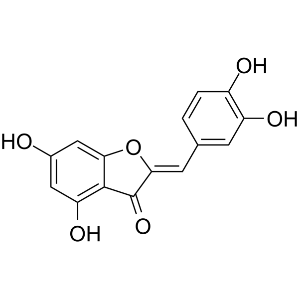
Aureusidin is a flavonoid compound that is isolated and extracted from Antirrhinum majus and exhibits oral activity. Aureusidin possesses antioxidant, neuroprotective, and anti-inflammatory properties. Aureusidin directly targets and binds MD2 and Caspase-3 with high affinity, synergistically inhibits the TLR4/NF-κB inflammatory pathway and GSDME-mediated pyroptosis, and activates the Nrf2/HO-1 antioxidant axis via the ROS/MAPKs pathway. Aureusidin is a bovine liver arginase inhibitor with an IC50 of 57.1 µM. Aureusidin is used for research on inflammation-related diseases (osteoarthritis), acute liver injury, and endothelial dysfunction.
Nur für Forschungszwecke. Wir verkaufen nicht an Patienten.
- Reinheit : 97.01%
- CAS. Nr.: 38216-54-5
- Formel: C15H10O6
- Molecular Weight:286.24
-
Speicherung:
4°C, protect from light
* In solvent : -80°C, 6 months; -20°C, 1 month (protect from light)

To place orders, for customer services and technical support, please contact: MedChemExpress USA
Tel: 609-228-6898 E-mail: [email protected] [email protected]


-
 Biologische Aktivität
-
Chemical Information
-
Lösungsmittel & Löslichkeit
- Protokoll
- Reinheit & Dokumentation
- Verweise
-
 Help & FAQs


Alle Endogenous Metabolite Isoform-spezifische Produkte anzeigen
MoreAlle Caspase Isoform-spezifische Produkte anzeigen
MoreAlle TNF Receptor Isoform-spezifische Produkte anzeigen
More

Biologische Aktivität
Beschreibung
|
NF-κB |
p38 MAP kinase |
Caspase-3 |
TLR4 |
HO-1 |
iNOS |
TNF-α |
Cellular Effect
|
Cell Line

|
Type | Value | Description | References |
|---|---|---|---|---|
| HeLa | IC50 |
18.5 μM
Compound: 36
|
Inhibition of HDAC in human HeLa cell extract after 15 mins by fluorescence assay
Inhibition of HDAC in human HeLa cell extract after 15 mins by fluorescence assay
|
[PMID: 25455492] |
| HT-22 | EC50 |
11.9 μM
Compound: 2
|
Neuroprotective activity against glutamate induced-neurotoxicity in mouse HT-22 cells assessed as cell viability preincubated for 4 hrs followed by glutamate stimulation and measured after 12 hrs by MTT assay
Neuroprotective activity against glutamate induced-neurotoxicity in mouse HT-22 cells assessed as cell viability preincubated for 4 hrs followed by glutamate stimulation and measured after 12 hrs by MTT assay
|
[PMID: 36126323] |
In Vitro
Aureusidin (20-60 μM; 1-6 h) promotes Nrf2 nuclear translocation, downregulates Keap1 expression, and upregulates HO-1 and NQO1 expression in RAW264.7 cells.[3]
Aureusidin (60 μM; 15-60 min) significantly increases Akt and MAPKs phosphorylation in RAW264.7 cells[3].
Aureusidin (2.5-20 mM; 5 min) directly binds MD2 protein in recombinant MD2 protein solution in a dose-dependent manner[4].
Aureusidin (10-100 µM; 1 h) inhibits purified bovine liver arginase with an IC50 of 57.1 µM[1].
Aureusidin (10 μM; 1 h) directly targets and binds to Caspase-3 protein in cell lysates of primary rat chondrocytes as a biotin-labeled probe via Pull-down assay[2].
MedChemExpress (MCE) has not independently confirmed the accuracy of these methods. They are for reference only.
-
Cell Line:RAW264.7
-
Concentration:1, 10, 20, 40, 60, 80, 100 μM
-
Incubation Time:24 h
-
Result:Did not inhibit the proliferation of RAW264.7 cells up to 60 μM.
Significantly inhibited the survival rate of RAW264.7 cells at 80 and 100 μM.
-
Cell Line:RAW264.7
-
Concentration:20, 40, 60 μM
-
Incubation Time:1 h (pretreatment); 24 h (co-treatment with LPS)
-
Result:Obviously inhibited PGE2 and TNF-α release induced by LPS in a dose-dependent manner.
-
Cell Line:RAW264.7
-
Concentration:20, 40, 60 μM
-
Incubation Time:1 h (pretreatment); 24 h (co-treatment with LPS)
-
Result:Obviously inhibited the LPS-induced expression of iNOS and COX-2 proteins.
-
Cell Line:RAW264.7
-
Concentration:20, 40, 60 μM
-
Incubation Time:1 h (pretreatment); 4 h (co-treatment with LPS)
-
Result:Significantly inhibited the gene expressions of iNOS, TNF-α, COX-2, IL-1β, and IL-6 in a dose-dependent manner.
-
Cell Line:RAW264.7
-
Concentration:60 μM
-
Incubation Time:1 h (pretreatment); 1 h (co-treatment with LPS)
-
Result:NF-κB was mainly concentrated in the cytoplasm after treatment.
-
Cell Line:RAW264.7
-
Concentration:20, 40, 60 μM
-
Incubation Time:1 h (pretreatment)
-
Result:Prevented the expression of p-IκBα induced by LPS.
Significantly inhibited the expression of NF-κB in the nucleus.
-
Cell Line:Kupffer cells (KCs)
-
Concentration:5, 10, 20 mM
-
Incubation Time:6 h (pretreatment); 3, 6, 12 h (co-treatment with LPS)
-
Result:LPS induction significantly decreased the viability of KCs, which was significantly suppressed by Aur in a dose-dependent pattern.
-
Cell Line:Kupffer cells (KCs)
-
Concentration:5, 10, 20 mM
-
Incubation Time:6 h (pretreatment); 12 h (co-treatment with LPS)
-
Result:Inhibited the apoptosis of KCs.
-
Cell Line:Kupffer cells (KCs)
-
Concentration:5, 10, 20 mM
-
Incubation Time:6 h (pretreatment); 12 h (co-treatment with LPS)
-
Result:LPS activated the TLR4 signal and upregulated the expression of TLR4, MyD88, MD2, and p-P65, while Aur treatment downregulated these protein levels.
-
Cell Line:Primary rat chondrocytes
-
Concentration:5, 10, 20 μM
-
Incubation Time:48 h
-
Result:Dose-dependently reversed IL-1β-induced aberrant expression changes of Col2a1, Acan, Adamts5, Mmp13, and Mmp3.
-
Cell Line:Primary rat chondrocytes
-
Concentration:5, 10, 20 μM
-
Incubation Time:48 h
-
Result:Dose-dependently alleviated pyroptotic morphological changes and reduced PI-positive cell numbers.
-
Cell Line:Primary rat chondrocytes
-
Concentration:5, 10, 20 μM
-
Incubation Time:48 h
-
Result:Dose-dependently reversed IL-1β-induced increase in LDH release.
-
Cell Line:Primary rat chondrocytes
-
Concentration:5, 10, 20 μM
-
Incubation Time:48 h
-
Result:Dose-dependently decreased the IL-1β-elevated PI-positive cell percentage.
-
Cell Line:Primary rat chondrocytes
-
Concentration:5, 10, 20 μM
-
Incubation Time:48 h
-
Result:Dose-dependently inhibited IL-1β-induced upregulation of GSDME-N, Caspase-3, and C-Caspase-3 expression.
-
Cell Line:Primary rat chondrocytes
-
Concentration:20 μM
-
Incubation Time:48 h
-
Result:Did not further modify the expression of Collagen II, Aggrecan, ADAMTS-5, MMP13, and MMP3 compared to Z-DEVD-FMK treatment alone.\nDid not further alter GSDME-N, Caspase-3, and C-Caspase-3 expression compared to Z-DEVD-FMK treatment alone.
In Vivo
MedChemExpress (MCE) has not independently confirmed the accuracy of these methods. They are for reference only.
-
Animal Model:C57BL/6 mice[4]
-
Dosage:20 mg/kg
-
Administration:i.g.; once daily; 7 consecutive days
-
Result:Attenuated tissue lesions, relieved inflammatory response and tissue edema.
Suppressed expression of MD2.
Significantly downregulated expression of AST and ALT.
Suppressed expression of inflammatory factors including IL-18, IL-1β, and TNF-α in peripheral blood and liver tissues.
Resulted in barely detectable expression levels of TLR4, MyD88, MD2, and p-P65, lower than the LPS/D-GalN group.
-
Animal Model:Sprague-Dawley rats (male, 8 weeks old, ACLT-induced osteoarthritis)[2]
-
Dosage:5, 10, and 20 mg/kg
-
Administration:i.g.; once daily; 6 weeks
-
Result:Alleviated cartilage damage and cartilage matrix degradation in a dose-dependent manner.
Reduced OARSI scores in a dose-dependent manner.
Reduced elevated serum CTX-II levels dose-dependently, with 20 mg/kg restoring levels to 38.98 pg/mL.
Reduced elevated serum COMP levels dose-dependently, with 20 mg/kg restoring levels to 3.40 ng/mL.
Reversed ACLT-induced downregulation of Col2a1 and Acan and upregulation of Adamts5, Mmp13, and Mmp3 in cartilage tissue in a dose-dependent manner.
Reduced C-Caspase-3 and GSDME-positive cell numbers in cartilage tissues dose-dependently.
Downregulated GSDME-N, Caspase-3, and C-Caspase-3 protein expression dose-dependently with no effect on GSDME-FL.
Reduced elevated IL-1β and IL-18 levels in cartilage and serum dose-dependently.


Chemical Information
-
CAS. Nr. 38216-54-5
-
Appearance Solid
-
Molecular Weight 286.24
-
Formel C15H10O6
-
Color Yellow to orange
-
SMILES
O=C1C2=C(O)C=C(O)C=C2O/C1=C\C3=CC(O)=C(C=C3)O
-
Structure Classification
-
Versand
Room temperature in continental US; may vary elsewhere.
-
Speicherung
4°C, protect from light
* In solvent : -80°C, 6 months; -20°C, 1 month (protect from light)


Lösungsmittel & Löslichkeit
In Vitro:
DMSO : 62.5 mg/mL (218.35 mM; ultrasonic and warming and heat to 60°C; Hygroscopic DMSO has a significant impact on the solubility of product, please use newly opened DMSO)
Please refer to the solubility information to select the appropriate solvent. Once prepared, please aliquot and store the solution to prevent product inactivation from repeated freeze-thaw cycles.
Storage method and period of stock solution: -80°C, 6 months; -20°C, 1 month (protect from light). When stored at -80°C, please use it within 6 months. When stored at -20°C, please use it within 1 month.
Please refer to the solubility information to select the appropriate solvent. Once prepared, please aliquot and store the solution to prevent product inactivation from repeated freeze-thaw cycles.
Storage method and period of stock solution: -80°C, 6 months; -20°C, 1 month (protect from light). When stored at -80°C, please use it within 6 months. When stored at -20°C, please use it within 1 month.
Konzentration (Stammlösung) × Volumen (Stammlösung) = Konzentration (Ziellösung) × Volumen (Ziellösung)
In Vivo:
Select the appropriate dissolution method based on your experimental animal and administration route.
- For the following dissolution methods, please ensure to first prepare a clear stock solution using an In Vitro approach and then sequentially add co-solvents:
- To ensure reliable experimental results, the clarified stock solution can be appropriately stored based on storage conditions. As for the working solution for In Vivo experiments, it is recommended to prepare freshly and use it on the same day.
- The percentages shown for the solvents indicate their volumetric ratio in the final prepared solution. If precipitation or phase separation occurs during preparation, heat and/or sonication can be used to aid dissolution.
Add each solvent one by one: 10% DMSO 40% PEG300 5% Tween-80 45% Saline
Solubility: ≥ 2.08 mg/mL (7.27 mM); Clear solution
This protocol yields a clear solution of ≥ 2.08 mg/mL (saturation unknown).
Taking 1 mL working solution as an example, add 100 μL DMSO stock solution (20.8 mg/mL) to 400 μL PEG300, and mix evenly; then add 50 μL Tween-80 and mix evenly; then add 450 μL Saline to adjust the volume to 1 mL.
Preparation of Saline: Dissolve 0.9 g sodium chloride in ddH₂O and dilute to 100 mL to obtain a clear Saline solution.
Add each solvent one by one: 10% DMSO 90% (20% SBE-β-CD in Saline)
Solubility: ≥ 2.08 mg/mL (7.27 mM); Clear solution
This protocol yields a clear solution of ≥ 2.08 mg/mL (saturation unknown).
Taking 1 mL working solution as an example, add 100 μL DMSO stock solution (20.8 mg/mL) to 900 μL 20% SBE-β-CD in Saline, and mix evenly.
Preparation of 20% SBE-β-CD in Saline (4°C, storage for one week): 2 g SBE-β-CD powder is dissolved in 10 mL Saline, completely dissolve until clear.
In Vivo Dissolution Calculator
Please enter the basic information of animal experiments:
-
-
-
-
Recommended: Prepare an additional quantity of animals to account for potential losses during experiments.
Please enter your animal formula composition:
-
%DMSO +
Recommended: Keep the proportion of DMSO in working solution below 2% if your animal is weak.
-
%+
-
+%Tween-80 + +
-
%Saline +
The co-solvents required include: DMSO, . All of co-solvents are available by MedChemExpress (MCE). , Tween 80. All of co-solvents are available by MedChemExpress (MCE).
Working solution concentration: 0.22 mg/mL
Method for preparing stock solution: mg drug dissolved in μL DMSO. Stock solution concentration: mg/mL. * In solvent : -80°C, 6 months; -20°C, 1 month (protect from light)
1. Take μL DMSO stock solution;
2. Add μL .
μL , mix evenly;
3. Then add μL Tween 80, mix evenly;
4. Then add μL
Please ensure that the stock solution in the first step is dissolved to a clear state, and add co-solvents in sequence. You can use ultrasonic heating (ultrasonic cleaner, recommended frequency 20-40 kHz), vortexing, etc. to assist dissolution.


Reinheit & Dokumentation
-
Data Sheet (303 KB)
-
SDS (251 KB)
- English - EN (251 KB)
- Français - FR (251 KB)
- Deutsch - DE (251 KB)
- Norwegian - NO (251 KB)
- Español - ES (251 KB)
- Swedish - SV (251 KB)
- Italian - IT (251 KB)
- Korean - KR (251 KB)
- Portuguese - PT (251 KB)
-
Handling Instructions (2659 KB)
Verweise
[1]. Arraki K, et al. Mammalian Arginase Inhibitory Activity of Methanolic Extracts and Isolated Compounds from Species. Molecules (Basel, Switzerland). 2021 Mar 18;26(6):1694. [Content Brief]
[2]. Feng S, et al. Aureusidin protects against osteoarthritis via Caspase-3/gasdermin E-mediated pyroptosis. Toxicology and applied pharmacology. 2026 Sep;514:117911. [Content Brief]


Complete Stock Solution Preparation Table
Please refer to the solubility information to select the appropriate solvent. Once prepared, please aliquot and store the solution to prevent product inactivation from repeated freeze-thaw cycles.
Storage method and period of stock solution: -80°C, 6 months; -20°C, 1 month (protect from light). When stored at -80°C, please use it within 6 months. When stored at -20°C, please use it within 1 month.
| Optional Solvent | Concentration Solvent Mass | 1 mg | 5 mg | 10 mg | 25 mg |
|---|---|---|---|---|---|
| DMSO | 1 mM | 3.4936 mL | 17.4679 mL | 34.9357 mL | 87.3393 mL |
| 5 mM | 0.6987 mL | 3.4936 mL | 6.9871 mL | 17.4679 mL | |
| 10 mM | 0.3494 mL | 1.7468 mL | 3.4936 mL | 8.7339 mL | |
| 15 mM | 0.2329 mL | 1.1645 mL | 2.3290 mL | 5.8226 mL | |
| 20 mM | 0.1747 mL | 0.8734 mL | 1.7468 mL | 4.3670 mL | |
| 25 mM | 0.1397 mL | 0.6987 mL | 1.3974 mL | 3.4936 mL | |
| 30 mM | 0.1165 mL | 0.5823 mL | 1.1645 mL | 2.9113 mL | |
| 40 mM | 0.0873 mL | 0.4367 mL | 0.8734 mL | 2.1835 mL | |
| 50 mM | 0.0699 mL | 0.3494 mL | 0.6987 mL | 1.7468 mL | |
| 60 mM | 0.0582 mL | 0.2911 mL | 0.5823 mL | 1.4557 mL | |
| 80 mM | 0.0437 mL | 0.2183 mL | 0.4367 mL | 1.0917 mL | |
| 100 mM | 0.0349 mL | 0.1747 mL | 0.3494 mL | 0.8734 mL |


Aureusidin Related Classifications
HY-N9834 Related Classifications
 Powered by Bioz
Keywords
- Aureusidin
- 38216-54-5
- Endogenous Metabolite
- Reactive Oxygen Species (ROS)
- Toll-like Receptor (TLR)
- Caspase
- Pyroptosis
- Apoptosis
- NF-κB
- Arginase
- Keap1-Nrf2
- Heme Oxygenase (HO)
- p38 MAPK
- PGE synthase
- TNF Receptor
- NO Synthase
- COX
- bovine liver arginase
- RAW264.7 cells
- MD2
- Kupffer cells
- TLR4/MD2-NF-κB signaling
- Nrf2 nuclear translocation
- primary rat chondrocytes
- HO-1
- IκBα phosphorylation
- NF-κB nuclear translocation
- Inhibitor
- inhibitor
- inhibit