



Neoandrographolide
Based on 1 publication(s) in Google Scholar
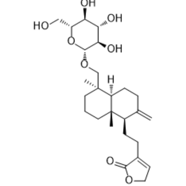
Neoandrographolide is a diterpenoid compound isolated from Andrographis paniculata. Neoandrographolide inhibits osteoclasts differentiation and bone resorption through inhibition of MAPK/NF-κB/PI3K/AKT/GSK3β/PPAR/CAMK signaling pathway. Neoandrographolide inhibits apoptosis in rat embryonic ventricular cardiomyocytes. Neoandrographolide inhibits iNOS and the generation of ROS, activates eNOS, exhibiting anti-inflammatory and hypolipidemic activity.
Nur für Forschungszwecke. Wir verkaufen nicht an Patienten.
- Reinheit : 99.41%
- CAS. Nr.: 27215-14-1
- Formel: C26H40O8
- Molecular Weight:480.59
-
Speicherung:
4°C, protect from light
* In solvent : -80°C, 2 years; -20°C, 1 year (protect from light)

To place orders, for customer services and technical support, please contact: MedChemExpress USA
Tel: 609-228-6898 E-mail: [email protected] [email protected]


-
 Biologische Aktivität
-
Chemical Information
-
Lösungsmittel & Löslichkeit
- Protokoll
- Reinheit & Dokumentation
- Verweise
-
 Help & FAQs


-

HY-15371
ForskolinAdenylate Cyclase Activator -
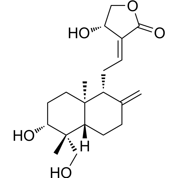
HY-N0191
AndrographolideNF-κB Inhibitor -
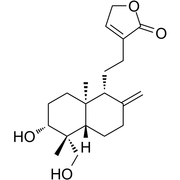
HY-N4323
14-DeoxyandrographolideCalcium Channel Blocker -
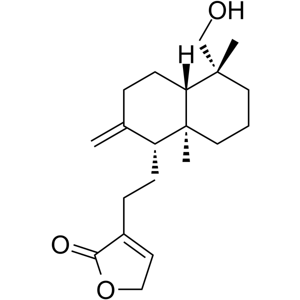
HY-N9388
AndrograpaninAnti-Inflammatory Agent -
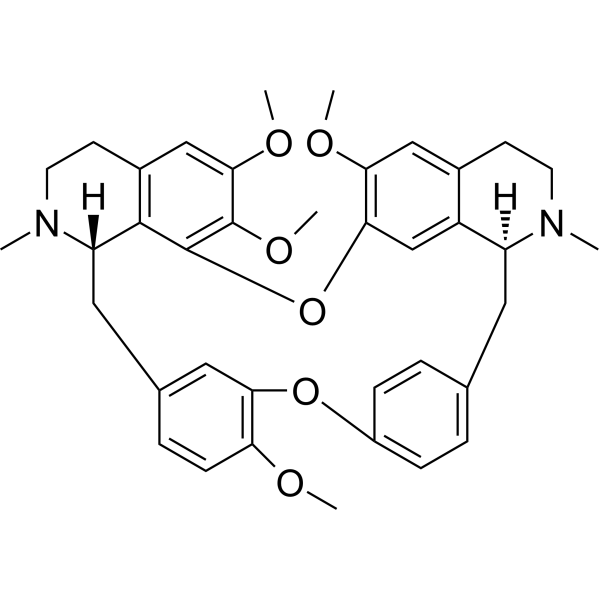
HY-13764
TetrandrineCalcium Channel Inhibitor -
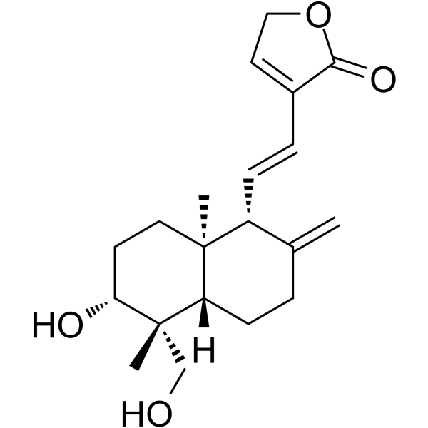
HY-N1490
14-Deoxy-11,12-didehydroandrographolideNF-κB Inhibitor -
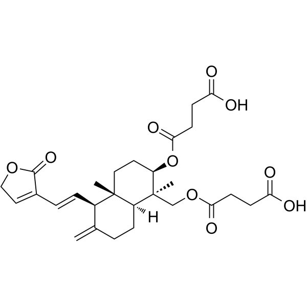
HY-N0677
Dehydroandrographolide succinateInfluenza Virus Inhibitor -

HY-N0721R
Neoandrographolide (Standard)Natural Product -
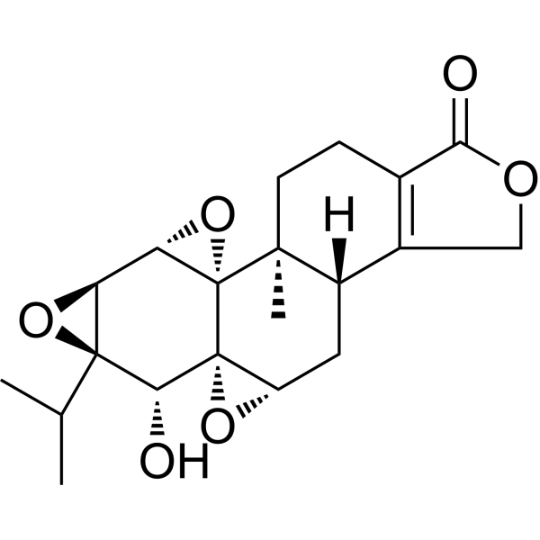
HY-32735
TriptolideImmunosuppressive Agent -
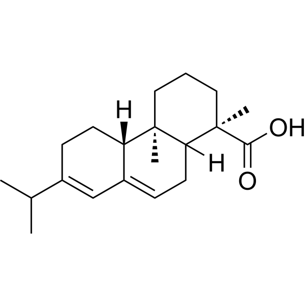
HY-N6871
Abietic acidBacterial Inhibitor; IKKβ Inhibitor -
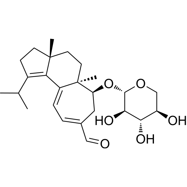
HY-111954
(+)-Erinacin AAnticancer Agent -
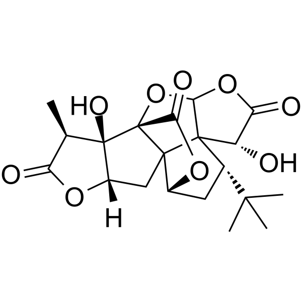
HY-B0355
Ginkgolide AGABA Antagonist
Publications Citing Use of MedChemExpress (MCE) Neoandrographolide
More

Biologische Aktivität
Beschreibung
Cellular Effect
|
Cell Line

|
Type | Value | Description | References |
|---|---|---|---|---|
| HL-60 | GI50 |
26.67 μM
Compound: 5
|
Antiproliferative activity against human HL60 cells by trypan blue assay
Antiproliferative activity against human HL60 cells by trypan blue assay
|
[PMID: 18357994] |
In Vitro
Neoandrographolide (0-30 μM, 6 days) inhibits osteoclast differentiation-related gene expression of NFATc1, c-Fos, CTSK and MMP9, inhibits RANKL-induced osteoclasts maturation in murine bone marrow macrophage[2].
Neoandrographolide (10-90 μM, 24 h) inhibits the expression of inflammatory factors TNF-α and IL-1β[3].
Neoandrographolide (7.5-150 μM, 24-36 h) inhibits PMA (HY-18739)-stimulated respiratory burst, inhibits LPS (HY-D1056)-induced TNF-α and NO production in cell RAW264.7[4].
MedChemExpress (MCE) has not independently confirmed the accuracy of these methods. They are for reference only.
-
Cell Line:murine bone marrow macrophage
-
Concentration:0-30 μM
-
Incubation Time:6 days
-
Result:Inhibited the expression of NFATc1, c-Fos, CTSK and MMP9.
In Vivo
Neoandrographolide (10 mg/kg, ip, once a day for 6 days) alleviates the ischemia-reperfusion injury in mouse models[3].
Neoandrographolide (50-150 mg/kg, po, single dose) inhibits Dimethyl benzene-induced ear edema, exhibits anti-inflammatory activity in mouse models[4].
MedChemExpress (MCE) has not independently confirmed the accuracy of these methods. They are for reference only.
-
Animal Model:OVX-induced osteoporosis in mouse models[2]
-
Dosage:15-30 mg/kg
-
Administration:ip, 3 times a week for 8 weeks
-
Result:Alleviated OVX-induced bone loss.
-
Animal Model:Mouse myocardial ischemia-reperfusion injury model[3]
-
Dosage:10 mg/kg
-
Administration:ip, once a day for 6 days
-
Result:Improved the cardiac contractile function, reduced the levels of myocardial injury markers (CK-MB and cTNI), reduced infarction area.
-
Animal Model:Dimethyl benzene-induced ear edema in mouse models[4]
-
Dosage:50-150 mg/kg
-
Administration:po, single dose
-
Result:Inhibited ear edema at a dose of 150 mg/kg.
- Detection of Apoptosis
- TUNEL staining for apoptotic DNA fragmentation
- Annexin V plus membrane-impermeant dye apoptosis staining
- ROS/oxidative-stress fluorescent staining
- Apoptosis Solutions
- Research Protocol for Inflammation-related Diseases
- Osteoclast differentiation from monocyte/macrophage precursors


Chemical Information
-
CAS. Nr. 27215-14-1
-
Appearance Solid
-
Molecular Weight 480.59
-
Formel C26H40O8
-
Color White to off-white
-
SMILES
C[C@@]([C@@H]1CCC2=CCOC2=O)(CCC3)[C@@](CCC1=C)([H])[C@]3(C)CO[C@@H]([C@@H]([C@@H](O)[C@@H]4O)O)O[C@@H]4CO
-
Synonyms
Neoandrographiside
-
Structure Classification
-
Versand
Room temperature in continental US; may vary elsewhere.
-
Speicherung
4°C, protect from light
* In solvent : -80°C, 2 years; -20°C, 1 year (protect from light)


Publications (1)
-
Journal Impact Factor
-
Most Recent
-
Vet Microbiol
The Chinese medicine monomer Schisandrin C inhibits PRRSV infection by regulating the OGT-PI3K/AKT/mTOR signaling pathway. [Abstract]2026 May:316:110992. PMID: 41865607


Lösungsmittel & Löslichkeit
In Vitro:
DMSO : 100 mg/mL (208.08 mM; Need ultrasonic; Hygroscopic DMSO has a significant impact on the solubility of product, please use newly opened DMSO)
Please refer to the solubility information to select the appropriate solvent. Once prepared, please aliquot and store the solution to prevent product inactivation from repeated freeze-thaw cycles.
Storage method and period of stock solution: -80°C, 2 years; -20°C, 1 year (protect from light). When stored at -80°C, please use it within 2 years. When stored at -20°C, please use it within 1 year.
Please refer to the solubility information to select the appropriate solvent. Once prepared, please aliquot and store the solution to prevent product inactivation from repeated freeze-thaw cycles.
Storage method and period of stock solution: -80°C, 2 years; -20°C, 1 year (protect from light). When stored at -80°C, please use it within 2 years. When stored at -20°C, please use it within 1 year.
Konzentration (Stammlösung) × Volumen (Stammlösung) = Konzentration (Ziellösung) × Volumen (Ziellösung)
In Vivo:
Select the appropriate dissolution method based on your experimental animal and administration route.
- For the following dissolution methods, please ensure to first prepare a clear stock solution using an In Vitro approach and then sequentially add co-solvents:
- To ensure reliable experimental results, the clarified stock solution can be appropriately stored based on storage conditions. As for the working solution for In Vivo experiments, it is recommended to prepare freshly and use it on the same day.
- The percentages shown for the solvents indicate their volumetric ratio in the final prepared solution. If precipitation or phase separation occurs during preparation, heat and/or sonication can be used to aid dissolution.
In Vivo Dissolution Calculator
Please enter the basic information of animal experiments:
-
-
-
-
Recommended: Prepare an additional quantity of animals to account for potential losses during experiments.
Please enter your animal formula composition:
-
%DMSO +
Recommended: Keep the proportion of DMSO in working solution below 2% if your animal is weak.
-
%+
-
+%Tween-80 + +
-
%Saline +
The co-solvents required include: DMSO, . All of co-solvents are available by MedChemExpress (MCE). , Tween 80. All of co-solvents are available by MedChemExpress (MCE).
Working solution concentration: 0.22 mg/mL
Method for preparing stock solution: mg drug dissolved in μL DMSO. Stock solution concentration: mg/mL. * In solvent : -80°C, 2 years; -20°C, 1 year (protect from light)
1. Take μL DMSO stock solution;
2. Add μL .
μL , mix evenly;
3. Then add μL Tween 80, mix evenly;
4. Then add μL
Please ensure that the stock solution in the first step is dissolved to a clear state, and add co-solvents in sequence. You can use ultrasonic heating (ultrasonic cleaner, recommended frequency 20-40 kHz), vortexing, etc. to assist dissolution.


Reinheit & Dokumentation
-
Data Sheet (279 KB)
-
SDS (394 KB)
- English - EN (394 KB)
- Français - FR (394 KB)
- Deutsch - DE (394 KB)
- Norwegian - NO (394 KB)
- Español - ES (394 KB)
- Swedish - SV (394 KB)
- Italian - IT (394 KB)
- Korean - KR (394 KB)
- Portuguese - PT (394 KB)
-
Handling Instructions (2659 KB)
Verweise
[2]. Tang K, et al., Neoandrographolide inhibits mature osteoclast differentiation to alleviate bone loss and treat osteoporosis. Front Pharmacol. 2025 Feb 11;16:1466057. [Content Brief]
[3]. Liu Y, et al., Amelioratory effect of neoandrographolide on myocardial ischemic-reperfusion injury by its anti-inflammatory and anti-apoptotic activities. Environ Toxicol. 2021 Dec;36(12):2367-2379. [Content Brief]
[4]. Liu J, et al., In vivo and in vitro anti-inflammatory activities of neoandrographolide. Am J Chin Med. 2007;35(2):317-28. [Content Brief]
[5]. Yang T, et al., Hypolipidemic effects of andrographolide and neoandrographolide in mice and rats. Phytother Res. 2013 Apr;27(4):618-23. [Content Brief]


Complete Stock Solution Preparation Table
Please refer to the solubility information to select the appropriate solvent. Once prepared, please aliquot and store the solution to prevent product inactivation from repeated freeze-thaw cycles.
Storage method and period of stock solution: -80°C, 2 years; -20°C, 1 year (protect from light). When stored at -80°C, please use it within 2 years. When stored at -20°C, please use it within 1 year.
| Optional Solvent | Concentration Solvent Mass | 1 mg | 5 mg | 10 mg | 25 mg |
|---|---|---|---|---|---|
| DMSO | 1 mM | 2.0808 mL | 10.4039 mL | 20.8078 mL | 52.0194 mL |
| 5 mM | 0.4162 mL | 2.0808 mL | 4.1616 mL | 10.4039 mL | |
| 10 mM | 0.2081 mL | 1.0404 mL | 2.0808 mL | 5.2019 mL | |
| 15 mM | 0.1387 mL | 0.6936 mL | 1.3872 mL | 3.4680 mL | |
| 20 mM | 0.1040 mL | 0.5202 mL | 1.0404 mL | 2.6010 mL | |
| 25 mM | 0.0832 mL | 0.4162 mL | 0.8323 mL | 2.0808 mL | |
| 30 mM | 0.0694 mL | 0.3468 mL | 0.6936 mL | 1.7340 mL | |
| 40 mM | 0.0520 mL | 0.2601 mL | 0.5202 mL | 1.3005 mL | |
| 50 mM | 0.0416 mL | 0.2081 mL | 0.4162 mL | 1.0404 mL | |
| 60 mM | 0.0347 mL | 0.1734 mL | 0.3468 mL | 0.8670 mL | |
| 80 mM | 0.0260 mL | 0.1300 mL | 0.2601 mL | 0.6502 mL | |
| 100 mM | 0.0208 mL | 0.1040 mL | 0.2081 mL | 0.5202 mL |


Neoandrographolide Related Classifications
HY-N0721 Related Classifications
 Powered by Bioz