



Flavokawain C
Based on 4 publication(s) in Google Scholar
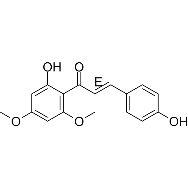
Flavokawain C is an orally active natural chalcone. Flavokawain C inhibits the proliferation of various cancer cells. Flavokawain C upregulates GADD153 in cancer cells, inhibits the phosphorylation of Akt and JNK, suppresses early ERK phosphorylation, activates late ERK phosphorylation, activates caspase related subtypes, induces PARP-1 cleavage, causes upregulation of p21 and p27, downregulation of mutant p53 and anti-apoptotic IAP proteins, elevates intracellular ROS levels, reduces SOD activity, and induces apoptosis. Flavokawain C downregulates FABP4, induces autophagy in cancer cells, and activates the AMPK/mTOR pathway. Flavokawain C decreases the expression of glycolysis-related proteins GLUT1 and HK2, and inhibits glycolysis in nasopharyngeal carcinoma cells. Flavokawain C inhibits the activation of the EGFR/PI3K/Akt/mTOR signaling pathway and reduces the expression of HSP90B1. Flavokawain C inhibits angiogenesis by decreasing the expression of angiogenic proteins Ang-1 and VEGF in human umbilical vein endothelial cells. Flavokawain C increases γ-H2AX levels in cells, inhibits the phosphorylation of FAK, PI3K and AKT in cells, and induces DNA damage in cells. Flavokawain C exerts anti-tumor activity in multiple tumor xenograft mouse models. Flavokawain C is applicable to research related to colorectal cancer, colon adenocarcinoma, nephroblastoma, nasopharyngeal carcinoma and liver cancer.
For research use only. We do not sell to patients.
- Purity: 99.53%
- CAS No.: 37308-75-1
- Formula: C17H16O5
- Molecular Weight:300.31
-
Storage:
4°C, protect from light
* In solvent : -80°C, 6 months; -20°C, 1 month (protect from light)


-
 Biological Activity
-
Chemical Information
-
Solvent & Solubility
- Purity & Documentation
- References
-
 Help & FAQs
-

Apoptosis Compound Library
HY-L003
-
Cell Cycle/DNA Damage Compound Library
HY-L004
-
Epigenetics Compound Library
HY-L005
-
Immunology/Inflammation Compound Library
HY-L007
-
JAK/STAT Compound Library
HY-L008
-
Kinase Inhibitor Library
HY-L009
-
MAPK Compound Library
HY-L010
-
Membrane Transporter/Ion Channel Compound Library
HY-L011
-
Metabolism/Protease Compound Library
HY-L012
-
NF-κB Signaling Compound Library
HY-L014
-
PI3K/Akt/mTOR Compound Library
HY-L015
-
Protein Tyrosine Kinase Compound Library
HY-L016
-
Stem Cell Signaling Compound Library
HY-L017
-
Natural Product Library
HY-L021
-
Anti-Cancer Compound Library
HY-L025
-
Autophagy Compound Library
HY-L029
-
Anti-Aging Compound Library
HY-L034
-
Antioxidant Compound Library
HY-L037
-
Differentiation Inducing Compound Library
HY-L038
-
Reprogramming Compound Library
HY-L039
-
Oxygen Sensing Compound Library
HY-L045
-
Ferroptosis Compound Library
HY-L051
-
Endoplasmic Reticulum Stress Compound Library
HY-L054
-
Phenols Library
HY-L057
-
Glycolysis Compound Library
HY-L058
-
Pyroptosis Compound Library
HY-L059
-
Cytoskeleton Compound Library
HY-L060
-
Orally Active Compound Library
HY-L061
-
Glutamine Metabolism Compound Library
HY-L064
-
Traditional Chinese Medicine Active Compound Library
HY-L065
-
Flavonoids Library
HY-L068
-
Anti-Hepatitis C Virus Compound Library
HY-L073
-
Anti-Breast Cancer Compound Library
HY-L074
-
Anti-Lung Cancer Compound Library
HY-L075
-
Anti-Pancreatic Cancer Compound Library
HY-L077
-
Anti-Blood Cancer Compound Library
HY-L079
-
Anti-Cancer Metabolism Compound Library
HY-L083
-
Anti-Parkinson's Disease Compound Library
HY-L085
-
Anti-Obesity Compound Library
HY-L087
-
Angiogenesis-Related Compound Library
HY-L088
-
Transcription Factor-Targeted Library
HY-L090
-
Lipid Metabolism Compound Library
HY-L091
-
Glucose Metabolism Compound Library
HY-L092
-
Anti-Liver Cancer Compound Library
HY-L101
-
Anti-Colorectal Cancer Compound Library
HY-L103
-
Anti-Cancer Natural Product Library
HY-L107
-
Antidepressant Compound Library
HY-L108
-
Protein-protein Interaction Inhibitor Library
HY-L109
-
Anti-inflammatory Traditional Chinese Medicine Active Compound Library
HY-L114
-
Plant-Sourced Natural Product Library
HY-L115
-
Human Metabolite Library
HY-L123
-
Anti-Prostate Cancer Compound Library
HY-L124
-
Anti-Pulmonary Fibrosis Compound Library
HY-L125
-
Cancer Stem Cells Compound Library
HY-L135
-
Pain-Related Compound Library
HY-L139
-
Metabolic Enzyme Compound Library
HY-L146
-
Membrane Protein-targeted Compound Library
HY-L149
-
Membrane Receptor-targeted Compound Library
HY-L150
-
Cytokine Inhibitors Library
HY-L161
-
Cell Death Library
HY-L162
-
Serine/Threonine Kinase Inhibitor Library
HY-L164
-
Extracellular Vesicles (EVs) Compound Library
HY-L168
-
Anti-Hematopathy Compound Library
HY-L171
-
Anti-Ovarian Cancer Compound Library
HY-L173
-
Multi-Target Compound Library
HY-L176
-
Radioprotector Library
HY-L178
-
Bioactive Compound Library Max
HY-L181
-
MCE Bioactive Compound Library
HY-L001V
-
Natural Product Library Plus
HY-L021P
-
Natural Product and Natural Product-Like Compound Library
HY-L021M
-
Bioactive Compound Library
HY-L001
-
Anti-Gastric Cancer Compound Library
HY-L184
-
Anti-Fibrosis Compound Library
HY-L185
-
Anti-Brain Cancer Compound Library
HY-L188
-
Protein Kinase Compound Library
HY-L196
-
Non-Alcoholic Fatty Liver Disease (NAFLD) Compound Library
HY-L199
-
RO5 Drug-like Natural Product Library
HY-L200
-
Cell Proliferation Compound Library
HY-L201
-
Lactic Acid Metabolic Compound Library
HY-L204
-
High-Throughput Bioactive Compound Library
HY-L205
-
High-Throughput Natural Product Library
HY-L206
-
Anti-Rheumatic Arthritis Compound Library
HY-L210
-
Mass Spectrometry Human Metabolite Library
HY-L215
-
Diarrhea-Related Traditional Chinese Medicine Active Compound Library
HY-L224
-
Mongolian Medicine Compound Library
HY-L238
-
Lactylation Compound Library
HY-L249
-
Mass Spectrometry Natural Product Library
HY-L262


Publications Citing Use of MedChemExpress (MCE) Flavokawain C
More Customer Validation & Images

-
Cell Proliferation/Viability Assay
-
Cell Proliferation/Viability Assay
-
WB
-
Cell Migration/Invasion Assay
-
Cell Autophagy Assay
All Caspase Isoforms
MoreAll AMPK Isoforms
MoreAll EGFR Isoforms
MoreAll VEGFR Isoforms
More

Biological Activity
|
Cell Line

|
Type | Value | Description | References |
|---|---|---|---|---|
| A549 | IC50 |
8.1 μg/mL
Compound: Flavokawain C
|
Inhibition of TNF-alpha-induced NF-kappaB expressed in human A549 cells treated 1 hr after TNFalpha challenge measured after 6 hrs by luciferase reporter gene assay
Inhibition of TNF-alpha-induced NF-kappaB expressed in human A549 cells treated 1 hr after TNFalpha challenge measured after 6 hrs by luciferase reporter gene assay
|
[PMID: 19716299] |
| B16-F10 | IC50 |
6.9 μM
Compound: 1d
|
Antimelanogenic activity in mouse B16F10 cells assessed as inhibition of melanin production after 4 days
Antimelanogenic activity in mouse B16F10 cells assessed as inhibition of melanin production after 4 days
|
[PMID: 25597012] |
| HeLa | IC50 |
19.2 μM
Compound: 5, flavokawin
|
Cytotoxicity against human HeLa cells by MTT assay after 72 hrs
Cytotoxicity against human HeLa cells by MTT assay after 72 hrs
|
[PMID: 18611049] |
| K562 | IC50 |
8 μM
Compound: Flavokawain C
|
Inhibition of NF-kappaB transactivation in TNF-alpha-stimulated human K562 cells preincubated for 2 hrs followed by TNF-alpha challenge measured after 6 hrs by dual luciferase reporter gene assay
Inhibition of NF-kappaB transactivation in TNF-alpha-stimulated human K562 cells preincubated for 2 hrs followed by TNF-alpha challenge measured after 6 hrs by dual luciferase reporter gene assay
|
[PMID: 24775915] |
| NIH3T3 | IC50 |
3.1 μM
Compound: 2c
|
Inhibition of cobalt chloride-induced HIF-1 activation expressed in mouse NIH3T3 cells after 8 hrs by luciferase reporter gene assay
Inhibition of cobalt chloride-induced HIF-1 activation expressed in mouse NIH3T3 cells after 8 hrs by luciferase reporter gene assay
|
[PMID: 21112783] |
Flavokawain C (60 μM; 6-48 h) upregulates the endoplasmic reticulum stress marker GADD153, inhibits Akt phosphorylation, suppresses early ERK phosphorylation and late JNK phosphorylation, and activates late ERK phosphorylation in HCT 116 cells[1].
Flavokawain C (40-80 μM; 6-72 h) reduces the viability of HT-29 cells, induces apoptotic morphological and nuclear changes, triggers DNA fragmentation, increases the proportion of apoptotic cells, decreases mitochondrial membrane potential, and induces G2/M cell cycle arrest[2].
Flavokawain C (40-80 μM; 0-48 h) activates caspase-3, -8, and -9, induces PARP-1 cleavage, triggers p53-independent upregulation of p21 and p27, downregulation of mutant p53, upregulation of the endoplasmic reticulum stress marker GADD153, and downregulation of anti-apoptotic IAP proteins (XIAP, c-IAP1, c-IAP2), increases intracellular ROS levels, and decreases SOD activity in HT-29 cells[2].
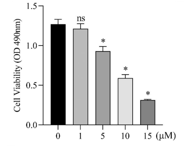
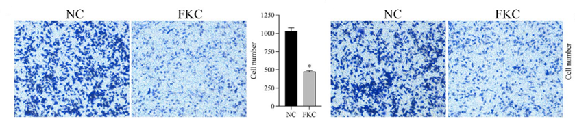
Flavokawain C (1-15 μM; 24-72 h) inhibits the viability and proliferation of G401 cells, suppresses their colony-forming ability, and inhibits cell migration and invasion[3].
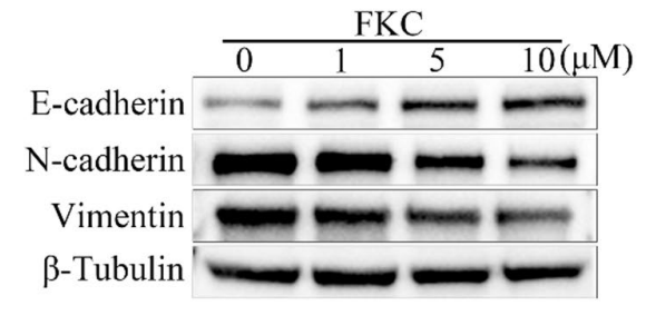
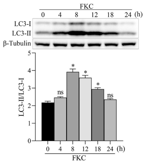
Flavokawain C (1-10 μM; 0-24 h) regulates the expression of EMT markers, downregulates FABP4, induces complete autophagic flux, and activates the AMPK/mTOR pathway in G401 cells[3].
Flavokawain C (0.5-4 μM; 48 h) inhibits the proliferation of human nasopharyngeal carcinoma HNE1 and CNE2 cells and increases the apoptotic rate of these cells[4].
Flavokawain C (4 μM; 48 h) inhibits glycolysis in human nasopharyngeal carcinoma HNE1 and CNE2 cells by reducing the extracellular acidification rate, glucose consumption and lactate production, as well as downregulating the expression of glycolysis-related proteins GLUT1 and HK2[4].
Flavokawain C (4 μM; 48 h) inhibits the activation of the EGFR/PI3K/Akt/mTOR signaling pathway and reduces the expression of HSP90B1 in human nasopharyngeal carcinoma HNE1 and CNE2 cells[4].
Flavokawain C (4 μM; 48 h) inhibits angiogenesis in human nasopharyngeal carcinoma HNE1 and CNE2 cells by impairing the tube formation and migration capacities of human umbilical vein endothelial cells (HUVECs) and reducing the expression of angiogenic proteins Ang-1 and VEGF in HUVECs[4].
Flavokawain C (48 h) selectively inhibits the viability of Huh-7, Hep3B and HepG2 hepatocellular carcinoma cells, with IC50 values ranging from 23.42 μM to 30.71 μM; while it exhibits low toxicity to normal MIHA hepatocytes, with an IC50 of 53.95 μM[5].
Flavokawain C (2.0-8.0 μM; 2 weeks) inhibits colony formation of Huh-7, Hep3B and HepG2 hepatocellular carcinoma cells[5].
Flavokawain C (4.0-16.0 μM; 48 h) inhibits DNA replication, induces apoptosis, downregulates the expression of anti-apoptotic protein Bcl2, upregulates the expression of pro-apoptotic protein Bax, and promotes apoptosis by reducing the Bcl2/Bax ratio in Huh-7 and Hep3B hepatocellular carcinoma cells[5].
Flavokawain C (4.0-16.0 μM; 48 h) increases the level of γ-H2AX, a marker of DNA damage response, in Huh-7 and Hep3B cells, inhibits the phosphorylation of FAK, PI3K and AKT in cells, and induces DNA damage[5].
Flavokawain C (4.0-16.0 μM; 48 h) reduces extracellular matrix adhesion of Huh-7 and Hep3B cells and inhibits cell migration[5].
MedChemExpress (MCE) has not independently confirmed the accuracy of these methods. They are for reference only.
-
Cell Line:HCT 116 human colon carcinoma cells
-
Concentration:60 μM
-
Incubation Time:6, 12, 18, 24, 48 h
-
Result:Induced time-dependent upregulation of GADD153 protein levels.
Showed no GADD153 expression was detected in untreated control cells.\nCaused a transient increase in phosphorylated Akt levels at 6 hours.
Induced time-dependent decrease in phosphorylated Akt levels through 48 hours.
Showed no significant changes in total Akt protein levels were observed.\nCaused time-dependent decreases in phosphorylated ERK levels at 6, 12, and 18 hours.
Induced dramatic increases in phosphorylated ERK levels at 24 and 48 hours.
Showed total ERK levels remained unchanged.
Caused small reductions in phosphorylated JNK levels at 24 and 48 hours.
Showed no significant changes in total JNK, phosphorylated p38, or total p38 levels.
-
Cell Line:human colon adenocarcinoma HT-29 cells
-
Concentration:40, 60, 80 μM
-
Incubation Time:0, 6, 12, 24, 48, 72 h
-
Result:Reduced HT-29 cell viability in a dose- and time-dependent manner.
Decreased cell viability from 644.51% (control) to 185.17%, 111.81%, and 104.94% at 40, 60, and 80 μM respectively after 72 h.
Showed comparable inhibitory effect between 60 and 80 μM.
-
Cell Line:human colon adenocarcinoma HT-29 cells
-
Concentration:40, 60, 80 μM
-
Incubation Time:48 h
-
Result:Induced dose-dependent apoptotic morphological changes, including cell shrinkage, surface blebbing, cytoplasmic vacuolation, chromatin condensation/fragmentation, and late apoptotic cells (pink fluorescence from propidium iodide staining).\nCaused a concentration-dependent increase in DNA fragmentation.
Increased TUNEL-positive cells to 11.6% at 80 μM relative to control.
-
Cell Line:human colon adenocarcinoma HT-29 cells
-
Concentration:40-80 μM
-
Incubation Time:24-48 h
-
Result:Caused a dose- and time-dependent increase in Annexin V-FITC-positive apoptotic cells.
-
Cell Line:human colon adenocarcinoma HT-29 cells
-
Concentration:40-80 μM
-
Incubation Time:24-48 h
-
Result:At 24 h, dose-dependently increased G2/M phase cell population (with a small G1 phase accumulation) and decreased S phase population.
At 48 h, caused a marked dose-dependent increase in G2/M phase cell population, with the highest increase at 80 μM.
-
Cell Line:G401 nephroblastoma cells
-
Concentration:10 μM
-
Incubation Time:12 h
-
Result:Increased the number of both yellow (autophagosomes) and red (autolysosomes) puncta per cell compared to control, indicating enhanced autophagic initiation and maturation.
-
Cell Line:Huh-7, Hep3B
-
Concentration:4.0 μM, 8.0 μM, 16.0 μM
-
Incubation Time:48 h
-
Result:Dose-dependently increased the number of 53BP1 foci (a marker of DNA damage response) in both cell lines.
Increased average 53BP1 foci per cell to >15 in both cell lines at 16.0 μM, compared to ~1.5 foci per cell in untreated controls.
Flavokawain C (3 mg/kg; i.p.; 4 weeks (subcutaneous tumor model); 8 weeks (liver metastasis model)) reduces subcutaneous nasopharyngeal carcinoma tumor volume and weight, inhibits liver metastasis, and downregulates glycolysis, angiogenesis, and EGFR/PI3K/Akt/mTOR pathway activation in vivo, with effects enhanced by HSP90B1 knockdown and reversed by HSP90B1 overexpression[4].
Flavokawain C (16 mg/kg; i.p.; daily; 14 days) significantly inhibits Huh-7 liver cancer xenograft growth in nude mice without causing notable body weight loss, via reduced tumor cell proliferation and induced DNA damage[5].
Flavokawain C (1-3 mg/kg; i.p.; thrice weekly; 19 days) dose-dependently inhibits HCT 116 colon carcinoma xenograft growth in BALB/c nude mice, with the 3 mg/kg dose achieving up to 52.17% tumor volume inhibition, via induction of apoptosis and reduction of cell proliferation, without causing significant organ toxicity[6].
MedChemExpress (MCE) has not independently confirmed the accuracy of these methods. They are for reference only.
-
Animal Model:BALB/c nude (5-week-old male, weight range 19.0g to 22.3g at euthanasia, subcutaneous nephroblastoma xenograft model)[3]
-
Dosage:3 mg/kg
-
Administration:i.p.; thrice weekly; 4 weeks
-
Result:Significantly reduced tumor volume and tumor weight compared to the control group.
Reduced expression of Ki67, FABP4, and Vimentin in tumor tissues.
Increased expression of E-cadherin in tumor tissues.
Induced loose tumor structure and increased inflammatory cell infiltration in tumor samples.
Had its tumor growth inhibition effect partially reversed by FABP4 overexpression.
-
Animal Model:BALB/C nude (male, 5-6 weeks old)[4]
-
Dosage:3 mg/kg
-
Administration:i.p.; 4 weeks (subcutaneous tumor model); 8 weeks (liver metastasis model)
-
Result:Reduced subcutaneous tumor volume and weight compared to vehicle control.
Downregulated the expression of HSP90B1, GLUT1, HK2, Ang-1, and VEGF in tumor tissues.
Inhibited the phosphorylation of EGFR, PI3K, Akt, and mTOR in tumor tissues.
Inhibited liver metastasis of NPC tumors, as measured by reduced bioluminescence signal from luciferase-expressing tumor cells.
Enhanced inhibitory effect on tumor growth and pathway suppression was observed with HSP90B1 knockdown.
Reversed inhibitory effect on tumor growth and pathway suppression was observed with HSP90B1 overexpression.
-
Animal Model:BALB/c nude mice (6-week-old)[5]
-
Dosage:16 mg/kg
-
Administration:i.p.; daily; 14 days
-
Result:Significantly reduced tumor growth rate, final tumor volume, and final tumor weight relative to controls.
Caused no significant changes in mouse body weight during treatment.
Induced cytoplasmic-nuclear separation and nuclear fragmentation (cell death) in tumor tissues via H&E staining.
Reduced Ki67 (proliferation marker) levels in treated tumor tissues.
Increased γ-H2AX (DNA damage marker) levels in treated tumor tissues.
-
Animal Model:BALB/c nude (female, 6 weeks old)[6]
-
Dosage:1 mg/kg; 3 mg/kg
-
Administration:i.p.; thrice weekly; 19 days
-
Result:Reduced final mean tumor volume to 658.19 mm3 (600% growth from initial 84.48 mm3) with 18.73-23.99% tumor volume inhibition (%T/C) over days 3-19 at 1 mg/kg.
Reduced final mean tumor volume to 411.31 mm3 (300% growth from initial 86.56 mm3) with 23.43-52.17% tumor volume inhibition (%T/C) over days 3-19 at 3 mg/kg.
Increased tumor necrotic area significantly (p < 0.05 vs control) at 3 mg/kg.
Increased TUNEL-positive cells significantly (p < 0.05 vs control) at 3 mg/kg.
Increased cleaved caspase-3 immunoreactivity score significantly (p < 0.05 vs control) at 3 mg/kg.
Decreased Ki67 immunoreactivity score significantly (p < 0.05 vs control) at 3 mg/kg.
Up-regulated Ig mu chain C region (secreted form) and down-regulated GRP78, hemopexin, kininogen-1, and apolipoprotein E compared to vehicle controls, with levels returning to near normal ranges at 3 mg/kg.
Caused no significant body weight loss in either dose group; kept serum liver (AST, ALT, ALP) and kidney (creatine, urea) function parameters within normal limits, with only mild urea elevation in the 1 mg/kg group; caused no pathological damage to major organs (heart, spleen, liver, lungs, kidneys).


Chemical Information
-
CAS No. 37308-75-1
-
Appearance Solid
-
Molecular Weight 300.31
-
Formula C17H16O5
-
Color Light yellow to orange
-
SMILES
O=C(C1=C(OC)C=C(OC)C=C1O)/C=C/C2=CC=C(O)C=C2
-
Structure Classification
-
Initial Source
-
Shipping
Room temperature in continental US; may vary elsewhere.
-
Storage
4°C, protect from light
* In solvent : -80°C, 6 months; -20°C, 1 month (protect from light)


Publications (4)
-
Journal Impact Factor
-
Most Recent
-
Food Chem
Effects of sun drying combined with baking processes on the flavor quality of Chongqing Tuocha raw tea. [Abstract]2025 Dec 30:497:146992. PMID: 41285060 -
Food Chem
Flavonoid-mediated metabolic underpinning quality variation in red bud-sport pear mutants. [Abstract]2025 May 31:489:144992. PMID: 40466530 -
Phytomedicine
The kava chalcone flavokawain B exerts inhibitory activity and synergizes with BCL-2 inhibition in malignant B-cell lymphoma. [Abstract]2023 Nov:120:155074. PMID: 37716033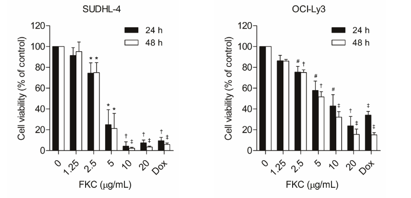
Flavokawain C purchased from MedChemExpress. Usage Cited in: Phytomedicine. 2023 Nov:120:155074. [Abstract]
SUDHL-4 and OCI-Ly3 cells were treated with FKC (Flavokawain C; HY-N2445; 1.25-20 µg/mL) or Doxorubicin (HY-15142A; 10 µM) for 24 and 48 h. Cell viability was measured using the MTS assay. *P < 0.05, #P < 0.01, †P < 0.001, and ‡P < 0.0001 versus control at corresponding time points.SUDHL-4 (A), OCI-Ly3 (B), Raji (C), and Jeko-1 (D) cells were treated with FKA (1.25-20 µg/mL) or doxorubicin (10 µM) for 24 and 48 h. Cell viability was measured using the MTS assay. *P < 0.05, #P < 0.01, †P < 0.001, and ‡P < 0.0001 versus control at corresponding time points.
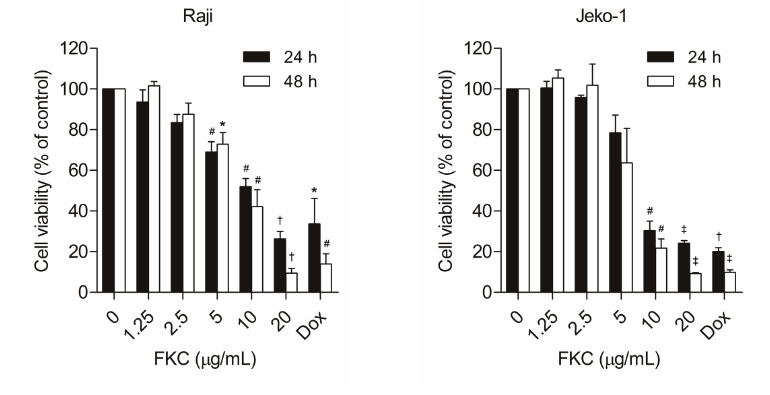
Flavokawain C purchased from MedChemExpress. Usage Cited in: Phytomedicine. 2023 Nov:120:155074. [Abstract]
Raji and Jeko-1 cells were treated with FKC (Flavokawain C; HY-N2445; 1.25-20 µg/mL) or Doxorubicin (HY-15142A; 10 µM) for 24 and 48 h. Cell viability was measured using the MTS assay. *P < 0.05, #P < 0.01, †P < 0.001, and ‡P < 0.0001 versus control at corresponding time points.SUDHL-4 (A), OCI-Ly3 (B), Raji (C), and Jeko-1 (D) cells were treated with FKA (1.25-20 µg/mL) or doxorubicin (10 µM) for 24 and 48 h. Cell viability was measured using the MTS assay. *P < 0.05, #P < 0.01, †P < 0.001, and ‡P < 0.0001 versus control at corresponding time points.
-
Sci Rep
Flavokawain C suppresses nephroblastoma growth by inducing autophagy-mediated downregulation of FABP4 via AMPK/mTOR pathway. [Abstract]2026 Mar 4. PMID: 41781551
Flavokawain C purchased from MedChemExpress. Usage Cited in: Sci Rep. 2026 Mar 4. [Abstract]
FKC (Flavokawain C) inhibited the growth of G401 cells. The cell viability of G401 treated with different concentrations of FKC (0, 1, 5, 10, and 15 μM) was detected by CCK-8 assay.
Flavokawain C purchased from MedChemExpress. Usage Cited in: Sci Rep. 2026 Mar 4. [Abstract]
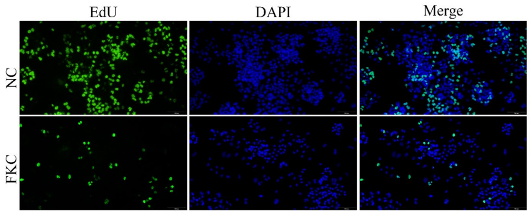
The proliferation of G401 cells treated with or without FKC (Flavokawain C; 10 μM) was examined by EdU assay.
Flavokawain C purchased from MedChemExpress. Usage Cited in: Sci Rep. 2026 Mar 4. [Abstract]
G401 cells were treated with different concentrations of FKC (Flavokawain C; 0, 1, 5, and 10 μM), and the expression of epithelial-mesenchymal transition (EMT) markers was detected by western blot analysis.
Flavokawain C purchased from MedChemExpress. Usage Cited in: Sci Rep. 2026 Mar 4. [Abstract]
The inhibitory effect of FKC (Flavokawain C; 10 μM) on the migration and invasion of G401 cells was detected by transwell assay. The bar graphs show the relative protein expression levels normalized to β-tubulin.
Flavokawain C purchased from MedChemExpress. Usage Cited in: Sci Rep. 2026 Mar 4. [Abstract]
FKC (Flavokawain C) induced autophagy in G401 cells. G401 cells were treated with 10 μM FKC for different time (0, 4, 8, 12, 18, and 24 h), and the expression of LC3 was detected by western blot.
Flavokawain C purchased from MedChemExpress. Usage Cited in: Sci Rep. 2026 Mar 4. [Abstract]
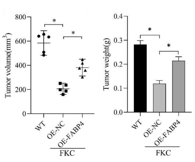
FKC (Flavokawain C) inhibited the growth and metastasis of nephroblastoma in vivo. Nude mice were subcutaneously injected with normal and oeNC or oeFABP4 infected G401 cells. One week after cell injection, mice in the treatment group were intraperitoneally administered 3 mg/kg FKC thrice a week for 4 weeks; control mice received the same volume of normal saline. The value of tumor volume and weight was quantified in each group.
Flavokawain C purchased from MedChemExpress. Usage Cited in: Sci Rep. 2026 Mar 4. [Abstract]
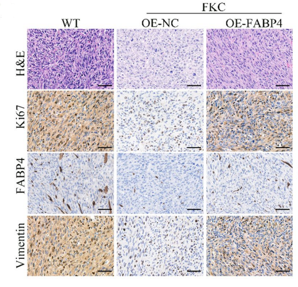
FKC (Flavokawain C) inhibited the growth and metastasis of nephroblastoma in vivo. Nude mice were subcutaneously injected with normal and oeNC or oeFABP4 infected G401 cells. One week after cell injection, mice in the treatment group were intraperitoneally administered 3 mg/kg FKC thrice a week for 4 weeks; control mice received the same volume of normal saline. Tumor tissue sections stained with hematoxylin–eosin, the expression of Ki67, FABP4 and Vimentin by immunohistochemistry.


Solvent & Solubility
DMSO : 125 mg/mL (416.24 mM; Need ultrasonic; Hygroscopic DMSO has a significant impact on the solubility of product, please use newly opened DMSO)
Please refer to the solubility information to select the appropriate solvent. Once prepared, please aliquot and store the solution to prevent product inactivation from repeated freeze-thaw cycles.
Storage method and period of stock solution: -80°C, 6 months; -20°C, 1 month (protect from light). When stored at -80°C, please use it within 6 months. When stored at -20°C, please use it within 1 month.
Please refer to the solubility information to select the appropriate solvent. Once prepared, please aliquot and store the solution to prevent product inactivation from repeated freeze-thaw cycles.
Storage method and period of stock solution: -80°C, 6 months; -20°C, 1 month (protect from light). When stored at -80°C, please use it within 6 months. When stored at -20°C, please use it within 1 month.
Concentration (start) × Volume (start) = Concentration (final) × Volume (final)
Select the appropriate dissolution method based on your experimental animal and administration route.
- For the following dissolution methods, please ensure to first prepare a clear stock solution using an In Vitro approach and then sequentially add co-solvents:
- To ensure reliable experimental results, the clarified stock solution can be appropriately stored based on storage conditions. As for the working solution for In Vivo experiments, it is recommended to prepare freshly and use it on the same day.
- The percentages shown for the solvents indicate their volumetric ratio in the final prepared solution. If precipitation or phase separation occurs during preparation, heat and/or sonication can be used to aid dissolution.
Add each solvent one by one: 10% DMSO 40% PEG300 5% Tween-80 45% Saline
Solubility: ≥ 1.25 mg/mL (4.16 mM); Clear solution
This protocol yields a clear solution of ≥ 1.25 mg/mL (saturation unknown).
Taking 1 mL working solution as an example, add 100 μL DMSO stock solution (12.5 mg/mL) to 400 μL PEG300, and mix evenly; then add 50 μL Tween-80 and mix evenly; then add 450 μL Saline to adjust the volume to 1 mL.
Preparation of Saline: Dissolve 0.9 g sodium chloride in ddH₂O and dilute to 100 mL to obtain a clear Saline solution.
Add each solvent one by one: 10% DMSO 90% (20% SBE-β-CD in Saline)
Solubility: ≥ 1.25 mg/mL (4.16 mM); Clear solution
This protocol yields a clear solution of ≥ 1.25 mg/mL (saturation unknown).
Taking 1 mL working solution as an example, add 100 μL DMSO stock solution (12.5 mg/mL) to 900 μL 20% SBE-β-CD in Saline, and mix evenly.
Preparation of 20% SBE-β-CD in Saline (4°C, storage for one week): 2 g SBE-β-CD powder is dissolved in 10 mL Saline, completely dissolve until clear.
Please enter the basic information of animal experiments:
-
-
-
-
Recommended: Prepare an additional quantity of animals to account for potential losses during experiments.
Please enter your animal formula composition:
-
%DMSO +
Recommended: Keep the proportion of DMSO in working solution below 2% if your animal is weak.
-
%+
-
+%Tween-80 + +
-
%Saline +
The co-solvents required include: DMSO, . All of co-solvents are available by MedChemExpress (MCE). , Tween 80. All of co-solvents are available by MedChemExpress (MCE).
Working solution concentration: 0.22 mg/mL
Method for preparing stock solution: mg drug dissolved in μL DMSO. Stock solution concentration: mg/mL. * In solvent : -80°C, 6 months; -20°C, 1 month (protect from light)
1. Take μL DMSO stock solution;
2. Add μL .
μL , mix evenly;
3. Then add μL Tween 80, mix evenly;
4. Then add μL
Please ensure that the stock solution in the first step is dissolved to a clear state, and add co-solvents in sequence. You can use ultrasonic heating (ultrasonic cleaner, recommended frequency 20-40 kHz), vortexing, etc. to assist dissolution.


Purity & Documentation
-
Data Sheet (295 KB)
-
SDS (393 KB)
- English - EN (393 KB)
- Français - FR (393 KB)
- Deutsch - DE (393 KB)
- Norwegian - NO (393 KB)
- Español - ES (393 KB)
- Swedish - SV (393 KB)
- Italian - IT (393 KB)
- Portuguese - PT (393 KB)
-
Handling Instructions (2659 KB)
References
[1]. Phang CW, et al. Flavokawain C Inhibits Cell Cycle and Promotes Apoptosis, Associated with Endoplasmic Reticulum Stress and Regulation of MAPKs and Akt Signaling Pathways in HCT 116 Human Colon Carcinoma Cells. PLoS One. 2016;11(2):e0148775. Published 2016 Feb 9. [Content Brief]
[2]. Phang CW, et al. Induction of Apoptosis and Cell Cycle Arrest by Flavokawain C on HT-29 Human Colon Adenocarcinoma via Enhancement of Reactive Oxygen Species Generation, Upregulation of p21, p27, and GADD153, and Inactivation of Inhibitor of Apoptosis Proteins. Pharmacogn Mag. 2017;13(Suppl 2):S321-S328. [Content Brief]
[3]. Liu Q, et al. Flavokawain C suppresses nephroblastoma growth by inducing autophagy-mediated downregulation of FABP4 via AMPK/mTOR pathway. Sci Rep. Published online March 4, 2026. [Content Brief]
[4]. Hu Y, et al. Flavokawain C inhibits glucose metabolism and tumor angiogenesis in nasopharyngeal carcinoma by targeting the HSP90B1/STAT3/HK2 signaling axis. Cancer Cell Int. 2024;24(1):158. Published 2024 May 6. [Content Brief]
[5]. Wang R, et al. Flavokawain C inhibits proliferation and migration of liver cancer cells through FAK/PI3K/AKT signaling pathway. J Cancer Res Clin Oncol. 2024;150(3):117. Published 2024 Mar 9. [Content Brief]
[6]. Phang CW, et al. Flavokawain C exhibits anti-tumor effects on in vivo HCT 116 xenograft and identification of its apoptosis-linked serum biomarkers via proteomic analysis. Biomed Pharmacother. 2021;137:110846. [Content Brief]
[7]. Phang CW, et al. Proteomic analysis of flavokawain C-induced cell death in HCT 116 colon carcinoma cell line. Eur J Pharmacol. 2019;853:388-399. [Content Brief]


Complete Stock Solution Preparation Table
Please refer to the solubility information to select the appropriate solvent. Once prepared, please aliquot and store the solution to prevent product inactivation from repeated freeze-thaw cycles.
Storage method and period of stock solution: -80°C, 6 months; -20°C, 1 month (protect from light). When stored at -80°C, please use it within 6 months. When stored at -20°C, please use it within 1 month.
| Optional Solvent | Concentration Solvent Mass | 1 mg | 5 mg | 10 mg | 25 mg |
|---|---|---|---|---|---|
| DMSO | 1 mM | 3.3299 mL | 16.6495 mL | 33.2989 mL | 83.2473 mL |
| 5 mM | 0.6660 mL | 3.3299 mL | 6.6598 mL | 16.6495 mL | |
| 10 mM | 0.3330 mL | 1.6649 mL | 3.3299 mL | 8.3247 mL | |
| 15 mM | 0.2220 mL | 1.1100 mL | 2.2199 mL | 5.5498 mL | |
| 20 mM | 0.1665 mL | 0.8325 mL | 1.6649 mL | 4.1624 mL | |
| 25 mM | 0.1332 mL | 0.6660 mL | 1.3320 mL | 3.3299 mL | |
| 30 mM | 0.1110 mL | 0.5550 mL | 1.1100 mL | 2.7749 mL | |
| 40 mM | 0.0832 mL | 0.4162 mL | 0.8325 mL | 2.0812 mL | |
| 50 mM | 0.0666 mL | 0.3330 mL | 0.6660 mL | 1.6649 mL | |
| 60 mM | 0.0555 mL | 0.2775 mL | 0.5550 mL | 1.3875 mL | |
| 80 mM | 0.0416 mL | 0.2081 mL | 0.4162 mL | 1.0406 mL | |
| 100 mM | 0.0333 mL | 0.1665 mL | 0.3330 mL | 0.8325 mL |


Flavokawain C Related Classifications
Cat. No.: HY-N2445
 Powered by Bioz
- Flavokawain C
- 37308-75-1
- Apoptosis
- Akt
- JNK
- PERK
- Caspase
- PARP
- MDM-2/p53
- IAP
- Reactive Oxygen Species (ROS)
- SOD
- FABP
- Autophagy
- AMPK
- mTOR
- GLUT
- EGFR
- PI3K
- HSP
- VEGFR
- FAK
- natural chalcone
- HCT 116 cells
- HT-29 cells
- G401 cells
- HNE1 cells
- CNE2 cells
- Huh-7 cells
- Hep3B cells
- HepG2cells
- BALB/c nude mice
- colorectal cancer
- colon adenocarcinoma
- nephroblastoma
- nasopharyngeal carcinoma
- liver cancer
- Inhibitor
- inhibitor
- inhibit