



Diazepinomicin
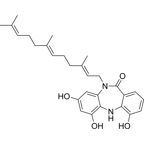
Diazepinomicin (ECO-4601) is a reagent with both anticancer and antibacterial activities.. Diazepinomicin decreases Bcl2 and BAX expression, induces dose-dependent cell death and apoptotic bodies, and inhibits Ras-MAPK signaling and tumor growth/proliferation. Diazepinomicin induces apoptosis through caspase 3 activation, PARP cleavage, cytochrome C release, loss of membrane potential, and DNA fragmentation, independent of PBR expression. Diazepinomicin binds PBR/TSPO and displaces PK11195 through its farnesyl side chain rather than the dibenzodiazepinone core. Diazepinomicin can be used in research on hepatocellular carcinoma, Gram-positive bacterial infections, and glioma.
Nos produits utilisent uniquement pour la recherche. Nous ne vendons pas aux patients.
- CAS No.: 733035-26-2
- Formule: C28H34N2O4
- Masse moléculaire:462.58
-
Stockage:
-20°C, protect from light, stored under nitrogen
* In solvent : -80°C, 6 months; -20°C, 1 month (protect from light, stored under nitrogen)

To place orders, for customer services and technical support, please contact: MedChemExpress USA
Tel: 609-228-6898 E-mail: [email protected] [email protected]


-
 Activité biologique
-
Chemical Information
- Protocole
- Pureté et documentation
- Références
-
 Help & FAQs




Voir tous les produits spécifiques à Isoform Caspase
More

Activité biologique
Description
|
BCL2 |
PBR |
Bax |
Ras |
p38 MAPK |
Caspase 3 |
PARP |
TSPO |
Cellular Effect
|
Cell Line

|
Type | Value | Description | References |
|---|---|---|---|---|
| HUVEC | IC50 |
0.43 μg/mL
Compound: 1, BU-4664L
|
Antiinvasive activity against HUVEC assessed as inhibition of cell migration after 6 hrs by Matrigel assay
Antiinvasive activity against HUVEC assessed as inhibition of cell migration after 6 hrs by Matrigel assay
|
[PMID: 20056543] |
| HUVEC | IC50 |
0.72 μg/mL
Compound: 1, BU-4664L
|
Antiangiogenic activity against HUVEC cells assessed as inhibition of vascular formation after 6 hrs by wound healing assay
Antiangiogenic activity against HUVEC cells assessed as inhibition of vascular formation after 6 hrs by wound healing assay
|
[PMID: 20056543] |
| HUVEC | IC50 |
2 μg/mL
Compound: 1, BU-4664L
|
Cytotoxicity against HUVEC cells
Cytotoxicity against HUVEC cells
|
[PMID: 20056543] |
| NCI-H460 | GI50 |
13.6 μM
Compound: 1; BU-4664L
|
Antiproliferative activity against human NCI-H460 cells assessed as cell growth inhibition by MTT assay
Antiproliferative activity against human NCI-H460 cells assessed as cell growth inhibition by MTT assay
|
[PMID: 34838651] |
| PC-3 | GI50 |
1.95 μM
Compound: 1; BU-4664L
|
Antiproliferative activity against human PC-3 cells assessed as cell growth inhibition by MTT assay
Antiproliferative activity against human PC-3 cells assessed as cell growth inhibition by MTT assay
|
[PMID: 34838651] |
| Renca | IC50 |
0.78 μg/mL
Compound: 1, BU-4664L
|
Antiinvasive activity against mouse Renca by Matrigel assay
Antiinvasive activity against mouse Renca by Matrigel assay
|
[PMID: 20056543] |
| Renca | IC50 |
8.3 μg/mL
Compound: 1, BU-4664L
|
Cytotoxicity against mouse Renca cells
Cytotoxicity against mouse Renca cells
|
[PMID: 20056543] |
In Vitro
Diazepinomicin (10-15 μM; 24 h) induces apoptotic nuclear condensation and apoptotic body formation in human hepatoma HepG2 cells[1].
Diazepinomicin (10-15 μM; 24 h) induces DNA fragmentation (a hallmark of apoptosis) in human hepatoma HepG2 cells[1].
Diazepinomicin (10-15 μM; 24 h) downregulates the expression of 25 kDa Bcl-2 and 20 kDa BAX proteins in human hepatoma HepG2 cells[1].
Diazepinomicin (10 µM; 0-6 h) induces cytochrome C release from mitochondria to cytosol between 2 and 4 h in both Jurkat WT and Jurkat-PBR cells[3].
Diazepinomicin (10 µM; 0-24 h) induces DNA nucleosomal fragmentation and more than 95% cell death by 24 h in both Jurkat WT and Jurkat-PBR cells[3].
MedChemExpress (MCE) has not independently confirmed the accuracy of these methods. They are for reference only.
-
Cell Line:human hepatoma HepG2 cells
-
Concentration:1; 2; 5; 10; 25 μM
-
Incubation Time:24 h
-
Result:Induced dose-dependent decreases in HepG2 cell proliferation, with increasing cell death observed at higher concentrations.
-
Cell Line:human hepatoma HepG2 cells
-
Concentration:10; 15 μM
-
Incubation Time:24 h
-
Result:Induced clumping of cells with slight distortion, as well as very condensed and fragmented nuclei (hallmark features of apoptosis).\nInduced condensed nuclei and apoptotic bodies, characteristic of apoptotic cells.\nInduced DNA fragmentation, visualized as characteristic DNA ladder bands on agarose gel electrophoresis.
-
Cell Line:human hepatoma HepG2 cells
-
Concentration:10; 15 μM
-
Incubation Time:24 h
-
Result:Reduced intensity of Bcl-2 (25 kDa) and BAX (20 kDa) protein bands, indicating downregulation of both anti-apoptotic Bcl-2 and pro-apoptotic BAX protein expression.
- Research Protocol for Infectious Diseases
- RNA extraction experimental
- Mammalian live/dead viability and cytotoxicity staining
- Annexin V plus membrane-impermeant dye apoptosis staining
- Patient-Derived Orthotopic Xenograft (PDOX)
- Liver Cancer Modeling
- Gram Staining of Tissue Sections
- Cell Viability Determination by MTT Colorimetric Assay
- Detection of Apoptosis
- TUNEL staining for apoptotic DNA fragmentation
- Cell Cytotoxicity Assay
- Fluorescent plasma-membrane potential dye assay
- Patient-Derived Xenograft (PDX)
- Apoptosis Solutions
- Subcutaneous Cell-Line-Derived Xenograft
- Orthotopic Cell-Line Xenograft


Chemical Information
-
CAS No. 733035-26-2
-
Appearance Solid
-
Masse moléculaire 462.58
-
Formule C28H34N2O4
-
Color Off-white to gray
-
SMILES
O=C1C2=CC=CC(O)=C2NC3=C(O)C=C(O)C=C3N1C/C=C(C)/CC/C=C(C)/CC/C=C(C)\C
-
Synonyms
ECO-4601; TLN-4601; BU 4664L
-
Livraison
Room temperature in continental US; may vary elsewhere.
-
Stockage
-20°C, protect from light, stored under nitrogen
* In solvent : -80°C, 6 months; -20°C, 1 month (protect from light, stored under nitrogen)


Pureté et documentation
-
Fiche technique (278 KB)
-
SDS (252 KB)
- English - EN (252 KB)
- Français - FR (252 KB)
- Deutsch - DE (252 KB)
- Norwegian - NO (252 KB)
- Español - ES (252 KB)
- Swedish - SV (252 KB)
- Italian - IT (252 KB)
- Korean - KR (252 KB)
- Portuguese - PT (252 KB)
-
Instruction de manipulation (2659 KB)
Références
[2]. Charan RD, et al. Diazepinomicin, a new antimicrobial alkaloid from a marine Micromonospora sp. J Nat Prod. 2004;67(8):1431-1433. [Content Brief]
[3]. Bertomeu T, et al. TLN-4601 peripheral benzodiazepine receptor (PBR/TSPO) binding properties do not mediate apoptosis but confer tumor-specific accumulation. Biochemical pharmacology. 2010 Nov 15;80(10):1572-9. [Content Brief]

Calculators
Concentration (start) × Volume (start) = Concentration (final) × Volume (final)


Diazepinomicin Related Classifications
HY-N6674 Related Classifications
 Powered by Bioz
Keywords
- Diazepinomicin
- 733035-26-2
- ECO-4601
- TLN-4601
- BU 4664L
- ECO4601
- ECO 4601
- TLN4601
- TLN 4601
- TLN-4601
- Apoptosis
- Bacterial
- Caspase
- PARP
- TSPO
- Bcl-2 Family
- Ras
- p38 MAPK
- poly ADP ribose polymerase
- Translocator Protein
- BAX
- Bcl2
- Gram-positive bacterial infection
- PBR/TSPO
- Ras-MAPK signaling
- apoptosis
- blood-brain barrier
- caspase 3
- glioma
- hepatocellular carcinoma
- Inhibitor
- inhibitor
- inhibit