



Pam2CSK4 TFA
Based on 1 publication(s) in Google Scholar
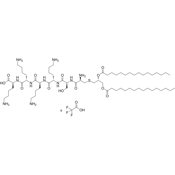
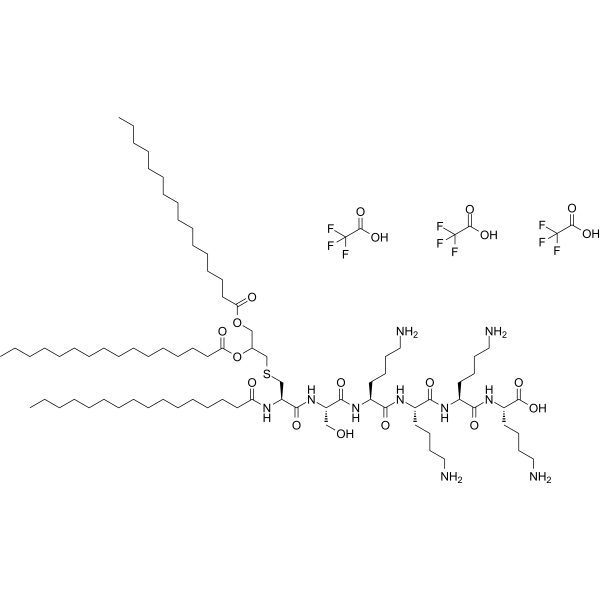
Pam2CSK4 TFA is a TLR2 agonist. Pam2CSK4 TFA induces the expression of iNOS and NO in macrophage cell lines via TBK1 and MyD88 molecules. Pam2CSK4 TFA activates the NF-κB and Bruton's tyrosine kinase signaling pathways in platelets, and promotes platelet-endothelial cell interactions. TLR2 activation triggered by Pam2CSK4 TFA expands myeloid-derived suppressor cells (MDSCs) and suppresses anti-tumor immune responses in the tumor microenvironment. Pam2CSK4 TFA acts as a Th2-polarizing adjuvant in mouse vaccine models against Leishmania major and Brugia malayi. Pam2CSK4 TFA can be used in the research of various diseases, including thromboinflammatory diseases, sepsis, atherosclerosis, heart failure, influenza, lymphoma, melanoma, cutaneous leishmaniasis and lymphatic filariasis.
Nur für Forschungszwecke. Wir verkaufen nicht an Patienten.
- Reinheit : 99.46%
- Formel: C65H126N10O12S.xC2HF3O2
- Molecular Weight:1271.82 (free base)
-
Speicherung:
Sealed storage, away from moisture and light.
Powder -80°C, 2 years , -20°C, 1 year* In solvent : -80°C, 6 months; -20°C, 1 month (sealed storage, away from moisture and light)

To place orders, for customer services and technical support, please contact: MedChemExpress USA
Tel: 609-228-6898 E-mail: [email protected] [email protected]


-
 Biologische Aktivität
-
Chemical Information
-
Lösungsmittel & Löslichkeit
- Protokoll
- Reinheit & Dokumentation
- Verweise
-
 Help & FAQs



Publications Citing Use of MedChemExpress (MCE) Pam2CSK4 TFA
More

Biologische Aktivität
Beschreibung
In Vitro
Pam2CSK4 (100 ng/mL; 6-24 h) TFA induces TLR2-mediated iNOS expression in RAW264.7 mouse macrophages, and this production depends on TBK1 and MyD88[1].
Pam2CSK4 (100 ng/mL; 6-24 h) TFA induces significant TLR2-mediated NO production in RAW264.7 murine macrophages, and this production is dependent on TBK1 and MyD88[1].
Pam2CSK4 (1-10 μg/mL; 25 min at 37°C) TFA induces concentration-dependent α-granule secretion and integrin αIIbβ3 activation in washed human platelets and platelets in citrated human whole blood[2].
Pam2CSK4 (10 μg/mL; 6-45 min at 37°C) TFA induces donor-dependent aggregation of washed human platelets under stirring conditions and enhances the adhesion of washed human platelets to collagen-coated surfaces[2].
Pam2CSK4 (10 μg/mL; 2-10 min at 37°C) TFA activates multiple signaling pathways in washed human platelets, including the Akt, PKC, NF-κB and MAPK (p38) pathways[2].
Pam2CSK4 (10 μg/mL) TFA induces dense granule secretion (ATP release) in washed human platelets, and this response depends on the TLR2, NF-κB and BTK signaling pathways[2].
Pam2CSK4 (10 μg/mL; 4 h at 37°C) TFA enhances the inflammatory response of HUVECs (characterized by upregulated ICAM-1 expression, increased IL-6 secretion and increased IL-8 secretion)[2] only when co-cultured with washed human platelets.
Pam2CSK4 (10 μg/mL; 10 min at 37°C platelet pre-incubation, 10 min co-culture) TFA enhances the adhesion of washed human platelets to untreated and TNF-α-stimulated HUVEC monolayers under static and physiologically relevant flow conditions[2].
Pam2CSK4 TFA is a potent dual TLR2 agonist for both human and murine sources. It induces NF-κB activity with an EC50 of 67 pM, and can stimulate the production of various cytokines and chemokines[3].
Pam2CSK4 (100 nM; 3 days) TFA treatment of CD11b+Gr1+ myeloid-derived suppressor cells (MDSCs) from EG7 tumor-bearing mice dose-dependently inhibited antigen-induced OT-IT cell proliferation[4].
MedChemExpress (MCE) has not independently confirmed the accuracy of these methods. They are for reference only. Further protocols information, click here.
-
Cell Line:RAW264.7
-
Concentration:100 ng/mL
-
Incubation Time:6 h
-
Result:Induced detectable inos gene expression.
Induced inos gene expression that was suppressed by pretreatment with TLR2 neutralizing monoclonal antibody.
-
Cell Line:RAW264.7
-
Concentration:100 ng/mL
-
Incubation Time:10 h
-
Result:Induced detectable iNOS protein expression.
Induced iNOS protein expression that was suppressed by pretreatment with TLR2 neutralizing monoclonal antibody.
-
Cell Line:Washed platelets
-
Concentration:10 μg/mL
-
Incubation Time:2, 5, 10 min
-
Result:Significantly increased tyrosine phosphorylation, increased 4G10 antibody expression.
Increased phosphorylation levels at Akt S473 and downstream substrates.
Increased phosphorylation levels of MAPK substrates and p38 T180Y182.
In Vivo
Pam2CSK4 (50 nmol; i.v.; single dose) TFA induces accumulation of CD11b+Gr1+ MDSCs in spleens of B16D8 melanoma-bearing mice[4].
Pam2CSK4 (10 μg; s.c.; two doses, 2 weeks apart) TFA as an adjuvant with ALM exacerbates cutaneous leishmaniasis disease severity in female C57BL/6 mice and drives a predominantly Th2-type immune response characterized by elevated antigen-specific IgG1 levels[6].
Pam2CSK4 (10 μg; s.c.; single dose) TFA as an adjuvant with BmMfE induces significant protective immunity against B. malayi L3 challenge in male BALB/c mice, reducing parasite burden by 41% and driving robust Th2 cytokine production and antigen-specific antibody responses[6].
MedChemExpress (MCE) has not independently confirmed the accuracy of these methods. They are for reference only.
-
Animal Model:C57BL/6J (B6 WT) female mice (8-12 weeks old); TLR2 knockout (TLR2KO) female mice (8-12 weeks old); C57BL6-Tg (CAG-EGFP) female mice (8-12 weeks old) EG7 cells (1x10⁶)[4]
-
Dosage:50 nM
-
Administration:i.v.; single dose
-
Result:Increased the proportion of CD11b+Gr1+ myeloid-derived suppressor cells (MDSCs) in CD45+ splenocytes to 12.9%.
Increased the proportion of CD11b+Gr1+ MDSCs in peripheral blood CD45+ cells to approximately 50%.
Increased the proportion of CD11b+Gr1+ MDSCs in tumor-bearing GFP+CD45+ host cells to approximately 27%.
Eliminated the increased proportion of CD11b+Gr1+ MDSCs in TLR2KO EG7 tumor-bearing mice.
Induced dose-dependent inhibition of OT-I splenocyte proliferation by CD11b+Gr1+ cells in tumor-bearing mice.
-
Animal Model:C57BL/6J (B6 WT) female mice (8-12 weeks old) ( B16D8 (6x10⁵)[4]
-
Dosage:50 nM
-
Administration:i.v.; single dose
-
Result:Increased the percentage of CD11b+Gr1+ MDSCs in CD45+ spleen cells to 21.1%.
-
Animal Model:C57BL/6 (female, 8-10 weeks old; vaccinated with autoclaved Leishmania major then challenged with L. major metacyclic promastigotes)[6]
-
Dosage:10 μg
-
Administration:s.c.; two doses, 2 weeks apart
-
Result:Increased lesion sizes significantly compared to PBS-vaccinated control mice from 4 weeks post-infection, with significantly increased area under the curve (AUC) values for lesion progression.
Elevated antigen-specific IgG1 responses significantly compared to PBS-vaccinated controls, and skewed the IgG1:IgG2c ratio toward a Th2 phenotype.
-
Animal Model:BALB/c (male, 8-10 weeks old; vaccinated with Brugia malayi microfilariae extract then challenged with B. malayi L3 larvae)[6]
-
Dosage:10 μg
-
Administration:s.c.; single dose
-
Result:Reduced parasite recovery by a significant mean 41%, equivalent to the protection induced by BmMfE + Alum.
Elevated antigen-specific IgG1 and IgG2a levels significantly , with IgG1 levels higher than those induced by BmMfE + Alum.
Induced significantly elevated production of antigen-specific IL-4, IL-5, and IL-13 (Th2 cytokines) in splenocytes upon re-stimulation with B.
malayi L3 extract, with no significant elevation of the Th1 cytokine IFNγ.


Chemical Information
-
Appearance Solid
-
Molecular Weight 1271.82 (free base)
-
Formel C65H126N10O12S.xC2HF3O2
-
Color White to off-white
-
Versand
Room temperature in continental US; may vary elsewhere.
-
Speicherung
Sealed storage, away from moisture and light
Powder -80°C 2 years -20°C 1 year * In solvent : -80°C, 6 months; -20°C, 1 month (sealed storage, away from moisture and light)


Publications (1)
-
Journal Impact Factor
-
Most Recent
-
PLoS Pathog
The UBC/SIRT5/DRP1 axis regulates mitochondrial dynamics to alleviate Staphylococcus aureus-induced oxidative stress and senescence in bovine mammary epithelial cells. [Abstract]2026 Feb 12;22(2):e1013975. PMID: 41678546


Lösungsmittel & Löslichkeit
In Vitro:
H2O : 100 mg/mL (Need ultrasonic)

Protokoll
-
Phagocytosis Functional Assay
A phagocytosis functional assay measures the ability of phagocytic cells, such as neutrophils, macrophages, monocytes, or microglia/macrophages, to bind and internalize particulate targets including bacteria, yeast particles, beads, or myelin particles. Fluorescent flow-cytometry assays detect target uptake as fluorescence associated with gated phagocytes, while pH-sensitive dyes such as pHrodo increase signal in acidic phagosomal compartments and therefore preferentially report internalized particles rather than particles remaining outside the cell. Microscopy or high-content imaging can be used to confirm intracellular localization and, in some protocols, to follow uptake kinetics.
-
RNA extraction experimental
By lysing cells, releasing RNA, and removing impurities such as proteins and DNA, high-purity RNA products are finally obtained. The commonly used traditional method is the guanidine isothiocyanate/phenol/chloroform method (Trizol), which is suitable for a variety of animal materials including animal tissues, microorganisms, cultured cells, etc., and most plant materials.
-
Multiplex immunofluorescence IHC
Multiplex immunofluorescence IHC detects multiple protein biomarkers in one tissue section by sequential antibody staining, HRP-mediated tyramide fluorophore deposition, heat-mediated antibody stripping, nuclear counterstaining, multispectral imaging, spectral unmixing, and digital cell phenotyping; TSA deposits fluorophore near the antigen so the fluorescence signal remains after primary and secondary antibodies are removed, enabling repeated staining cycles, including with antibodies from the same host species. Classic FFPE tumor immune-profiling applications use panels such as CD3, CD8, CD68/CD163, FOXP3, PD-1, PD-L1, pancytokeratin, Ki67, and DAPI to identify tumor cells, immune-cell subsets, checkpoint-marker expression, co-expression phenotypes, cell density, and spatial relationships in the tumor microenvironment.
-
LPS-Induced Endotoxemia/Systemic Inflammation
Lipopolysaccharide (LPS)-induced endotoxemia is a widely used in vivo model of acute systemic inflammation in which LPS, a Gram-negative bacterial endotoxin, activates innate immune signaling primarily through TLR4, leading to rapid and transient induction of pro-inflammatory cytokines such as TNF-α, IL-6, and IL-1β in circulation and tissues. This cytokine surge is commonly used as a measurable readout of systemic inflammatory activation and immune dysregulation, and is typically assessed within hours after intraperitoneal LPS administration in mouse models of endotoxemia. The model captures key features of systemic inflammatory response syndrome, including cytokine release, immune cell activation, and downstream tissue responses, and has been used to evaluate anti-inflammatory interventions such as cytokine modulation, lipid mediators, and immune cell-targeting therapies.
-
Naïve CD4+ T-cell subset differentiation/polarization
Naïve CD4+ T-cell subset differentiation/polarization is an in vitro assay in which purified naïve CD4+ T cells are activated through TCR and CD28 costimulation and cultured with defined cytokines and neutralizing antibodies to generate Th0, Th1, Th2, Th17, or induced Treg-like populations. Differentiation is detected by subset-associated cytokines and transcription factors: IFN-γ/T-bet for Th1, IL-4/GATA3 for Th2, IL-17A/RORγt for Th17, and Foxp3 for induced Treg cells. The assay readout is usually generated by intracellular cytokine staining after restimulation, transcription-factor staining by flow cytometry, ELISA of secreted cytokines, or gene-expression analysis. The result reflects cytokine-directed lineage commitment or polarization rather than antigen-specific immune protection by itself.
-
Multiplex immunohistochemistry
Multiplex immunohistochemistry (mIHC), also known as tyramide dignal amplification (TSA), is an enzymatic detection method that uses horseradish peroxidase (HRP) to perform high-density in-situ labeling of target proteins or nucleic acids.
-
Research Protocol for Cardiovascular Diseases
Cardiovascular disease can be modeled as maladaptive cardiac remodeling, where ischemic injury or pressure overload activates inflammatory signaling, fibroblast activation, extracellular-matrix deposition, cardiomyocyte hypertrophy, vascular remodeling, and progressive ventricular dysfunction. The TGF-β/SMAD axis is a central profibrotic pathway after myocardial injury and pressure overload, while innate immune and cytokine pathways regulate leukocyte recruitment, scar formation, and adverse remodeling. Key unresolved questions include which inflammatory signals are reparative versus harmful, when fibrosis is protective versus maladaptive, and whether pathway inhibition improves function without weakening necessary infarct healing or compensatory remodeling.
-
Research Protocol for Inflammation-related Diseases
The NLRP3 inflammasome is a cytosolic innate immune signaling platform that integrates priming signals and danger-signal activation to promote caspase-1 activation, maturation of IL-1β and IL-18, and gasdermin D-mediated pyroptotic cell death. The core experimental logic is to determine whether inflammatory disease phenotypes are driven by increased NLRP3 expression, ASC-containing inflammasome assembly, caspase-1 cleavage, GSDMD cleavage, and extracellular release of IL-1β/IL-18 rather than by nonspecific cell injury alone. The pathway is strongly linked to inflammation-related disease phenotypes because monosodium urate crystals activate NALP3/NLRP3 inflammasome signaling in gout-like crystal inflammation, cholesterol crystals activate NLRP3 inflammasomes in atherogenesis models, and DSS-induced intestinal inflammation has been reported to involve NLRP3 inflammasome activity. However, experimental colitis studies also show context-dependent protective effects of NLRP3 inflammasome co
-
Research Protocol for Cancer Immunology
Cancer immunology studies how the immune system recognizes, suppresses, edits, or fails to eliminate malignant cells through tumor antigen release, antigen presentation, T-cell priming, immune trafficking, tumor-cell killing, and feedback inhibition in the tumor microenvironment. The cancer-immunity cycle links tumor antigenicity, dendritic-cell priming, CD8+ T-cell infiltration, cytotoxic function, and immune-checkpoint regulation to tumor rejection or immune escape. Immune-checkpoint pathways such as PD-1/PD-L1 and CTLA-4 suppress antitumor T-cell activity and can be therapeutically blocked, but many tumors remain resistant because of poor antigen presentation, weak T-cell infiltration, suppressive myeloid cells, regulatory T cells, and tumor-intrinsic immune-exclusion programs. Unresolved questions include which immune-cell states predict response, how tumor-intrinsic pathways exclude immune cells, how myeloid suppression limits checkpoint blockade, and which combination strategies


Reinheit & Dokumentation
-
Data Sheet (284 KB)
-
SDS (252 KB)
- English - EN (252 KB)
- Français - FR (252 KB)
- Deutsch - DE (252 KB)
- Norwegian - NO (252 KB)
- Español - ES (252 KB)
- Swedish - SV (252 KB)
- Italian - IT (252 KB)
- Korean - KR (252 KB)
- Portuguese - PT (252 KB)
-
Handling Instructions (2659 KB)
Verweise
[1]. Kulsantiwong P, et al. Pam2CSK4 and Pam3CSK4 induce iNOS expression via TBK1 and MyD88 molecules in mouse macrophage cell line RAW264.7. Inflamm Res. 2017 Oct;66(10):843-853. [Content Brief]
[2]. Parra-Izquierdo I, et al. The Toll-Like Receptor 2 Ligand Pam2CSK4 Activates Platelet Nuclear Factor-κB and Bruton's Tyrosine Kinase Signaling to Promote Platelet-Endothelial Cell Interactions. Front Immunol. 2021 Aug 30;12:729951. [Content Brief]
[3]. Kaur A, et al. An efficient and scalable synthesis of potent TLR2 agonistic PAM2CSK4. RSC Adv. 2018 Mar 5;8(18):9587-9596. [Content Brief]
[4]. Maruyama A, et al. Pam2 lipopeptides systemically increase myeloid-derived suppressor cells through TLR2 signaling. Biochem Biophys Res Commun. 2015;457(3):445-450. [Content Brief]
[5]. Shukla NM, et al. Recent Advances and Perspectives in Small-molecule TLR Ligands and Their Modulators. ACS Med Chem Lett. 2018;9(12):1156-1159. Published 2018 Dec 3. [Content Brief]
[6]. Halliday A, et al. The TLR2/6 ligand Pam2CSK4 TFA is a Th2 polarizing adjuvant in Leishmania major and Brugia malayi murine vaccine models. Parasit Vectors. 2016 Feb 20;9:96. [Content Brief]

Calculators
Konzentration (Stammlösung) × Volumen (Stammlösung) = Konzentration (Ziellösung) × Volumen (Ziellösung)


Pam2CSK4 TFA Related Classifications
HY-P1181A Related Classifications
 Powered by Bioz