



Kuwanon G
Based on 1 publication(s) in Google Scholar
Kuwanon G is a flavonoid compound and an antagonist of the bombesin receptor. Kuwanon G has multiple activities such as bactericidal, anti-tumor, anti-inflammatory, antioxidant, anti-atherosclerotic, and neuroprotective effects. Kuwanon G exhibits strong antibacterial activity against oral pathogens, especially cariogenic bacteria and periodontal pathogens. Kuwanon G can induce apoptosis and inhibit proliferation, migration, and invasion of tumor cells. Kuwanon G can be used in the research of diseases such as gastric cancer and atherosclerosis.
For research use only. We do not sell to patients.
- Purity : 99.45%
- CAS No.: 75629-19-5
- Formula: C40H36O11
- Molecular Weight:692.71
-
Storage:
4°C, protect from light
* In solvent : -80°C, 6 months; -20°C, 1 month (protect from light)

To place orders, for customer services and technical support, please contact: MedChemExpress USA
Tel: 609-228-6898 E-mail: [email protected] [email protected]


-
 Biological Activity
-
Chemical Information
-
Solvent & Solubility
- Protocols
- Purity & Documentation
- References
-
 Help & FAQs


-
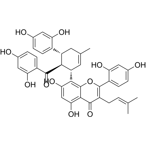
HY-N2600
Kuwanon HBombesin Receptor Antagonist/Platelet Activation Inhibitor/GRPR Antagonist -
HY-N3513
MulberrinOATP2B1 Inhibitor -
HY-N2181
AcetylshikoninAnti-inflammatory agent, AChE Inhibitor, P450 Inhibitor, Neuroprotective agents -
HY-N2393
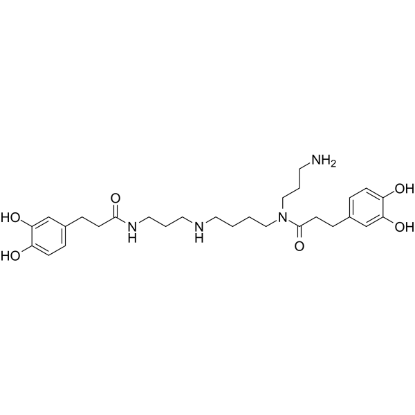
Kukoamine BActive Compound -

HY-N4247R
Kuwanon G (Standard)Standard of Kuwanon G -
HY-12302
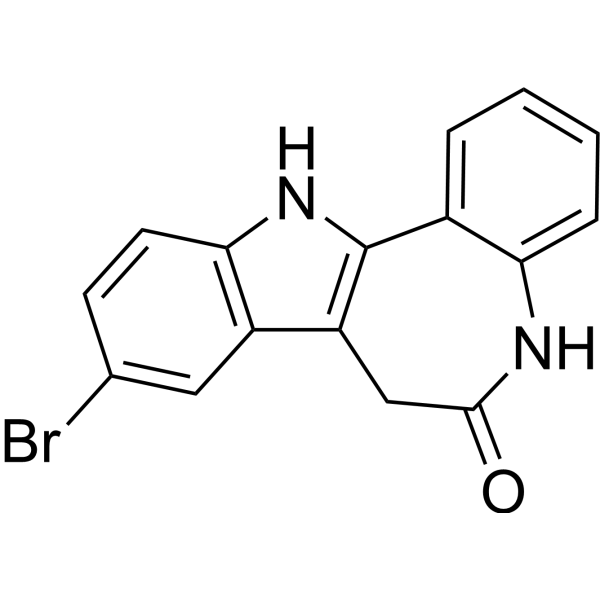
KenpaulloneCDK1/GSK-3β Inhibitor
Publications Citing Use of MedChemExpress (MCE) Kuwanon G
More

Biological Activity
Description
Cellular Effect
|
Cell Line

|
Type | Value | Description | References |
|---|---|---|---|---|
| Platelet | IC50 |
13.2 x 10⁻⁵ M
Compound: kumanon G
|
Inhibition of thromboxane B2 formation in Wistar King platelets
Inhibition of thromboxane B2 formation in Wistar King platelets
|
[PMID: 3097265] |
| Platelet | IC50 |
8.93 x 10⁻⁵ M
Compound: kumanon G
|
Inhibition of 12-hydroxy-5,8,10-heptadecatrienoic acid formation in Wistar King platelets
Inhibition of 12-hydroxy-5,8,10-heptadecatrienoic acid formation in Wistar King platelets
|
[PMID: 3097265] |
In Vitro
Kuwanon G (5-70 μM; 48 h) can inhibit the apoptosis of HT22 cells induced by Advanced glycation end products (HY-NP165) and the elevation of MDA and ROS levels, and restore the intracellular Ach level. The mechanism involves the PI3K/Akt/GSK3αβ signaling pathway[1].
Kuwanon G (0-20 μg/mL; 0-10 min) has an MIC of 8.0 μg/mL for Streptococcus mutans[2].
Kuwanon G (0-160 μM; 1-24 h) can inhibit the proliferation, migration and invasion, promote cell apoptosis, and inhibit the expression of MMP2 and MMP9 in gastric cancer cells. The mechanism involves the inhibition of the PI3K/AKT/mTOR pathway[3].
Kuwanon G (2-20 μM; 24 h) can inhibit lipid accumulation and the mRNA levels of inflammatory factors in RAW 264.7 cells treated with ox-LDL (HY-NP135). The mechanism involves the activation of the LXRα-ABCA1/ABCG1 pathway and the inhibition of NF-κB[4].
MedChemExpress (MCE) has not independently confirmed the accuracy of these methods. They are for reference only. Further protocols information, click here.
-
Cell Line:MGC 803, HGC 27, AGS and SGC-7901 cells
-
Concentration:0, 1.25, 5, 10, 20, 40, 80 and 160 μM
-
Incubation Time:24 h
-
Result:Inhibited the proliferation of gastric cancer cells in a concentration-dependent manner.
-
Cell Line:RAW264.7 cells treated ox-LDL (HY-NP135)
-
Concentration:20 μM
-
Incubation Time:24 h
-
Result:Increased the levels of ABCA1, ABCG1 and LXRα.
In Vivo
Kuwanon G (5 mg/kg; intraperitoneal injection; once every other day; 16 weeks) has an ameliorative effect in the atherosclerotic mouse model of ApoE-/- fed a high-fat diet[4].
MedChemExpress (MCE) has not independently confirmed the accuracy of these methods. They are for reference only.
-
Animal Model:Male ApoE-deficient (ApoE-/-) mice treated high-fat diet[4]
-
Dosage:5 mg/kg
-
Administration:Intraperitoneal injection; once every other day; 16 weeks
-
Result:Remarkably reduced the atherosclerotic lesion areas and macrophage content.
Reduced hyperlipidemia and serum inflammatory cytokines in vivo.


Chemical Information
-
CAS No. 75629-19-5
-
Appearance Solid
-
Molecular Weight 692.71
-
Formula C40H36O11
-
Color Light yellow to yellow
-
SMILES
O=C1C(C/C=C(C)\C)=C(C2=CC=C(O)C=C2O)OC3=C([C@H]4C=C(C)C[C@@H](C5=CC=C(O)C=C5O)[C@@H]4C(C6=CC=C(O)C=C6O)=O)C(O)=CC(O)=C13
-
Structure Classification
-
Initial Source
-
Shipping
Room temperature in continental US; may vary elsewhere.
-
Storage
4°C, protect from light
* In solvent : -80°C, 6 months; -20°C, 1 month (protect from light)


Publications (1)
-
Journal Impact Factor
-
Most Recent


Solvent & Solubility
In Vitro:
DMSO : 50 mg/mL (72.18 mM; Need ultrasonic; Hygroscopic DMSO has a significant impact on the solubility of product, please use newly opened DMSO)
Please refer to the solubility information to select the appropriate solvent. Once prepared, please aliquot and store the solution to prevent product inactivation from repeated freeze-thaw cycles.
Storage method and period of stock solution: -80°C, 6 months; -20°C, 1 month (protect from light). When stored at -80°C, please use it within 6 months. When stored at -20°C, please use it within 1 month.
Please refer to the solubility information to select the appropriate solvent. Once prepared, please aliquot and store the solution to prevent product inactivation from repeated freeze-thaw cycles.
Storage method and period of stock solution: -80°C, 6 months; -20°C, 1 month (protect from light). When stored at -80°C, please use it within 6 months. When stored at -20°C, please use it within 1 month.
Concentration (start) × Volume (start) = Concentration (final) × Volume (final)

Protocols
-
Research Protocol for Infectious Diseases
Infectious-disease experiments test how pathogens interact with host barriers, innate immune receptors, inflammatory signaling, pathogen replication, and tissue injury; pattern-recognition receptors such as TLRs, RIG-I-like receptors, NOD-like receptors, and inflammasomes detect microbial molecules and activate NF-κB, interferon, and cytokine responses. The central hypothesis is that infection severity reflects the balance between pathogen burden and host response: protective inflammation restricts pathogen growth, whereas excessive or mislocalized inflammation contributes to tissue damage and disease phenotype. Unresolved questions include which host pathways are protective versus pathogenic, why some infection models fail to translate to human disease, and which combined readouts best predict clinically relevant infection outcomes.
-
Apoptosis
Apoptosis, also called programmed cell death, is generally characterized by distinct morphological characteristics.
-
TUNEL staining for apoptotic DNA fragmentation
TUNEL staining detects DNA strand breaks by using terminal deoxynucleotidyl transferase to add labeled nucleotides to exposed 3′-OH DNA termini, generating either microscopic staining in fixed cells or tissue sections, or fluorescence/cytometric signal in cell suspensions. TUNEL positivity reflects DNA fragmentation but should not be interpreted alone as definitive apoptosis, because TUNEL can also label necrotic, autolytic, mechanically damaged, or DNA-repair-associated DNA breaks.
-
Annexin V plus membrane-impermeant dye apoptosis staining
Annexin V-based apoptosis assays rely on the detection of phosphatidylserine (PS) externalization from the inner leaflet of the plasma membrane to the outer leaflet, an early biochemical hallmark of apoptosis. Fluorescently labeled Annexin V binds PS in a calcium-dependent manner, enabling identification of early apoptotic cells by flow cytometry or fluorescence microscopy. When combined with a membrane-impermeant DNA-binding dye (e. g. , propidium iodide), this approach allows discrimination between viable (Annexin V−/dye−), early apoptotic (Annexin V+/dye−), and late apoptotic or necrotic (Annexin V+/dye+) cell populations by assessing membrane integrity and PS exposure.
-
Cell migration
Cell migration is a method that plays an important role in wound healing, cell differentiation, embryonic development, etc.
-
Apoptosis Solutions
Apoptosis is a regulated, generally non-lytic cell-death pathway that removes unwanted, damaged, infected, or abnormal cells through coordinated morphological changes, caspase activation, DNA fragmentation, and membrane remodeling. The intrinsic apoptosis pathway is controlled mainly by mitochondrial outer membrane permeabilization, BCL-2 family proteins, cytochrome c release, apoptosome formation, caspase-9 activation, and downstream executioner caspase-3/7 activation. The extrinsic apoptosis pathway is initiated by death receptors such as Fas, TNFR, and TRAIL receptors, which recruit adaptor proteins and activate caspase-8 before engaging executioner caspases or mitochondrial amplification through BID cleavage. Apoptosis is linked to many phenotypes, including cancer cell killing, tissue homeostasis, immune regulation, neurodegeneration, infection response, and treatment-induced cytotoxicity; unresolved questions include how apoptosis interacts with necroptosis, pyroptosis, ferroptos
-
Cell invasion
Cell invasion is the ability of cells to migrate from one area to another via the extracellular matrix. Cell invasion is the response of normal and cancer cells to chemical and mechanical stimuli. Before migrating to a new region, the extracellular matrix is degraded by proteases within the cell. Cell invasion often occurs during wound repair, vascularization and inflammation, abnormal tissue invasion, and tumor cell metastasis.
-
Research Protocol for Cardiovascular Diseases
Cardiovascular disease can be modeled as maladaptive cardiac remodeling, where ischemic injury or pressure overload activates inflammatory signaling, fibroblast activation, extracellular-matrix deposition, cardiomyocyte hypertrophy, vascular remodeling, and progressive ventricular dysfunction. The TGF-β/SMAD axis is a central profibrotic pathway after myocardial injury and pressure overload, while innate immune and cytokine pathways regulate leukocyte recruitment, scar formation, and adverse remodeling. Key unresolved questions include which inflammatory signals are reparative versus harmful, when fibrosis is protective versus maladaptive, and whether pathway inhibition improves function without weakening necessary infarct healing or compensatory remodeling.
-
Research Protocol for Inflammation-related Diseases
The NLRP3 inflammasome is a cytosolic innate immune signaling platform that integrates priming signals and danger-signal activation to promote caspase-1 activation, maturation of IL-1β and IL-18, and gasdermin D-mediated pyroptotic cell death. The core experimental logic is to determine whether inflammatory disease phenotypes are driven by increased NLRP3 expression, ASC-containing inflammasome assembly, caspase-1 cleavage, GSDMD cleavage, and extracellular release of IL-1β/IL-18 rather than by nonspecific cell injury alone. The pathway is strongly linked to inflammation-related disease phenotypes because monosodium urate crystals activate NALP3/NLRP3 inflammasome signaling in gout-like crystal inflammation, cholesterol crystals activate NLRP3 inflammasomes in atherogenesis models, and DSS-induced intestinal inflammation has been reported to involve NLRP3 inflammasome activity. However, experimental colitis studies also show context-dependent protective effects of NLRP3 inflammasome co
-
Bacterial live/dead nucleic-acid viability staining
The LIVE/DEAD bacterial viability staining method is based on differential permeability of nucleic-acid-binding fluorescent dyes, most commonly SYTO 9 and propidium iodide (PI), which enables discrimination of bacterial populations with intact versus compromised cytoplasmic membranes. SYTO 9 penetrates both intact and damaged bacterial membranes and binds nucleic acids to produce green fluorescence, whereas propidium iodide penetrates only cells with compromised membranes and fluoresces red while also reducing SYTO 9 signal through competitive binding and fluorescence interactions. The resulting fluorescence pattern is interpreted as a proxy for membrane integrity, which is widely used as an indicator of bacterial viability in microscopy, flow cytometry, and spectroscopic platforms. However, mechanistic studies show that SYTO 9 and PI interactions involve displacement and fluorescence resonance energy transfer effects, which can influence signal interpretation depending on dye ratios a
-
Cell Viability Determination by MTT Colorimetric Assay
The following protocol uses the MTT colorimetric assay as a classic literature-established method for assessing cell viability/metabolic activity in cultured mammalian cells. MTT[3-(4,5-dimethylthiazol-2-yl)-2,5-diphenyltetrazolium bromide] is reduced by metabolically active cells to a colored formazan product; the amount of formazan is quantified spectrophotometrically and provides an indirect measure of metabolically active viable cells. Importantly, MTT reduction reflects cellular oxidoreductase/metabolic activity rather than an absolute direct count of living cells, so changes in cellular metabolism can alter the signal independently of cell number.


Purity & Documentation
-
Data Sheet (282 KB)
-
SDS (251 KB)
- English - EN (251 KB)
- Français - FR (251 KB)
- Deutsch - DE (251 KB)
- Norwegian - NO (251 KB)
- Español - ES (251 KB)
- Swedish - SV (251 KB)
- Italian - IT (251 KB)
- Korean - KR (251 KB)
- Portuguese - PT (251 KB)
-
Handling Instructions (2659 KB)
References
[1]. Gan WJ, et al. Kuwanon G protects HT22 cells from advanced glycation end product-induced damage. Exp Ther Med. 2021 May;21(5):425. [Content Brief]
[2]. Park KM, et al. Kuwanon G: an antibacterial agent from the root bark of Morus alba against oral pathogens. J Ethnopharmacol. 2003 Feb;84(2-3):181-5. [Content Brief]
[3]. Geng Z, et al. [Kuwanon G inhibits growth, migration and invasion of gastric cancer cells by regulating the PI3K/AKT/mTOR pathway]. Nan Fang Yi Ke Da Xue Xue Bao. 2024 Aug 20;44(8):1476-1484. [Content Brief]
[4]. Liu XX, et al. Kuwanon G attenuates atherosclerosis by upregulation of LXRα-ABCA1/ABCG1 and inhibition of NFκB activity in macrophages. Toxicol Appl Pharmacol. 2018 Feb 15;341:56-63. [Content Brief]
[5]. Mihara S, et al. Non-peptide bombesin receptor antagonists, kuwanon G and H, isolated from mulberry. Biochem Biophys Res Commun. 1995 Aug 15;213(2):594-9. [Content Brief]


Complete Stock Solution Preparation Table
Please refer to the solubility information to select the appropriate solvent. Once prepared, please aliquot and store the solution to prevent product inactivation from repeated freeze-thaw cycles.
Storage method and period of stock solution: -80°C, 6 months; -20°C, 1 month (protect from light). When stored at -80°C, please use it within 6 months. When stored at -20°C, please use it within 1 month.
| Optional Solvent | Concentration Solvent Mass | 1 mg | 5 mg | 10 mg | 25 mg |
|---|---|---|---|---|---|
| DMSO | 1 mM | 1.4436 mL | 7.2180 mL | 14.4361 mL | 36.0901 mL |
| 5 mM | 0.2887 mL | 1.4436 mL | 2.8872 mL | 7.2180 mL | |
| 10 mM | 0.1444 mL | 0.7218 mL | 1.4436 mL | 3.6090 mL | |
| 15 mM | 0.0962 mL | 0.4812 mL | 0.9624 mL | 2.4060 mL | |
| 20 mM | 0.0722 mL | 0.3609 mL | 0.7218 mL | 1.8045 mL | |
| 25 mM | 0.0577 mL | 0.2887 mL | 0.5774 mL | 1.4436 mL | |
| 30 mM | 0.0481 mL | 0.2406 mL | 0.4812 mL | 1.2030 mL | |
| 40 mM | 0.0361 mL | 0.1805 mL | 0.3609 mL | 0.9023 mL | |
| 50 mM | 0.0289 mL | 0.1444 mL | 0.2887 mL | 0.7218 mL | |
| 60 mM | 0.0241 mL | 0.1203 mL | 0.2406 mL | 0.6015 mL |


Kuwanon G Related Classifications
HY-N4247 Related Classifications
 Powered by Bioz