



Phoenixin-20

Based on 1 Customer Validation
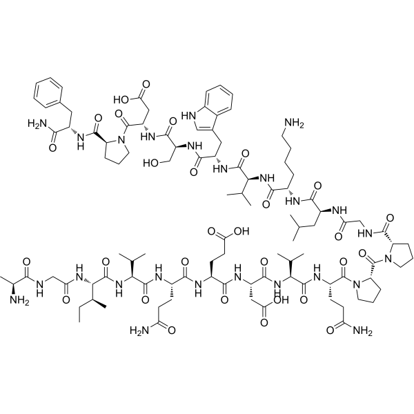
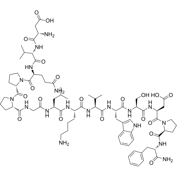
Phoenixin-20 (PNX-20) is a neuropeptide that activates GPR173. Phoenixin-20 inhibits LPS-induced TLR-4/MyD88/NF-κB signaling, pro-inflammatory cytokines, adhesion molecules, MMPs, and cytotoxicity. Phoenixin-20 increases SIRT1 and inhibits NLRP3/ASC/caspase-1, oxidative stress, and IL-1β/IL-18. Phoenixin-20 alleviates ox-LDL-induced endothelial dysfunction, monocyte adhesion, adhesion molecules, cytokines, NADPH oxidase/NOX-4, and ROS. Phoenixin-20 promotes GPR173/STAT6-mediated anti-inflammatory polarization of microglia and neuroprotection. Phoenixin-20 can be used in research on dental pulp infection, pulmonary arterial hypertension, atherosclerosis, neuroinflammation, anxiety, and neurodegenerative diseases.
For research use only. We do not sell to patients.
- Purity : 99.66%
- CAS No.: 1415039-77-8
- Formula: C101H153N25O29
- Molecular Weight:2181.45
-
Storage:
Sealed storage, away from moisture and light.
Powder -80°C, 2 years , -20°C, 1 year* In solvent : -80°C, 6 months; -20°C, 1 month (sealed storage, away from moisture and light)

To place orders, for customer services and technical support, please contact: MedChemExpress USA
Tel: 609-228-6898 E-mail: [email protected] [email protected]


-
 Biological Activity
-
Chemical Information
-
Solvent & Solubility
- Protocols
- Purity & Documentation
- References
-
 Help & FAQs


All Caspase Isoforms
More

Biological Activity
Description
|
GPR173 |
TLR4 |
NF-κB |
MyD88 |
SIRT1 |
NLRP3 |
Caspase-1 |
ASC |
IL-1β |
IL-18 |
NOX4 |
STAT6 |
In Vitro
Phoenixin-20 (15-30 nM; 24 h) attenuates LPS-induced cytotoxicity by reducing LDH release in hDPCs[1].
Phoenixin-20 (15-30 nM; 24 h) inhibits LPS-induced IL-6 and MCP-1 production in hDPCs at both mRNA and protein levels[1].
Phoenixin-20 (10-20 nM) reduces inflammatory cytokine secretion in hypoxia-treated human PMECs[2].
Phoenixin-20 (10-20 nM; 24 h) inhibits LPS-induced IL-1β and IL-18 secretion in BV2 microglial cells[6].
Phoenixin-20 (20 nM; 24 h) inhibits LPS-induced IL-1β and IL-18 production in BV2 microglia, a process that requires SIRT1 activity, and this effect can be abolished by Nicotinamide (1 mM)[6].
Phoenixin-20 (15-30 nM; 24 h) inhibits LPS-induced VCAM-1 and ICAM-1 expression in hDPCs[1].
Phoenixin-20 (15-30 nM; 24 h) inhibits LPS-induced MMP-2 and MMP-9 expression in hDPCs[1].
Phoenixin-20 (1-100 nM; 1-24 h) upregulated vitellogenin, esr2a, esr2b, and shbg mRNA expression in zebrafish liver (ZFL) cells, supporting a vitellogenic effect[9].
Phoenixin-20 (10-100 ng/mL; 24 h) stimulates oocyte maturation in zebrafish stage III follicles by increasing germinal vesicle breakdown[9].
Phoenixin-20 (5-20 nM; 48 h) dose-dependently induces PGC-1α mRNA and protein in M17 cells[10].
Phoenixin-20 (5-20 nM; 48 h) dose-dependently induces NRF-1 and TFAM mRNA and protein in M17 cells[10].
Phoenixin-20 (48 h) enhances mitochondrial respiration rate and ATP production in M17 cells[10].
Phoenixin-20 increases GPR173 mRNA approximately threefold and GPR173 protein approximately 2.5-fold in M17 cells[10].
Phoenixin-20 (15-30 nM; 24 h) inhibits LPS-induced TLR-4 and MyD88 expression in hDPCs[1].
Phoenixin-20 (15-30 nM; 24 h) inhibits LPS-induced NF-κB activation by suppressing p65 nuclear translocation and NF-κB reporter gene activity in hDPCs[1].
Phoenixin-20 (15-30 nM; 24 h) lost its inhibitory effect on LPS-induced MMP-2, MMP-9, and NF-κB activation in GPR173-silenced hDPCs, indicating that this effect requires GPR173[1].
Phoenixin-20-induced M2 polarization of BV2 microglia depends on GPR173, as GPR173 knockdown abolishes the induced expression of M2 markers[7].
Phoenixin-20 (10-20 nM) reduces MDA levels and increases SOD activity in hypoxia-treated human PMECs[2].
Phoenixin-20 (10-20 nM; 24 h) inhibits LPS-induced NOX-4 and TxNIP mRNA and restores SIRT1 mRNA in BV2 microglial cells[6].
Phoenixin-20 (10-20 nM; 24 h) inhibits LPS-induced protein expression of NOX-4, TxNIP, NLRP3, ASC, and cleaved caspase-1 (P10) and restores SIRT1 protein in BV2 microglial cells[6].
Phoenixin-20 (10-20 nM; 24 h) dose-dependently inhibits LPS-induced intracellular ROS production in BV2 microglial cells[6].
Phoenixin-20 (10-20 nM) inhibits NLRP3/ASC signaling and upregulates SIRT1 in hypoxia-treated human PMECs[2].
Phoenixin-20 (20 nM; 24 h) inhibition of NLRP3/ASC in hypoxia-treated human PMECs is mediated by SIRT1[2].
Phoenixin-20 (20 nM; 24 h) inhibits LPS-induced NLRP3 expression in BV2 microglial cells, an effect that depends on SIRT1 activity and can be abolished by Nicotinamide (HY-B0150) (1 mM)[6].
Phoenixin-20 (10-20 nM; 24 h) reduces ox-LDL-induced adhesion of THP-1 monocytes to HAECs, with a more pronounced reduction at 20 nM[3].
Phoenixin-20 (10-20 nM; 24 h) reduces ox-LDL-induced ICAM-1 and VCAM-1 expression in HAECs, with 20 nM producing a stronger inhibitory effect[3].
Phoenixin-20 (10-20 nM; 24 h) ameliorates ox-LDL-induced IL-1β, IL-8, and MCP-1 expression and secretion in HAECs[3].
Phoenixin-20 (10-20 nM; 24 h) reduces ox-LDL-induced ROS production and NOX-4 expression in HAECs[3].
Phoenixin-20 (10-20 nM; 24 h) inhibits ox-LDL-induced NF-κB activation in HAECs, and 20 nM Phoenixin-20 also reduces p-IκBα and restores total IκBα levels[3].
Phoenixin-20 (20 nM; 6 h) promotes M2 polarization in BV2 microglia, increasing Fizz1, Arg-1, YM1, IL-10, and p-STAT6[7].
Phoenixin-20 (1-100 nM; 1-2 h) regulates the mRNA expression of genes involved in glucose transport, glycolysis, gluconeogenesis, and glycogen metabolism in zebrafish liver (ZFL) cells[8].
Phoenixin-20 (100 nM; 1.5 h) increases the glycolysis-related ATP production rate in zebrafish liver (ZFL) cells without altering the total ATP production rate[8].
Phoenixin-20 (10 nM; 48 h) promotes mitochondrial biogenesis in M17 cells by increasing mtDNA/nDNA, Tomm22/Timm50/Atp5d/Ndufs3 mRNA, and NDUFB8 protein[10].
Phoenixin-20 (5-20 nM; 48 h) activates CREB, and inhibition of CREB by H89 (10 μM) prevents its effects on mitochondrial regulators and biogenesis in M17 cells[10].
MedChemExpress (MCE) has not independently confirmed the accuracy of these methods. They are for reference only. Further protocols information, click here.
-
Cell Line:human dental pulp cells (hDPCs)
-
Concentration:15 and 30 nM
-
Incubation Time:24 h (with 20 μg/mL LPS)
-
Result:At 15 and 30 nM, decreased LPS-induced LDH release in a dose-responsive manner.
-
Cell Line:Human pulmonary microvascular endothelial cells (PMECs)
-
Concentration:20 nM
-
Incubation Time:24 h
-
Result:Hypoxia-induced increases in NLRP3 and ASC were repressed at 20 nM, but this repression was reversed by SIRT1 knockdown.
-
Cell Line:BV2 microglial cells
-
Concentration:10 nM and 20 nM
-
Incubation Time:24 h
-
Result:LPS-induced NOX-4 mRNA was dose-responsively suppressed.
LPS-induced TxNIP mRNA was dose-responsively inhibited.
LPS-induced repression of SIRT1 mRNA was robustly ameliorated.
20 nM produced the greatest inhibition where noted.
-
Cell Line:BV2 microglial cells
-
Concentration:10 nM and 20 nM
-
Incubation Time:24 h
-
Result:LPS-induced NOX-4 protein was suppressed.
LPS-induced TxNIP protein was dose-responsively inhibited.
LPS-induced NLRP3, ASC, and cleaved caspase-1 (P10) were significantly suppressed.
20 nM showed the greatest inhibitory effect.
LPS-induced repression of SIRT1 protein was robustly ameliorated.
-
Cell Line:BV2 microglial cells
-
Concentration:10 nM and 20 nM
-
Incubation Time:24 h
-
Result:LPS-induced production of IL-1β and IL-18 was inhibited.
-
Cell Line:BV2 microglial cells
-
Concentration:20 nM (Phoenixin-20); 1 μg/mL (LPS); 1 mM (nicotinamide)
-
Incubation Time:24 h
-
Result:SIRT1 activation blockage by Nicotinamide completely abolished the inhibitory effect on LPS-induced NLRP3 expression.
-
Cell Line:BV2 microglial cells
-
Concentration:20 nM (Phoenixin-20); 1 μg/mL (LPS); 1 mM (nicotinamide)
-
Incubation Time:24 h
-
Result:SIRT1 activation blockage completely abolished the inhibitory effect on LPS-induced IL-1β and IL-18 production.
-
Cell Line:BV2 murine microglial cells
-
Concentration:20 nM (with 10 ng/mL IL-4)
-
Incubation Time:6 h
-
Result:Increased Fizz1, Arg-1, YM1, and IL-10 expression.
Increased p-STAT6 levels.
In Vivo
Phoenixin-20 (4 mg/kg; s.c.) elicits repetitive scratching behavior in male Swiss-Webster mice with pruritus[5].
Phoenixin-20 (2 µg per mouse; i.c.v. into the lateral cerebral ventricle; consecutive injection before and after induction; initiated 24 h before MCAO and maintained during the experiment) reduces brain infarction and neurological deficit and promotes an M2-like microglial marker profile in C57BL/6 MCAO mice[7].
Phoenixin-20 (100 ng/g body weight; injected; daily; 28 days) attenuates hypoxia-induced PAH in male rats, reducing hemodynamic and remodeling parameters and suppressing inflammation, oxidative stress, and NLRP3 signaling in lung tissue[2].
MedChemExpress (MCE) has not independently confirmed the accuracy of these methods. They are for reference only.
-
Animal Model:male, 7-9 weeks[2]
-
Dosage:100 ng/g body weight
-
Administration:injected; daily; 28 days
-
Result:RVSP was maintained around 20 mmHg.
mPAP was 19.17 mmHg.
RV/LV+S was 0.27.
W% was 22.16%.
TNF-α was 15.02 pg/mL.
IL-6 was around 37 pg/mL.
MCP-1 was 11.96 pg/mL.
MDA was 1.09 nmol/mg protein.
SOD activity was 67.3 U/mg protein.
-
Animal Model:male, 7-9 weeks[2]
-
Dosage:100 ng/g body weight
-
Administration:injected; daily; 28 days
-
Result:RVSP was 26.53 mmHg.
mPAP was 25.24 mmHg.
RV/LV+S was 0.31.
W% was 30.33%.
TNF-α was 22.96 pg/mL.
IL-6 was 45.32 pg/mL.
MCP-1 was 14.25 pg/mL.
MDA was 1.32 nmol/mg protein.
SOD activity was 56.4 U/mg protein.
-
Animal Model:Swiss-Webster (male, 20-25 g)[5]
-
Dosage:4 mg/kg
-
Administration:s.c.; to nape of the neck; 30 min
-
Result:Evoked 57 scratching bouts (n = 7) across 30 min.
Latency was 3-5 min and duration was 10-15 min.
-
Animal Model:C57BL/6 (male, 8-12 weeks, 25-30 g)[7]
-
Dosage:2 µg per mouse (25-30 g body weight)
-
Administration:i.c.v. into the lateral cerebral ventricle; consecutive injection before and after induction; initiated 24 h before MCAO and maintained during the experiment
-
Result:Brain infarction volume was approximately 17%, compared with about 33% in placebo MCAO mice.
Neurological deficit score averaged 17%, compared with about 33% in placebo MCAO mice.
After three days, peri-infarct brain tissue had lower CD11b, CD86, iNOS, TNF-α, and IL-6 expression and higher Fizz1, Arg-1, YM1, and IL-10 expression than placebo MCAO mice.


Chemical Information
-
CAS No. 1415039-77-8
-
Appearance Solid
-
Molecular Weight 2181.45
-
Formula C101H153N25O29
-
Color White to off-white
-
Synonyms
PNX-20
-
Sequence
Ala-Gly-Ile-Val-Gln-Glu-Asp-Val-Gln-Pro-Pro-Gly-Leu-Lys-Val-Trp-Ser-Asp-Pro-Phe-NH2
-
Sequence Shortening
AGIVQEDVQPPGLKVWSDPF-NH2
-
Shipping
Room temperature in continental US; may vary elsewhere.
-
Storage
Sealed storage, away from moisture and light
Powder -80°C 2 years -20°C 1 year * In solvent : -80°C, 6 months; -20°C, 1 month (sealed storage, away from moisture and light)


Solvent & Solubility
In Vitro:
DMSO : 50 mg/mL (22.92 mM; Need ultrasonic; Hygroscopic DMSO has a significant impact on the solubility of product, please use newly opened DMSO)
Please refer to the solubility information to select the appropriate solvent. Once prepared, please aliquot and store the solution to prevent product inactivation from repeated freeze-thaw cycles.
Storage method and period of stock solution: -80°C, 6 months; -20°C, 1 month (sealed storage, away from moisture and light). When stored at -80°C, please use it within 6 months. When stored at -20°C, please use it within 1 month.
Please refer to the solubility information to select the appropriate solvent. Once prepared, please aliquot and store the solution to prevent product inactivation from repeated freeze-thaw cycles.
Storage method and period of stock solution: -80°C, 6 months; -20°C, 1 month (sealed storage, away from moisture and light). When stored at -80°C, please use it within 6 months. When stored at -20°C, please use it within 1 month.
Concentration (start) × Volume (start) = Concentration (final) × Volume (final)

Protocols
-
Research Protocol for Infectious Diseases
Infectious-disease experiments test how pathogens interact with host barriers, innate immune receptors, inflammatory signaling, pathogen replication, and tissue injury; pattern-recognition receptors such as TLRs, RIG-I-like receptors, NOD-like receptors, and inflammasomes detect microbial molecules and activate NF-κB, interferon, and cytokine responses. The central hypothesis is that infection severity reflects the balance between pathogen burden and host response: protective inflammation restricts pathogen growth, whereas excessive or mislocalized inflammation contributes to tissue damage and disease phenotype. Unresolved questions include which host pathways are protective versus pathogenic, why some infection models fail to translate to human disease, and which combined readouts best predict clinically relevant infection outcomes.
-
LPS-Induced Endotoxemia/Systemic Inflammation
Lipopolysaccharide (LPS)-induced endotoxemia is a widely used in vivo model of acute systemic inflammation in which LPS, a Gram-negative bacterial endotoxin, activates innate immune signaling primarily through TLR4, leading to rapid and transient induction of pro-inflammatory cytokines such as TNF-α, IL-6, and IL-1β in circulation and tissues. This cytokine surge is commonly used as a measurable readout of systemic inflammatory activation and immune dysregulation, and is typically assessed within hours after intraperitoneal LPS administration in mouse models of endotoxemia. The model captures key features of systemic inflammatory response syndrome, including cytokine release, immune cell activation, and downstream tissue responses, and has been used to evaluate anti-inflammatory interventions such as cytokine modulation, lipid mediators, and immune cell-targeting therapies.
-
Cell Cytotoxicity Assay
Cytotoxicity assays are usually based on the assessment of cell membrane damage, which can also be indirectly detected by measuring cell viability. Detection methods include MTT assay, CKK-8 assay, LDH assay and ATP assay, etc.
-
Mammalian live/dead viability and cytotoxicity staining
Live/dead viability and cytotoxicity staining assays are based on the simultaneous detection of intracellular esterase activity in metabolically active (viable) cells and membrane integrity loss in non-viable cells. In commonly used dual-staining approaches, membrane-permeant fluorogenic substrates are converted by intracellular esterases into fluorescent products in live cells, while impermeant DNA-binding dyes selectively enter cells with compromised plasma membranes and label nucleic acids in dead or dying cells, enabling discrimination between viable and non-viable populations by fluorescence microscopy or flow cytometry.
-
ROS/oxidative-stress fluorescent staining
ROS/oxidative-stress fluorescent staining uses cell-permeant fluorogenic probes that become fluorescent after oxidation inside cells or tissues; commonly used examples include DCFH-DA/DCFDA for broad cellular oxidant detection, DHE for superoxide-related signal detection, MitoSOX for mitochondrial superoxide-related signal detection, and CellROX probes for oxidative-stress-associated fluorescence readouts. The assay detects probe oxidation rather than a single ROS species unless the probe and analysis method have been chemically validated for that species. DCFH-DA enters cells, is deacetylated by intracellular esterases to DCFH, and produces fluorescent DCF after oxidation, so the readout is used as an operational measure of total cellular oxidative stress rather than a species-specific ROS measurement. DHE and MitoSOX can report superoxide-related oxidation, but red fluorescence alone can include non-specific ethidium-like oxidation products; HPLC or optimized spectral approaches are
-
Apoptosis Solutions
Apoptosis is a regulated, generally non-lytic cell-death pathway that removes unwanted, damaged, infected, or abnormal cells through coordinated morphological changes, caspase activation, DNA fragmentation, and membrane remodeling. The intrinsic apoptosis pathway is controlled mainly by mitochondrial outer membrane permeabilization, BCL-2 family proteins, cytochrome c release, apoptosome formation, caspase-9 activation, and downstream executioner caspase-3/7 activation. The extrinsic apoptosis pathway is initiated by death receptors such as Fas, TNFR, and TRAIL receptors, which recruit adaptor proteins and activate caspase-8 before engaging executioner caspases or mitochondrial amplification through BID cleavage. Apoptosis is linked to many phenotypes, including cancer cell killing, tissue homeostasis, immune regulation, neurodegeneration, infection response, and treatment-induced cytotoxicity; unresolved questions include how apoptosis interacts with necroptosis, pyroptosis, ferroptos
-
Research Protocol for Cardiovascular Diseases
Cardiovascular disease can be modeled as maladaptive cardiac remodeling, where ischemic injury or pressure overload activates inflammatory signaling, fibroblast activation, extracellular-matrix deposition, cardiomyocyte hypertrophy, vascular remodeling, and progressive ventricular dysfunction. The TGF-β/SMAD axis is a central profibrotic pathway after myocardial injury and pressure overload, while innate immune and cytokine pathways regulate leukocyte recruitment, scar formation, and adverse remodeling. Key unresolved questions include which inflammatory signals are reparative versus harmful, when fibrosis is protective versus maladaptive, and whether pathway inhibition improves function without weakening necessary infarct healing or compensatory remodeling.
-
Research Protocol for Inflammation-related Diseases
The NLRP3 inflammasome is a cytosolic innate immune signaling platform that integrates priming signals and danger-signal activation to promote caspase-1 activation, maturation of IL-1β and IL-18, and gasdermin D-mediated pyroptotic cell death. The core experimental logic is to determine whether inflammatory disease phenotypes are driven by increased NLRP3 expression, ASC-containing inflammasome assembly, caspase-1 cleavage, GSDMD cleavage, and extracellular release of IL-1β/IL-18 rather than by nonspecific cell injury alone. The pathway is strongly linked to inflammation-related disease phenotypes because monosodium urate crystals activate NALP3/NLRP3 inflammasome signaling in gout-like crystal inflammation, cholesterol crystals activate NLRP3 inflammasomes in atherogenesis models, and DSS-induced intestinal inflammation has been reported to involve NLRP3 inflammasome activity. However, experimental colitis studies also show context-dependent protective effects of NLRP3 inflammasome co
-
Research Protocol for Neurological Diseases
PINK1/Parkin-mediated mitophagy pathway is a mitochondrial quality-control signaling axis in which mitochondrial depolarization stabilizes PINK1 on damaged mitochondria, activates Parkin recruitment and E3 ubiquitin ligase activity, promotes ubiquitination of outer mitochondrial membrane proteins, recruits selective autophagy adaptors, and drives lysosomal degradation of damaged mitochondria. In neurological disease research, this pathway is experimentally important because neurons, especially dopaminergic neurons, are highly dependent on mitochondrial integrity, and defective mitochondrial turnover can lead to mitochondrial dysfunction, oxidative stress, impaired neuronal survival, α-synuclein accumulation, and neuroinflammatory damage-associated signals. The genetic disease link is strongest in Parkinson’s disease because mutations in PRKN/parkin cause autosomal recessive juvenile parkinsonism, mutations in PINK1 cause hereditary early-onset Parkinson’s disease, and Drosophila studie
-
Cell Viability Determination by MTT Colorimetric Assay
The following protocol uses the MTT colorimetric assay as a classic literature-established method for assessing cell viability/metabolic activity in cultured mammalian cells. MTT[3-(4,5-dimethylthiazol-2-yl)-2,5-diphenyltetrazolium bromide] is reduced by metabolically active cells to a colored formazan product; the amount of formazan is quantified spectrophotometrically and provides an indirect measure of metabolically active viable cells. Importantly, MTT reduction reflects cellular oxidoreductase/metabolic activity rather than an absolute direct count of living cells, so changes in cellular metabolism can alter the signal independently of cell number.
-
Pyroptosis Solutions
Pyroptosis is a lytic inflammatory cell-death pathway executed by gasdermin pores, most classically through inflammasome-mediated activation of caspase-1, cleavage of gasdermin D, membrane pore formation, LDH release, and secretion of IL-1β and IL-18. The canonical pathway is commonly modeled by priming cells with an inflammatory signal such as LPS to induce pro-IL-1β and inflammasome components, followed by an activation signal such as ATP or nigericin to activate NLRP3, ASC speck formation, caspase-1 cleavage, GSDMD cleavage, cytokine release, and pyroptotic membrane rupture. The non-canonical pathway is triggered when cytosolic LPS activates mouse caspase-11 or human caspase-4/5, leading to GSDMD cleavage and pyroptosis, and this can secondarily activate NLRP3-dependent IL-1β release. Pyroptosis is linked to inflammatory injury, infection, cancer, liver disease, ocular disease, placental inflammation, and other disease phenotypes, but unresolved questions include which gasdermin fam


Purity & Documentation
-
Data Sheet (324 KB)
-
SDS (252 KB)
- English - EN (252 KB)
- Français - FR (252 KB)
- Deutsch - DE (252 KB)
- Norwegian - NO (252 KB)
- Español - ES (252 KB)
- Swedish - SV (252 KB)
- Italian - IT (252 KB)
- Korean - KR (252 KB)
- Portuguese - PT (252 KB)
-
Handling Instructions (2659 KB)
References
[5]. Cowan A, et al. Phoenixin: A candidate pruritogen in the mouse. Neuroscience. 2015 Dec 03;310:541-8.
[9]. Rajeswari JJ, et al. Phoenixin-20 Stimulates mRNAs Encoding Hypothalamo-Pituitary-Gonadal Hormones, is Pro-Vitellogenic, and Promotes Oocyte Maturation in Zebrafish. Scientific reports. 2020 Apr 14;10(1):6264. [Content Brief]
[10]. Yang Y, et al. Phoenixin 20 promotes neuronal mitochondrial biogenesis via CREB-PGC-1α pathway. Journal of molecular histology. 2020 Apr;51(2):173-181. [Content Brief]


Complete Stock Solution Preparation Table
Please refer to the solubility information to select the appropriate solvent. Once prepared, please aliquot and store the solution to prevent product inactivation from repeated freeze-thaw cycles.
Storage method and period of stock solution: -80°C, 6 months; -20°C, 1 month (sealed storage, away from moisture and light). When stored at -80°C, please use it within 6 months. When stored at -20°C, please use it within 1 month.
| Optional Solvent | Concentration Solvent Mass | 1 mg | 5 mg | 10 mg | 25 mg |
|---|---|---|---|---|---|
| DMSO | 1 mM | 0.4584 mL | 2.2921 mL | 4.5841 mL | 11.4603 mL |
| 5 mM | 0.0917 mL | 0.4584 mL | 0.9168 mL | 2.2921 mL | |
| 10 mM | 0.0458 mL | 0.2292 mL | 0.4584 mL | 1.1460 mL | |
| 15 mM | 0.0306 mL | 0.1528 mL | 0.3056 mL | 0.7640 mL | |
| 20 mM | 0.0229 mL | 0.1146 mL | 0.2292 mL | 0.5730 mL |


Phoenixin-20 Related Classifications
HY-P5763 Related Classifications
 Powered by Bioz
Keywords
- Phoenixin-20
- 1415039-77-8
- PNX-20
- Phoenixin20
- Phoenixin 20
- PNX20
- PNX 20
- Toll-like Receptor (TLR)
- MyD88
- NF-κB
- Sirtuin
- NOD-like Receptor (NLR)
- ASCT
- Caspase
- Interleukin Related
- NADPH Oxidase
- Reactive Oxygen Species (ROS)
- STAT
- GPR173
- NLRP3
- SIRT1
- TLR-4
- atherosclerosis
- mitochondrial biogenesis
- neuroinflammation
- oxidative stress
- Inhibitor
- inhibitor
- inhibit