



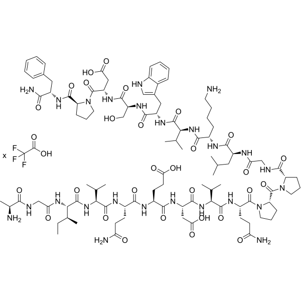
Phoenixin-20 TFA
Phoenixin-20 TFA (PNX-20 TFA) is a neuropeptide that activates GPR173. Phoenixin-20 TFA inhibits LPS-induced TLR-4/MyD88/NF-κB signaling, pro-inflammatory cytokines, adhesion molecules, MMPs, and cytotoxicity. Phoenixin-20 TFA increases SIRT1 and inhibits NLRP3/ASC/caspase-1, oxidative stress, and IL-1β/IL-18. Phoenixin-20 TFA alleviates ox-LDL-induced endothelial dysfunction, monocyte adhesion, adhesion molecules, cytokines, NADPH oxidase/NOX-4, and ROS. Phoenixin-20 TFA promotes GPR173/STAT6-mediated anti-inflammatory polarization of microglia and neuroprotection. Phoenixin-20 TFA can be used in research on dental pulp infection, pulmonary arterial hypertension, atherosclerosis, neuroinflammation, anxiety, and neurodegenerative diseases.
For research use only. We do not sell to patients.
- Formula: C101H153N25O29.xC2HF3O2
- Molecular Weight:2181.45 (free base)
-
Storage:
Please store the product under the recommended conditions in the Certificate of Analysis.

To place orders, for customer services and technical support, please contact: MedChemExpress USA
Tel: 609-228-6898 E-mail: [email protected] [email protected]


-
 Biological Activity
-
Chemical Information
- Protocols
- Purity & Documentation
- References
-
 Help & FAQs


All Caspase Isoforms
More

Biological Activity
Description
|
GPR173 |
TLR4 |
NF-κB |
MyD88 |
SIRT1 |
NLRP3 |
Caspase-1 |
ASC |
IL-1β |
IL-18 |
NOX4 |
STAT6 |
In Vitro
Phoenixin-20 (20 nM; 24 h) TFA inhibition of NLRP3/ASC in hypoxia-treated human PMECs is mediated by SIRT1[2].
Phoenixin-20 (20 nM; 24 h) TFA inhibits LPS-induced NLRP3 expression in BV2 microglial cells, an effect that depends on SIRT1 activity and can be abolished by Nicotinamide (HY-B0150) (1 mM)[6].
Phoenixin-20 (10-20 nM) TFA reduces inflammatory cytokine secretion in hypoxia-treated human PMECs[2].
Phoenixin-20 (10-20 nM; 24 h) TFA inhibits LPS-induced IL-1β and IL-18 secretion in BV2 microglial cells[6].
Phoenixin-20 (20 nM; 24 h) TFA inhibits LPS-induced IL-1β and IL-18 production in BV2 microglia, a process that requires SIRT1 activity, and this effect can be abolished by Nicotinamide (1 mM)[6].
Phoenixin-20 (15-30 nM; 24 h) TFA inhibits LPS-induced NF-κB activation by suppressing p65 nuclear translocation and NF-κB reporter gene activity in hDPCs[1].
Phoenixin-20 (15-30 nM; 24 h) TFA lost its inhibitory effect on LPS-induced MMP-2, MMP-9, and NF-κB activation in GPR173-silenced hDPCs, indicating that this effect requires GPR173[1].
Phoenixin-20 TFA-induced M2 polarization of BV2 microglia depends on GPR173, as GPR173 knockdown abolishes the induced expression of M2 markers[7].
Phoenixin-20 (10-20 nM; 24 h) TFA inhibits LPS-induced NOX-4 and TxNIP mRNA and restores SIRT1 mRNA in BV2 microglial cells[6].
Phoenixin-20 (10-20 nM; 24 h) TFA inhibits LPS-induced protein expression of NOX-4, TxNIP, NLRP3, ASC, and cleaved caspase-1 (P10) and restores SIRT1 protein in BV2 microglial cells[6].
Phoenixin-20 (10-20 nM; 24 h) TFA dose-dependently inhibits LPS-induced intracellular ROS production in BV2 microglial cells[6].
Phoenixin-20 (100 nM; 1.5 h) TFA increases the glycolysis-related ATP production rate in zebrafish liver (ZFL) cells without altering the total ATP production rate[8].
Phoenixin-20 (5-20 nM; 48 h) TFA activates CREB, and inhibition of CREB by H89 (10 μM) prevents its effects on mitochondrial regulators and biogenesis in M17 cells[10].
Phoenixin-20 (15-30 nM; 24 h) TFA inhibits LPS-induced MMP-2 and MMP-9 expression in hDPCs[1].
Phoenixin-20 (1-100 nM; 1-24 h) TFA upregulated vitellogenin, esr2a, esr2b, and shbg mRNA expression in zebrafish liver (ZFL) cells, supporting a vitellogenic effect[9].
Phoenixin-20 (10-100 ng/mL; 24 h) TFA stimulates oocyte maturation in zebrafish stage III follicles by increasing germinal vesicle breakdown[9].
Phoenixin-20 (5-20 nM; 48 h) TFA dose-dependently induces PGC-1α mRNA and protein in M17 cells[10].
Phoenixin-20 (5-20 nM; 48 h) TFA dose-dependently induces NRF-1 and TFAM mRNA and protein in M17 cells[10].
Phoenixin-20 (48 h) TFA enhances mitochondrial respiration rate and ATP production in M17 cells[10].
Phoenixin-20 TFA increases GPR173 mRNA approximately threefold and GPR173 protein approximately 2.5-fold in M17 cells[10].
MedChemExpress (MCE) has not independently confirmed the accuracy of these methods. They are for reference only.
-
Cell Line:human dental pulp cells (hDPCs)
-
Concentration:15 and 30 nM
-
Incubation Time:24 h (with 20 μg/mL LPS)
-
Result:At 15 and 30 nM, decreased LPS-induced LDH release in a dose-responsive manner.
-
Cell Line:Human pulmonary microvascular endothelial cells (PMECs)
-
Concentration:20 nM
-
Incubation Time:24 h
-
Result:Hypoxia-induced increases in NLRP3 and ASC were repressed at 20 nM, but this repression was reversed by SIRT1 knockdown.
-
Cell Line:BV2 microglial cells
-
Concentration:10 nM and 20 nM
-
Incubation Time:24 h
-
Result:LPS-induced NOX-4 mRNA was dose-responsively suppressed.
LPS-induced TxNIP mRNA was dose-responsively inhibited.
LPS-induced repression of SIRT1 mRNA was robustly ameliorated.
20 nM produced the greatest inhibition where noted.
-
Cell Line:BV2 microglial cells
-
Concentration:10 nM and 20 nM
-
Incubation Time:24 h
-
Result:LPS-induced NOX-4 protein was suppressed.
LPS-induced TxNIP protein was dose-responsively inhibited.
LPS-induced NLRP3, ASC, and cleaved caspase-1 (P10) were significantly suppressed.
20 nM showed the greatest inhibitory effect.
LPS-induced repression of SIRT1 protein was robustly ameliorated.
-
Cell Line:BV2 microglial cells
-
Concentration:10 nM and 20 nM
-
Incubation Time:24 h
-
Result:LPS-induced production of IL-1β and IL-18 was inhibited.
-
Cell Line:BV2 microglial cells
-
Concentration:20 nM (Phoenixin-20); 1 μg/mL (LPS); 1 mM (nicotinamide)
-
Incubation Time:24 h
-
Result:SIRT1 activation blockage by Nicotinamide completely abolished the inhibitory effect on LPS-induced NLRP3 expression.
-
Cell Line:BV2 microglial cells
-
Concentration:20 nM (Phoenixin-20); 1 μg/mL (LPS); 1 mM (nicotinamide)
-
Incubation Time:24 h
-
Result:SIRT1 activation blockage completely abolished the inhibitory effect on LPS-induced IL-1β and IL-18 production.
-
Cell Line:BV2 murine microglial cells
-
Concentration:20 nM (with 10 ng/mL IL-4)
-
Incubation Time:6 h
-
Result:Increased Fizz1, Arg-1, YM1, and IL-10 expression.
Increased p-STAT6 levels.
In Vivo
Phoenixin-20 (4 mg/kg; s.c.) TFA elicits repetitive scratching behavior in male Swiss-Webster mice with pruritus[5].
Phoenixin-20 (2 µg per mouse; i.c.v. into the lateral cerebral ventricle; consecutive injection before and after induction; initiated 24 h before MCAO and maintained during the experiment) TFA reduces brain infarction and neurological deficit and promotes an M2-like microglial marker profile in C57BL/6 MCAO mice[7].
MedChemExpress (MCE) has not independently confirmed the accuracy of these methods. They are for reference only.
-
Animal Model:male, 7-9 weeks[2]
-
Dosage:100 ng/g body weight
-
Administration:injected; daily; 28 days
-
Result:RVSP was maintained around 20 mmHg.
mPAP was 19.17 mmHg.
RV/LV+S was 0.27.
W% was 22.16%.
TNF-α was 15.02 pg/mL.
IL-6 was around 37 pg/mL.
MCP-1 was 11.96 pg/mL.
MDA was 1.09 nmol/mg protein.
SOD activity was 67.3 U/mg protein.
-
Animal Model:male, 7-9 weeks[2]
-
Dosage:100 ng/g body weight
-
Administration:injected; daily; 28 days
-
Result:RVSP was 26.53 mmHg.
mPAP was 25.24 mmHg.
RV/LV+S was 0.31.
W% was 30.33%.
TNF-α was 22.96 pg/mL.
IL-6 was 45.32 pg/mL.
MCP-1 was 14.25 pg/mL.
MDA was 1.32 nmol/mg protein.
SOD activity was 56.4 U/mg protein.
-
Animal Model:Swiss-Webster (male, 20-25 g)[5]
-
Dosage:4 mg/kg
-
Administration:s.c.; to nape of the neck; 30 min
-
Result:Evoked 57 scratching bouts (n = 7) across 30 min.
Latency was 3-5 min and duration was 10-15 min.
-
Animal Model:C57BL/6 (male, 8-12 weeks, 25-30 g)[7]
-
Dosage:2 µg per mouse (25-30 g body weight)
-
Administration:i.c.v. into the lateral cerebral ventricle; consecutive injection before and after induction; initiated 24 h before MCAO and maintained during the experiment
-
Result:Brain infarction volume was approximately 17%, compared with about 33% in placebo MCAO mice.
Neurological deficit score averaged 17%, compared with about 33% in placebo MCAO mice.
After three days, peri-infarct brain tissue had lower CD11b, CD86, iNOS, TNF-α, and IL-6 expression and higher Fizz1, Arg-1, YM1, and IL-10 expression than placebo MCAO mice.
- Phagocytosis Functional Assay
- Research Protocol for Infectious Diseases
- LPS-Induced Endotoxemia/Systemic Inflammation
- Cell Cytotoxicity Assay
- Mammalian live/dead viability and cytotoxicity staining
- ROS/oxidative-stress fluorescent staining
- Apoptosis Solutions
- Research Protocol for Cardiovascular Diseases
- Research Protocol for Inflammation-related Diseases
- Research Protocol for Neurological Diseases
- Cell Viability Determination by MTT Colorimetric Assay
- Pyroptosis Solutions


Chemical Information
-
Molecular Weight 2181.45 (free base)
-
Formula C101H153N25O29.xC2HF3O2
-
Synonyms
PNX-20 TFA
-
Sequence
Ala-Gly-Ile-Val-Gln-Glu-Asp-Val-Gln-Pro-Pro-Gly-Leu-Lys-Val-Trp-Ser-Asp-Pro-Phe-NH2
-
Sequence Shortening
AGIVQEDVQPPGLKVWSDPF-NH2
-
Shipping
Room temperature in continental US; may vary elsewhere.
-
Storage
Please store the product under the recommended conditions in the Certificate of Analysis.


Purity & Documentation
References
[5]. Cowan A, et al. Phoenixin: A candidate pruritogen in the mouse. Neuroscience. 2015 Dec 03;310:541-8.
[9]. Rajeswari JJ, et al. Phoenixin-20 Stimulates mRNAs Encoding Hypothalamo-Pituitary-Gonadal Hormones, is Pro-Vitellogenic, and Promotes Oocyte Maturation in Zebrafish. Scientific reports. 2020 Apr 14;10(1):6264. [Content Brief]
[10]. Yang Y, et al. Phoenixin 20 promotes neuronal mitochondrial biogenesis via CREB-PGC-1α pathway. Journal of molecular histology. 2020 Apr;51(2):173-181. [Content Brief]

Calculators
Concentration (start) × Volume (start) = Concentration (final) × Volume (final)


Phoenixin-20 TFA Related Classifications
HY-P5763A Related Classifications
 Powered by Bioz
Keywords
- Phoenixin-20
- PNX-20
- Phoenixin20
- Phoenixin 20
- PNX20
- PNX 20
- Epigenetic Reader Domain
- PGC-1α
- Toll-like Receptor (TLR)
- MyD88
- NF-κB
- Sirtuin
- NOD-like Receptor (NLR)
- ASCT
- Caspase
- Interleukin Related
- NADPH Oxidase
- Reactive Oxygen Species (ROS)
- STAT
- GPR173
- NLRP3
- SIRT1
- TLR-4
- atherosclerosis
- mitochondrial biogenesis
- neuroinflammation
- oxidative stress
- Inhibitor
- inhibitor
- inhibit