



Deoxyandrographolide
Based on 1 publication(s) in Google Scholar
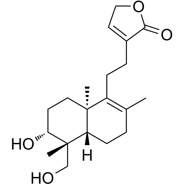
Deoxyandrographolide is an orally active lactone found in the Andrographis paniculata Nees. Deoxyandrographolide shows a KD of 38.4 μM of HDAC1. Deoxyandrographolide enhances GLUT4 plasma membrane translocation, activates PI3K and AMPK-dependent signaling pathways, suppresses fasting blood glucose, serum insulin, triglycerides, and LDL-cholesterol levels. Deoxyandrographolide enhances HDAC1 expression via inhibited ubiquitination degradation, represses H3K4me3, improves chromosome stability, and restrains aging biomarkers p16, p21, γH2A.X, p53 and ROS production. Deoxyandrographolide interacts with Foot-and-Mouth Disease Virus 3Cpro active site, inhibits protease and IFN-antagonist activity, derepresses ISG expression, and inhibits viral replication. Deoxyandrographolide can be used for the researches of type 2 diabetes mellitus, vascular senescence and virus infection.
Para uso exclusivo en investigación. No vendemos a pacientes.
- Pureza : 99.80%
- No. CAS: 79233-15-1
- Fòrmula: C20H30O4
- Peso molecular:334.45
-
Almacenamiento:
-20°C, protect from light
* In solvent : -80°C, 6 months; -20°C, 1 month (protect from light)

To place orders, for customer services and technical support, please contact: MedChemExpress USA
Tel: 609-228-6898 E-mail: [email protected] [email protected]


-
 Actividad biológica
-
Chemical Information
-
Solvente y solubilidad
- Protocolo
- Pureza y Documentación
- Referencias
-
 Help & FAQs


Publications Citing Use of MedChemExpress (MCE) Deoxyandrographolide
MoreVer todos los productos específicos de isoformas AMPK
More

Actividad biológica
Descripciòn
IC50 & Target
[1]|
GLUT4 |
HDAC1 38.4 μM () |
In Vitro
Deoxyandrographolide (2.5-25 μM; 1-24 h) dose- and time-dependently stimulates glucose uptake in L6 myotubes, and its effects are additive to Insulin without reducing cell viability[1].
Deoxyandrographolide (10 μM; 16 h) does not alter total cellular GLUT4 or GLUT1 protein levels in L6 myotubes, either alone or in combination with Insulin[1].
Deoxyandrographolide (10-25 μM; 16 h) dose-dependently promotes GLUT4 translocation to the plasma membrane in L6-GLUT4myc myotubes, with a 1.23-fold increase at 25 μM for 16 h, and potentiates Insulin-induced GLUT4 translocation[1].
Deoxyandrographolide (10 μM; 16 h) stimulates glucose uptake in L6 myotubes via a PI-3-K-dependent pathway, and activates downstream signaling by increasing Akt and insulin receptor-β phosphorylation, with synergistic effects when combined with insulin[1].
Deoxyandrographolide (10-25 μM; 16 h) activates the AMPK pathway in L6 myotubes, as shown by increased AMPKα and ACC phosphorylation, and this pathway contributes to its ability to promote GLUT4 translocation[1].
Deoxyandrographolide (7.94-250 μM; 24 h) is non-toxic to rat aorta endothelial cells at concentrations up to 200 μM and to human microvascular endothelial cells at concentrations up to 125 μM after 24 h of incubation[2].
Deoxyandrographolide (50-150 μM; 48 h) inhibits Angiotensin II-induced upregulation of p16 and p21 mRNA in rat aorta endothelial cells and human microvascular endothelial cells[2].
Deoxyandrographolide (25-150 μM; 48 h) inhibits Angiotensin II-induced p53 upregulation in rat aorta endothelial cells, restores angiotensin II-reduced HDAC1 levels in human microvascular endothelial cells, and reduces Angiotensin II-induced H3K4me3 upregulation in rat aorta endothelial cells[2].
Deoxyandrographolide (50-100 μM; 48-60 h) at 50 and 100 μM reduces angiotensin II-induced γH2A.X and p21 upregulation, and at 100 μM reduces angiotensin II-induced ubiquitin upregulation in rat aorta endothelial cells[2].
Deoxyandrographolide (6.75-200 μM) binds directly to recombinant human HDAC1 protein with a KD of 38.4 μM, as measured by bio-layer interferometry[2].
Deoxyandrographolide (50-150 μM; 24 h) reduces angiotensin II-induced reactive oxygen species accumulation in wild-type rat aorta endothelial cells, but this effect is abolished in HDAC1-knockdown rat aorta endothelial cells[2].
Deoxyandrographolide (50-150 μM; 48 h) fails to inhibit angiotensin II-induced γH2A.X upregulation in HDAC1-knockdown rat aorta endothelial cells[2].
Deoxyandrographolide (100 μM; 48 h) modulates gene expression related to chromosome stability, cell cycle, senescence, and inflammation in angiotensin II-treated rat aorta endothelial cells[2].
Deoxyandrographolide (1-150 μM; 24 h) inhibits replication of FMDV serotype A in BHK-21 cells with an EC50 of 36.47 μM and a selective index of 9.22[3].
Deoxyandrographolide (1-100 μM; 16 h) inhibits the protease activity of FMDV 3Cpro in HEK 293T cells with an IC50 of 25.58 μM and IC90 of 122.88 μM[3].
Deoxyandrographolide (25.58-122.88 μM; 24 h) interferes with the IFN-antagonist activity of FMDV 3Cpro in HEK 293T cells, significantly upregulating the expression of interferon-stimulating genes ISG15, ISG56, Mx-1, OAS-1, and PKR[3].
Deoxyandrographolide (0.1-250 μM; 24 h) exhibits mild cytotoxicity in BHK-21 cells (CC50 = 332.3 μM) and HEK 293T cells (CC50 = 651.40 μM) with non-cytotoxic CC10 concentrations of 81.05 μM and 155.55 μM, respectively[3].
MedChemExpress (MCE) has not independently confirmed the accuracy of these methods. They are for reference only. Further protocols information, click here.
-
Cell Line:L6 myotubes
-
Concentration:10 μM
-
Incubation Time:16 h
-
Result:Increased p-Akt and P-TRβ expression.
Increased p-AMPK and P-ACC expression.
-
Cell Line:rat aorta endothelial cells (RaECs), human microvascular endothelial cells (HMEC-1)
-
Concentration:12.5, 25, 50, 10, 200 μM (RaECs); 7.94, 15.88, 31.75, 62.5, 125, 250 μM (HMEC-1)
-
Incubation Time:24 h
-
Result:Showed no significant cytotoxicity in RaECs at concentrations up to 200 μM.
Showed no significant cytotoxicity in HMEC-1 at concentrations up to 125 μM; cytotoxicity was observed at 250 μM.
-
Cell Line:rat aorta endothelial cells (RaECs), human microvascular endothelial cells (HMEC-1)
-
Concentration:50, 100, 150 μM
-
Incubation Time:48 h (co-incubated with 2 μM Ang II)
-
Result:Dose-dependently inhibited the Ang II-induced elevation of p16 and p21 mRNA levels in both RaECs and HMEC-1.
-
Cell Line:rat aorta endothelial cells (RaECs), human microvascular endothelial cells (HMEC-1)
-
Concentration:50, 100, 150 μM (p53 and HDAC1 assays); 25, 50, 100, 150 μM (H3K4me3 assay)
-
Incubation Time:48 h (co-incubated with 2 μM Ang II)
-
Result:Inhibited Ang II-induced upregulation of p53 protein in RaECs at 50, 100, and 150 μM.
Dose-dependently restored HDAC1 protein levels reduced by Ang II in HMEC-1 at 50, 100, and 150 μM.
Dose-dependently downregulated H3K4me3 levels increased by Ang II in RaECs at 25, 50, 100, and 150 μM.
-
Cell Line:rat aorta endothelial cells (RaECs)
-
Concentration:50, 100 μM (γH2A.X and p21 assays); 100 μM (ubiquitin assay)
-
Incubation Time:48 h (γH2A.X and p21 assays; co-incubated with 2 μM Ang II); 60 h (ubiquitin assay; co-incubated with 2 μM Ang II)
-
Result:Reduced Ang II-induced elevation of γH2A.X and p21 fluorescence intensity at 50 and 100 μM.
Decreased Ang II-induced elevation of total ubiquitin fluorescence intensity at 100 μM.
-
Cell Line:BHK-21, HEK 293T
-
Concentration:0.1,
1, 5, 10, 25, 50, 75, 100, 200, and 250 μM -
Incubation Time:24 h
-
Result:In BHK-21 cells, reached a CC50 of 332.3 μM and a CC10 of 81.05 μM.
In HEK 293T cells, reached a CC50 of 651.40 μM and a CC10 of 155.55 μM, indicating mild cytotoxicity only at high concentrations.
In Vivo
Deoxyandrographolide (100 mg/kg; p.o.; daily; 15 days) significantly reduces blood glucose, improves glucose tolerance, and normalizes lipid and insulin profiles in genetically diabetic db/db mice, including a 36.6% improvement in fasting blood glucose[1].
MedChemExpress (MCE) has not independently confirmed the accuracy of these methods. They are for reference only.
-
Animal Model:Sprague Dawley (male, 160 g, Streptozotocin-induced diabetic)[1]
-
Dosage:100 mg/kg
-
Administration:p.o.; single dose
-
Result:Reduced blood glucose by 20.2% at 5 hours post-dose.
Reduced blood glucose by 24.3% at 24 hours post-dose, as measured by AUC comparison to vehicle controls.
-
Animal Model:C57BL/KsJ-db/db (male, 40 g, genetically diabetic)[1]
-
Dosage:100 mg/kg
-
Administration:p.o.; daily; 15 days
-
Result:Reduced blood glucose significantly starting on day 11, continuing through day 14.
Improved glucose tolerance by 16.1% on day 10 and 23.4% on day 15 via OGTT.
Improved fasting blood glucose by 36.6%.
Reduced serum insulin by 29.9%.
Decreased serum triglycerides by 15.3%.
Decreased LDL-cholesterol by 13.1%.
Increased HDL-cholesterol by 23.4%.
Caused no significant effect on total cholesterol or body weight.


Chemical Information
-
No. CAS 79233-15-1
-
Appearance Solid
-
Peso molecular 334.45
-
Fòrmula C20H30O4
-
Color White to off-white
-
SMILES
C[C@@](C(CCC1=CCOC1=O)=C(C)CC2)(CC[C@H]3O)[C@@]2([H])[C@]3(C)CO
-
Structure Classification
-
Envío
Room temperature in continental US; may vary elsewhere.
-
Almacenamiento
-20°C, protect from light
* In solvent : -80°C, 6 months; -20°C, 1 month (protect from light)


Publications (1)
-
Journal Impact Factor
-
Most Recent
-
MedComm
Discovery of deoxyandrographolide and its novel effect on vascular senescence by targeting HDAC1. [Abstract]2023 Aug 17;4(5):e338. PMID: 37600507


Solvente y solubilidad
In Vitro:
DMSO : 100 mg/mL (299.00 mM; Need ultrasonic; Hygroscopic DMSO has a significant impact on the solubility of product, please use newly opened DMSO)
Please refer to the solubility information to select the appropriate solvent. Once prepared, please aliquot and store the solution to prevent product inactivation from repeated freeze-thaw cycles.
Storage method and period of stock solution: -80°C, 6 months; -20°C, 1 month (protect from light). When stored at -80°C, please use it within 6 months. When stored at -20°C, please use it within 1 month.
Please refer to the solubility information to select the appropriate solvent. Once prepared, please aliquot and store the solution to prevent product inactivation from repeated freeze-thaw cycles.
Storage method and period of stock solution: -80°C, 6 months; -20°C, 1 month (protect from light). When stored at -80°C, please use it within 6 months. When stored at -20°C, please use it within 1 month.
Concentration (start) × Volume (start) = Concentration (final) × Volume (final)
In Vivo:
Select the appropriate dissolution method based on your experimental animal and administration route.
- For the following dissolution methods, please ensure to first prepare a clear stock solution using an In Vitro approach and then sequentially add co-solvents:
- To ensure reliable experimental results, the clarified stock solution can be appropriately stored based on storage conditions. As for the working solution for In Vivo experiments, it is recommended to prepare freshly and use it on the same day.
- The percentages shown for the solvents indicate their volumetric ratio in the final prepared solution. If precipitation or phase separation occurs during preparation, heat and/or sonication can be used to aid dissolution.
Add each solvent one by one: 10% DMSO 40% PEG300 5% Tween-80 45% Saline
Solubility: ≥ 2.5 mg/mL (7.47 mM); Clear solution
This protocol yields a clear solution of ≥ 2.5 mg/mL (saturation unknown).
Taking 1 mL working solution as an example, add 100 μL DMSO stock solution (25.0 mg/mL) to 400 μL PEG300, and mix evenly; then add 50 μL Tween-80 and mix evenly; then add 450 μL Saline to adjust the volume to 1 mL.
Preparation of Saline: Dissolve 0.9 g sodium chloride in ddH₂O and dilute to 100 mL to obtain a clear Saline solution.
Add each solvent one by one: 10% DMSO 90% (20% SBE-β-CD in Saline)
Solubility: ≥ 2.5 mg/mL (7.47 mM); Clear solution
This protocol yields a clear solution of ≥ 2.5 mg/mL (saturation unknown).
Taking 1 mL working solution as an example, add 100 μL DMSO stock solution (25.0 mg/mL) to 900 μL 20% SBE-β-CD in Saline, and mix evenly.
Preparation of 20% SBE-β-CD in Saline (4°C, storage for one week): 2 g SBE-β-CD powder is dissolved in 10 mL Saline, completely dissolve until clear.
In Vivo Dissolution Calculator
Please enter the basic information of animal experiments:
-
-
-
-
Recommended: Prepare an additional quantity of animals to account for potential losses during experiments.
Please enter your animal formula composition:
-
%DMSO +
Recommended: Keep the proportion of DMSO in working solution below 2% if your animal is weak.
-
%+
-
+%Tween-80 + +
-
%Saline +
The co-solvents required include: DMSO, . All of co-solvents are available by MedChemExpress (MCE). , Tween 80. All of co-solvents are available by MedChemExpress (MCE).
Working solution concentration: 0.22 mg/mL
Method for preparing stock solution: mg drug dissolved in μL DMSO. Stock solution concentration: mg/mL. * In solvent : -80°C, 6 months; -20°C, 1 month (protect from light)
1. Take μL DMSO stock solution;
2. Add μL .
μL , mix evenly;
3. Then add μL Tween 80, mix evenly;
4. Then add μL
Please ensure that the stock solution in the first step is dissolved to a clear state, and add co-solvents in sequence. You can use ultrasonic heating (ultrasonic cleaner, recommended frequency 20-40 kHz), vortexing, etc. to assist dissolution.

Protocolo
-
RNA extraction experimental
By lysing cells, releasing RNA, and removing impurities such as proteins and DNA, high-purity RNA products are finally obtained. The commonly used traditional method is the guanidine isothiocyanate/phenol/chloroform method (Trizol), which is suitable for a variety of animal materials including animal tissues, microorganisms, cultured cells, etc., and most plant materials.
-
Research Protocol for Infectious Diseases
Infectious-disease experiments test how pathogens interact with host barriers, innate immune receptors, inflammatory signaling, pathogen replication, and tissue injury; pattern-recognition receptors such as TLRs, RIG-I-like receptors, NOD-like receptors, and inflammasomes detect microbial molecules and activate NF-κB, interferon, and cytokine responses. The central hypothesis is that infection severity reflects the balance between pathogen burden and host response: protective inflammation restricts pathogen growth, whereas excessive or mislocalized inflammation contributes to tissue damage and disease phenotype. Unresolved questions include which host pathways are protective versus pathogenic, why some infection models fail to translate to human disease, and which combined readouts best predict clinically relevant infection outcomes.
-
ROS/oxidative-stress fluorescent staining
ROS/oxidative-stress fluorescent staining uses cell-permeant fluorogenic probes that become fluorescent after oxidation inside cells or tissues; commonly used examples include DCFH-DA/DCFDA for broad cellular oxidant detection, DHE for superoxide-related signal detection, MitoSOX for mitochondrial superoxide-related signal detection, and CellROX probes for oxidative-stress-associated fluorescence readouts. The assay detects probe oxidation rather than a single ROS species unless the probe and analysis method have been chemically validated for that species. DCFH-DA enters cells, is deacetylated by intracellular esterases to DCFH, and produces fluorescent DCF after oxidation, so the readout is used as an operational measure of total cellular oxidative stress rather than a species-specific ROS measurement. DHE and MitoSOX can report superoxide-related oxidation, but red fluorescence alone can include non-specific ethidium-like oxidation products; HPLC or optimized spectral approaches are
-
Research Protocol for Endocrine Diseases
Endocrine diseases often arise from disrupted hormone production, hormone signaling, or target-tissue responsiveness; for diabetes-focused endocrine disease models, insulin signaling regulates glucose uptake, hepatic glucose output, lipid metabolism, and β-cell compensation. Type 2 diabetes develops through interacting defects in insulin resistance, β-cell dysfunction, adipose inflammation, hepatic glucose overproduction, altered incretin signaling, and ectopic lipid metabolism. A major unresolved question is whether endocrine dysfunction is driven primarily by target-tissue insulin resistance, intrinsic β-cell failure, immune/inflammatory stress, or combined multi-organ failure that differs by disease stage.
-
Research Protocol for Metabolic Diseases
AMP-activated protein kinase, AMPK, is a conserved cellular energy sensor that responds to reduced cellular energy status and coordinates metabolism by increasing ATP-generating catabolic pathways while suppressing ATP-consuming anabolic processes. In metabolic disease research, the AMPK pathway is experimentally relevant because it regulates hepatic lipid synthesis, fatty acid oxidation, glucose production, skeletal-muscle glucose disposal, mTORC1-linked biosynthesis, autophagy, mitochondrial homeostasis, and whole-body energy balance. The central pathway logic is that energy stress, metformin, exercise-like stimulation, or direct AMPK activators increase AMPKα Thr172 phosphorylation and downstream substrate phosphorylation, including ACC and RAPTOR. Phosphorylation of ACC suppresses lipogenesis and supports fatty acid oxidation, whereas phosphorylation of RAPTOR suppresses mTORC1 signaling and links cellular energy status to growth and protein synthesis control. The pathway is linked
-
Senescence-associated β-galactosidase staining
Senescence-associated β-galactosidase staining detects β-galactosidase activity that is histochemically visible at pH 6. 0 in senescent cells, where X-gal cleavage produces an insoluble blue precipitate observable by bright-field microscopy. This activity reflects increased lysosomal β-galactosidase/lysosomal mass rather than a senescence-essential enzyme, because GLB1 depletion or genetic lysosomal β-galactosidase deficiency can abolish SA-β-gal staining while cells still undergo senescence. SA-β-gal was originally reported in senescent but not presenescent fibroblasts and keratinocytes, absent from quiescent fibroblasts and terminally differentiated keratinocytes, and increased with donor age in human skin samples. Because SA-β-gal can also appear in some non-senescent or tissue-specific contexts, interpretation should be paired with experimental controls and, when possible, independent senescence markers.


Pureza y Documentación
-
Ficha de datos (292 KB)
-
SDS (252 KB)
- English - EN (252 KB)
- Français - FR (252 KB)
- Deutsch - DE (252 KB)
- Norwegian - NO (252 KB)
- Español - ES (252 KB)
- Swedish - SV (252 KB)
- Italian - IT (252 KB)
- Korean - KR (252 KB)
- Portuguese - PT (252 KB)
-
Instrucciones de manejo (2659 KB)
Referencias
[1]. Arha D, et al. Deoxyandrographolide promotes glucose uptake through glucose transporter-4 translocation to plasma membrane in L6 myotubes and exerts antihyperglycemic effect in vivo. Eur J Pharmacol. 2015;768:207-216. [Content Brief]
[2]. Lin Z, et al. Discovery of deoxyandrographolide and its novel effect on vascular senescence by targeting HDAC1. MedComm (2020). 2023;4(5):e338. Published 2023 Aug 17. [Content Brief]
[3]. Theerawatanasirikul S, et al. Andrographolide and deoxyandrographolide inhibit protease and IFN-antagonist activities of foot-and-mouth disease virus 3Cpro[J]. Animals, 2022, 12(15): 1995. [Content Brief]


Complete Stock Solution Preparation Table
Please refer to the solubility information to select the appropriate solvent. Once prepared, please aliquot and store the solution to prevent product inactivation from repeated freeze-thaw cycles.
Storage method and period of stock solution: -80°C, 6 months; -20°C, 1 month (protect from light). When stored at -80°C, please use it within 6 months. When stored at -20°C, please use it within 1 month.
| Optional Solvent | Concentration Solvent Mass | 1 mg | 5 mg | 10 mg | 25 mg |
|---|---|---|---|---|---|
| DMSO | 1 mM | 2.9900 mL | 14.9499 mL | 29.8998 mL | 74.7496 mL |
| 5 mM | 0.5980 mL | 2.9900 mL | 5.9800 mL | 14.9499 mL | |
| 10 mM | 0.2990 mL | 1.4950 mL | 2.9900 mL | 7.4750 mL | |
| 15 mM | 0.1993 mL | 0.9967 mL | 1.9933 mL | 4.9833 mL | |
| 20 mM | 0.1495 mL | 0.7475 mL | 1.4950 mL | 3.7375 mL | |
| 25 mM | 0.1196 mL | 0.5980 mL | 1.1960 mL | 2.9900 mL | |
| 30 mM | 0.0997 mL | 0.4983 mL | 0.9967 mL | 2.4917 mL | |
| 40 mM | 0.0747 mL | 0.3737 mL | 0.7475 mL | 1.8687 mL | |
| 50 mM | 0.0598 mL | 0.2990 mL | 0.5980 mL | 1.4950 mL | |
| 60 mM | 0.0498 mL | 0.2492 mL | 0.4983 mL | 1.2458 mL | |
| 80 mM | 0.0374 mL | 0.1869 mL | 0.3737 mL | 0.9344 mL | |
| 100 mM | 0.0299 mL | 0.1495 mL | 0.2990 mL | 0.7475 mL |


Deoxyandrographolide Related Classifications
HY-N0857 Related Classifications
 Powered by Bioz
Keywords
- Deoxyandrographolide
- 79233-15-1
- GLUT
- HDAC
- Virus Protease
- PI3K
- AMPK
- Akt
- Histone Demethylase
- MDM-2/p53
- IFNAR
- Reactive Oxygen Species (ROS)
- HEK 293T cells
- rat aorta endothelial cells
- GLUT4
- Foot-and-Mouth Disease Virus 3Cpro
- L6 myotubes
- PI-3-K
- human microvascular endothelial cells
- BHK-21 cells
- HDAC1
- Inhibitor
- inhibitor
- inhibit