



Hecogenin acetate

Based on 1 Customer Validation
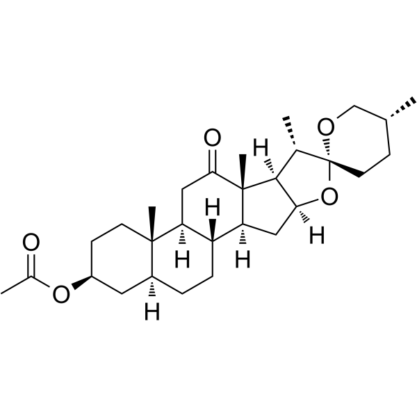
Hecogenin acetate is an orally active steroid saponin aglycone with extensive biological activities. Hecogenin acetate inhibits the phosphorylation of NF-κB and p38 MAPK signaling pathways, antagonizes TRPA1/TRPM8 channels, inhibits the production of pro-inflammatory cytokines, and has anti-inflammatory and analgesic effects. Hecogenin acetate inhibits the production of ROS and the activation of NLRP3 inflammasome; downregulates the expression of MMP-2, and has neuroprotective and anti-tumor activities. Hecogenin acetate enhances gastric mucosal defense and promotes ulcer healing. Hecogenin acetate can be used in combination with certain antibiotics to regulate bacterial efflux pumps and restore antibiotic sensitivity.
For research use only. We do not sell to patients.
- Purity : 98.57%
- CAS No.: 915-35-5
- Formula: C29H44O5
- Molecular Weight:472.66
-
Storage:Powder -20°C, 3 years , 4°C, 2 years ; In solvent -80°C, 6 months , -20°C, 1 month

To place orders, for customer services and technical support, please contact: MedChemExpress USA
Tel: 609-228-6898 E-mail: [email protected] [email protected]


-
 Biological Activity
-
Chemical Information
-
Solvent & Solubility
- Protocols
- Purity & Documentation
- References
-
 Help & FAQs


-
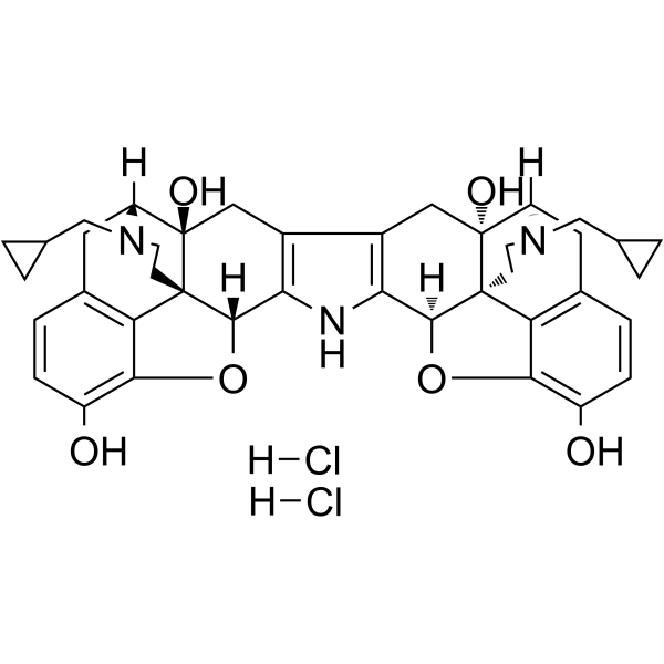
HY-100903
Norbinaltorphimine dihydrochlorideκ-opioid receptor Antagonist -
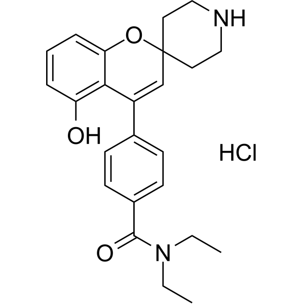
HY-13044
ADL-5859 hydrochlorideOpioid Receptor Agonist -

HY-15997B
(±)-U-50488 hydrochlorideKOR Agonist -
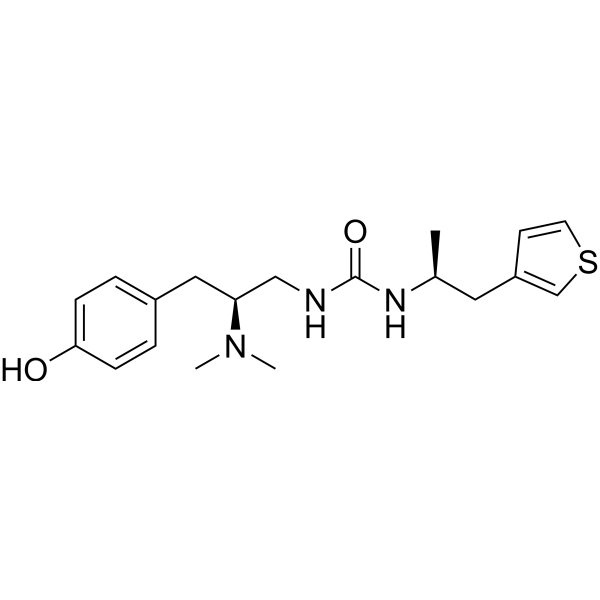
HY-101386
PZM21μ-opioid Receptor Agonist -
HY-N0398
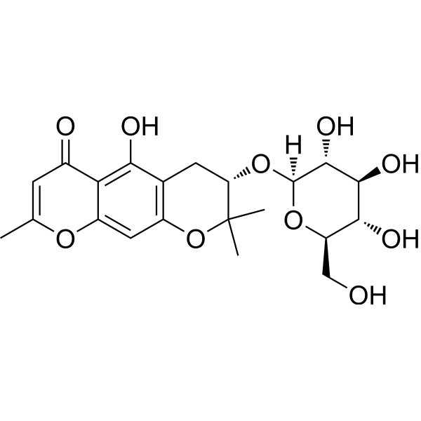
Sec-O-GlucosylhamaudolOpioid Receptor Inhibitor -
HY-115066
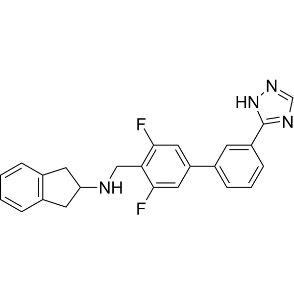
GSK1521498 free baseμ-opioid Receptor Antagonist -

HY-15997
(-)-U-50488 hydrochlorideKOR Agonist -
HY-10487
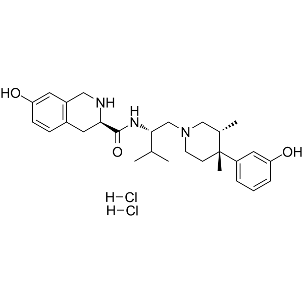
JDTic dihydrochloride -
HY-19627A
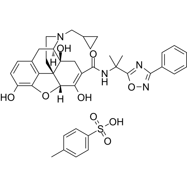
Naldemedine tosylateμ-opioid Receptor Antagonist -
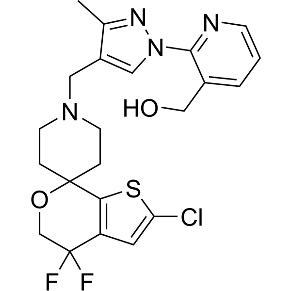
HY-114452
LY2940094NOP Antagonist -
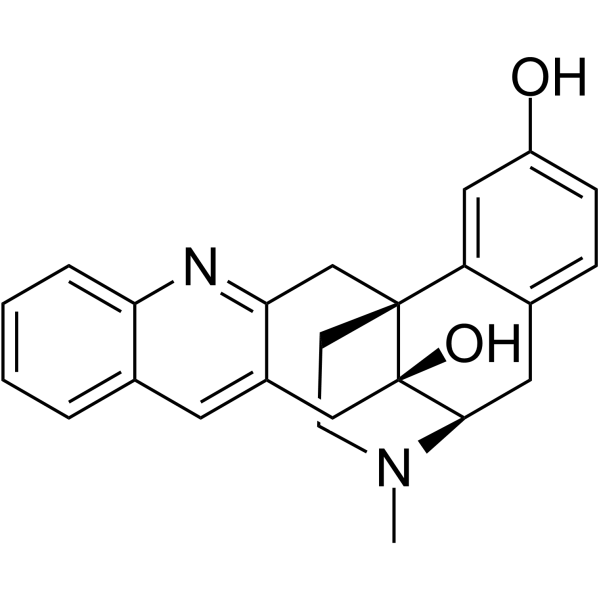
HY-120511
KNT-127δ-opioid Agonist -
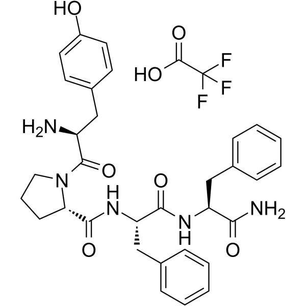
HY-P0186A
Endomorphin 2 TFAμ-opioid Receptor Agonist


Biological Activity
Description
|
p38 MAPK |
NF-κB |
ERK1 |
ERK2 |
MMP-2 |
NLRP3 inflammasome |
IL-1β |
IL-6 |
In Vitro
Hecogenin acetate has only a very weak direct antibacterial activity with MIC values for S. aureus ATCC and E. coli ATCC of 645 μg/mL and 512 μg/mL respectively[3].
Hecogenin acetate (1-150 μM, 24 h) has no significant effect on the viability of A549 cells when the concentration was lower than 75 μM, but at higher concentrations it reduces the viability of A549 cells[4].
Hecogenin acetate (50 μM, 24 h) reverses the cell membrane damage, ROS production, and increased activities of catalase (CAT) and superoxide dismutase (SOD) caused by H₂O₂ in A549 cells[4].
Hecogenin acetate (25-100 μM, 24-72 h) induces G0/G1 phase cell cycle arrest and cellular senescence by inhibiting the ERK1/2 signaling pathway and reducing the expression of MMP-2[4].
MedChemExpress (MCE) has not independently confirmed the accuracy of these methods. They are for reference only. Further protocols information, click here.
-
Cell Line:A549 cells
-
Concentration:25, 50, 75 and 100 μM
-
Incubation Time:72 h
-
Result:Dose-dependently arrested A549 cells in the G0/G1 phase.
-
Cell Line:A549 cells
-
Concentration:50 μM
-
Incubation Time:24 h
-
Result:Completely blocked the ERK1/2 phosphorylation induced by H₂O₂.
Inhibited the upregulation of matrix metalloproteinase-2 (MMP-2) expression caused by H₂O₂.
In Vivo
Hecogenin acetate (5-25 mg/kg, i.p., single dose or once daily for 7 days) inhibits the expression of the aldosterone synthase (CYP11B2) gene, thereby exerting a diuretic effect in rats[2].
Hecogenin acetate (2.5-10 mg/kg, p.o., single dose or once daily for 7 days) exhibits significant protective effects against acute gastric mucosal injury induced by various chemical substances (anhydrous ethanol, acidified ethanol) and pathological processes (ischemia-reperfusion) in rodent models[5].
MedChemExpress (MCE) has not independently confirmed the accuracy of these methods. They are for reference only.
-
Animal Model:Gastric injury induced by ischemia-reperfusion established in 2-month male Swiss mice weighing between 25 and 30 g[5]
-
Dosage:20 mg/kg
-
Administration:Intragastric administration (i.g.), once daily for 8 days (chronic pain model) or 15 days (neuropathic pain model model)
-
Result:Exhibited potent and long-lasting anti-allodynic effects in both chronic and neuropathic pain models.
Inhibited the activation of spinal microglia, reduce the release of pro-inflammatory cytokines (TNF-α, IL-1β, IL-6), and inhibit the activation of the NF-κB and p38 MAPK signaling pathways.
Antagonized TRPM8 and TRPA1 ion channels, reducing the sensitization of peripheral nociceptive receptors.
-
Animal Model:Acute and subacute diuretic assays established in Sprague-Dawley rats (180-240 g)[2]
-
Dosage:5, 10 and 25 mg/kg
-
Administration:Intraperitoneal injection (i.p.), single dose or once daily for 7 days
-
Result:Had a dose-dependent diuretic effect, which can significantly increase urine output and the excretion of sodium and chloride ions. Showed the characteristics of sodium and chlorine excretion while maintaining relatively high potassium levels. Down-regulated the expression of the aldosterone synthase (CYP11B2) gene in the adrenal gland to inhibit aldosterone synthesis.
-
Animal Model:Acute gastric ulcers induced by anhydrous ethanol and acidified ethanol established in 2-month male Swiss mice eighing between 25 and 30 g[5]
-
Dosage:5, 10, 20 mg/kg
-
Administration:Oral administration (p.o.), single dose
-
Result:Had a strong protective effect against acute gastric mucosal injury caused by anhydrous ethanol.
Effectively resisted more severe damage to the gastric mucosa caused by the combined attack of ethanol and gastric acid.
-
Animal Model:Gastric injury induced by ischemia-reperfusion and chronic gastric ulcers induced by acetic acid established in male Sprague-Dawley rats (250-300 g)[5]
-
Dosage:5, 10, 20 mg/kg
-
Administration:Oral administration (p.o.), single dose (gastric injury model) or once daily for 7 days (chronic gastric ulcers mmodel)
-
Result:Had a strong protective effect against ischemia-reperfusion gastric injury mediated by oxidative stress.
Effectively promoted the healing of chronic gastric ulcers.


Chemical Information
-
CAS No. 915-35-5
-
Appearance Solid
-
Molecular Weight 472.66
-
Formula C29H44O5
-
Color White to yellow
-
SMILES
[H][C@]1(O[C@@]2(OC[C@H](C)CC2)[C@@H](C)[C@@]1([C@]34C)[H])C[C@@]3([H])[C@]5([H])CC[C@@]6([H])C[C@@H](OC(C)=O)CC[C@]6(C)[C@@]5([H])CC4=O
-
Structure Classification
-
Initial Source
-
Shipping
Room temperature in continental US; may vary elsewhere.
-
Storage
Powder -20°C 3 years 4°C 2 years In solvent -80°C 6 months -20°C 1 month


Solvent & Solubility
In Vitro:
THF : 57.5 mg/mL (121.65 mM; Need ultrasonic)
Please refer to the solubility information to select the appropriate solvent. Once prepared, please aliquot and store the solution to prevent product inactivation from repeated freeze-thaw cycles.
Storage method and period of stock solution: -80°C, 6 months; -20°C, 1 month. When stored at -80°C, please use it within 6 months. When stored at -20°C, please use it within 1 month.
Please refer to the solubility information to select the appropriate solvent. Once prepared, please aliquot and store the solution to prevent product inactivation from repeated freeze-thaw cycles.
Storage method and period of stock solution: -80°C, 6 months; -20°C, 1 month. When stored at -80°C, please use it within 6 months. When stored at -20°C, please use it within 1 month.
Concentration (start) × Volume (start) = Concentration (final) × Volume (final)

Protocols
-
Kinase activity and phosphorylation assays
Kinase activity assays measure the ability of kinases to transfer phosphate groups from ATP to specific substrates, while phosphorylation assays detect the presence and levels of phosphorylated proteins. Common methods include radiolabeled ATP incorporation (e. g. ,), ADP release detection via bioluminescence (e. g. ,[3]), enzyme-linked immunosorbent assays (ELISA) for phospho-specific epitopes (e. g. ,[6]), and microtiter-based formats for high-throughput screening (e. g. ,[8]). The ADP-Glo assay quantifies kinase activity by measuring ADP produced during phosphorylation using a luciferase-based system. Radiometric assays involve autoradiography or scintillation counting after incorporation of 32P-labeled ATP into substrate proteins. ELISA-based approaches rely on phospho-specific antibodies to detect activated kinases in cell lysates or purified samples.
-
Western Blot
Western blotting (WB) is a commonly used experimental method in molecular biology, biochemistry, and immunogenetics for identifying and quantifying target proteins. It combines gel electrophoresis with immunoassay, enabling researchers to analyze protein expression, post-translational modifications, and molecular weight.
-
RNA extraction experimental
By lysing cells, releasing RNA, and removing impurities such as proteins and DNA, high-purity RNA products are finally obtained. The commonly used traditional method is the guanidine isothiocyanate/phenol/chloroform method (Trizol), which is suitable for a variety of animal materials including animal tissues, microorganisms, cultured cells, etc., and most plant materials.
-
Mitochondrial membrane-potential fluorescent assay
Mitochondrial membrane potential fluorescent assays estimate ΔΨm in living cells using lipophilic cationic dyes such as TMRM, TMRE, rhodamine 123, and JC-1, which accumulate in mitochondria according to membrane polarization; loss of signal after FCCP or CCCP treatment is interpreted as mitochondrial depolarization. TMRM/TMRE and rhodamine 123 are commonly used for semi-quantitative live-cell microscopy or flow cytometry, while JC-1 can report a shift from red aggregate fluorescence to green monomer fluorescence during depolarization; interpretation requires controls because dye concentration, quenching mode, cell type, dye efflux, and mitochondrial mass can affect fluorescence independently of ΔΨm.
-
LPS-Induced Endotoxemia/Systemic Inflammation
Lipopolysaccharide (LPS)-induced endotoxemia is a widely used in vivo model of acute systemic inflammation in which LPS, a Gram-negative bacterial endotoxin, activates innate immune signaling primarily through TLR4, leading to rapid and transient induction of pro-inflammatory cytokines such as TNF-α, IL-6, and IL-1β in circulation and tissues. This cytokine surge is commonly used as a measurable readout of systemic inflammatory activation and immune dysregulation, and is typically assessed within hours after intraperitoneal LPS administration in mouse models of endotoxemia. The model captures key features of systemic inflammatory response syndrome, including cytokine release, immune cell activation, and downstream tissue responses, and has been used to evaluate anti-inflammatory interventions such as cytokine modulation, lipid mediators, and immune cell-targeting therapies.
-
ROS/oxidative-stress fluorescent staining
ROS/oxidative-stress fluorescent staining uses cell-permeant fluorogenic probes that become fluorescent after oxidation inside cells or tissues; commonly used examples include DCFH-DA/DCFDA for broad cellular oxidant detection, DHE for superoxide-related signal detection, MitoSOX for mitochondrial superoxide-related signal detection, and CellROX probes for oxidative-stress-associated fluorescence readouts. The assay detects probe oxidation rather than a single ROS species unless the probe and analysis method have been chemically validated for that species. DCFH-DA enters cells, is deacetylated by intracellular esterases to DCFH, and produces fluorescent DCF after oxidation, so the readout is used as an operational measure of total cellular oxidative stress rather than a species-specific ROS measurement. DHE and MitoSOX can report superoxide-related oxidation, but red fluorescence alone can include non-specific ethidium-like oxidation products; HPLC or optimized spectral approaches are
-
Mitochondrial membrane-potential and mitochondrial mass staining
Mitochondrial membrane potential staining measures the electrochemical polarization across the mitochondrial inner membrane in live cells using lipophilic cationic fluorescent probes; early rhodamine-based work showed that selective mitochondrial dye accumulation is lost when the mitochondrial transmembrane potential is dissipated. JC-1 reports mitochondrial polarization by shifting from green monomer fluorescence to red J-aggregate fluorescence as dye concentration increases within energized mitochondria; therefore, the red/green fluorescence ratio is used as a relative readout of mitochondrial membrane potential. TMRE or TMRM staining provides a single-channel relative readout because these cationic rhodamine esters accumulate in polarized mitochondria, and lower fluorescence indicates reduced mitochondrial polarization when acquisition and dye-loading conditions are controlled. Mitochondrial mass staining is commonly performed with MitoTracker Green FM or related MitoTracker dyes as
-
Protocol for Kinase activity and phosphorylation assays
Kinase activity assays measure transfer of phosphate from ATP to a protein or peptide substrate, generating phosphorylated substrate, ADP, or incorporated radiolabeled phosphate as the readout; phosphorylation assays measure site-specific phosphorylation in cells or tissues as a proxy for kinase-pathway activation, inhibition, or substrate regulation. Phosphorylation can be detected by phospho-specific Western blot, immunoprecipitation kinase assay, phospho-immunofluorescence, phospho-flow cytometry, luminescent ADP detection, radiolabeled ATP incorporation, or reporter-based pathway assays, and these readouts can be applied to cancer cells, primary neurons, mouse tumors, organoids, inflammatory macrophages, ferroptosis studies, and mitophagy studies when the kinase target is biologically relevant.
-
Research Protocol for Inflammation-related Diseases
The NLRP3 inflammasome is a cytosolic innate immune signaling platform that integrates priming signals and danger-signal activation to promote caspase-1 activation, maturation of IL-1β and IL-18, and gasdermin D-mediated pyroptotic cell death. The core experimental logic is to determine whether inflammatory disease phenotypes are driven by increased NLRP3 expression, ASC-containing inflammasome assembly, caspase-1 cleavage, GSDMD cleavage, and extracellular release of IL-1β/IL-18 rather than by nonspecific cell injury alone. The pathway is strongly linked to inflammation-related disease phenotypes because monosodium urate crystals activate NALP3/NLRP3 inflammasome signaling in gout-like crystal inflammation, cholesterol crystals activate NLRP3 inflammasomes in atherogenesis models, and DSS-induced intestinal inflammation has been reported to involve NLRP3 inflammasome activity. However, experimental colitis studies also show context-dependent protective effects of NLRP3 inflammasome co
-
Pyroptosis Solutions
Pyroptosis is a lytic inflammatory cell-death pathway executed by gasdermin pores, most classically through inflammasome-mediated activation of caspase-1, cleavage of gasdermin D, membrane pore formation, LDH release, and secretion of IL-1β and IL-18. The canonical pathway is commonly modeled by priming cells with an inflammatory signal such as LPS to induce pro-IL-1β and inflammasome components, followed by an activation signal such as ATP or nigericin to activate NLRP3, ASC speck formation, caspase-1 cleavage, GSDMD cleavage, cytokine release, and pyroptotic membrane rupture. The non-canonical pathway is triggered when cytosolic LPS activates mouse caspase-11 or human caspase-4/5, leading to GSDMD cleavage and pyroptosis, and this can secondarily activate NLRP3-dependent IL-1β release. Pyroptosis is linked to inflammatory injury, infection, cancer, liver disease, ocular disease, placental inflammation, and other disease phenotypes, but unresolved questions include which gasdermin fam


Purity & Documentation
-
Data Sheet (285 KB)
-
SDS (251 KB)
- English - EN (251 KB)
- Français - FR (251 KB)
- Deutsch - DE (251 KB)
- Norwegian - NO (251 KB)
- Español - ES (251 KB)
- Swedish - SV (251 KB)
- Italian - IT (251 KB)
- Korean - KR (251 KB)
- Portuguese - PT (251 KB)
-
Handling Instructions (2659 KB)
References
[1]. Santos Passos FR, et al. Role of peripheral and central sensitization in the anti-hyperalgesic effect of hecogenin acetate, an acetylated sapogenin, complexed with β-cyclodextrin: Involvement of NFκB and p38 MAPK pathways. Neuropharmacology. 2021 Mar 15;186:108395. [Content Brief]
[2]. Alamoudi AJ, et al. Diuretic effects of Hecogenin and Hecogenin acetate via aldosterone synthase inhibition. Saudi Pharm J. 2024 Jul;32(7):102105. [Content Brief]
[3]. Santos Araújo NJ, et al. Evaluation of the antibacterial activity of hecogenin acetate and its inhibitory potential of NorA and MepA efflux pumps from Staphylococcus aureus. Microb Pathog. 2023 Jan;174:105925. [Content Brief]
[4]. Gasparotto J, et al. Hecogenin acetate inhibits reactive oxygen species production and induces cell cycle arrest and senescence in the A549 human lung cancer cell line. Anticancer Agents Med Chem. 2014;14(8):1128-35. [Content Brief]
[5]. Sousa AJC, et al. Antiulcerogenic and healing activity of hecogenin acetate in rodents. Naunyn Schmiedebergs Arch Pharmacol. 2023 Apr;396(4):759-769. [Content Brief]


Complete Stock Solution Preparation Table
Please refer to the solubility information to select the appropriate solvent. Once prepared, please aliquot and store the solution to prevent product inactivation from repeated freeze-thaw cycles.
Storage method and period of stock solution: -80°C, 6 months; -20°C, 1 month. When stored at -80°C, please use it within 6 months. When stored at -20°C, please use it within 1 month.
| Optional Solvent | Concentration Solvent Mass | 1 mg | 5 mg | 10 mg | 25 mg |
|---|---|---|---|---|---|
| THF | 1 mM | 2.1157 mL | 10.5784 mL | 21.1569 mL | 52.8921 mL |
| 5 mM | 0.4231 mL | 2.1157 mL | 4.2314 mL | 10.5784 mL | |
| 10 mM | 0.2116 mL | 1.0578 mL | 2.1157 mL | 5.2892 mL | |
| 15 mM | 0.1410 mL | 0.7052 mL | 1.4105 mL | 3.5261 mL | |
| 20 mM | 0.1058 mL | 0.5289 mL | 1.0578 mL | 2.6446 mL | |
| 25 mM | 0.0846 mL | 0.4231 mL | 0.8463 mL | 2.1157 mL | |
| 30 mM | 0.0705 mL | 0.3526 mL | 0.7052 mL | 1.7631 mL | |
| 40 mM | 0.0529 mL | 0.2645 mL | 0.5289 mL | 1.3223 mL | |
| 50 mM | 0.0423 mL | 0.2116 mL | 0.4231 mL | 1.0578 mL | |
| 60 mM | 0.0353 mL | 0.1763 mL | 0.3526 mL | 0.8815 mL | |
| 80 mM | 0.0264 mL | 0.1322 mL | 0.2645 mL | 0.6612 mL | |
| 100 mM | 0.0212 mL | 0.1058 mL | 0.2116 mL | 0.5289 mL |


Hecogenin acetate Related Classifications
HY-126941 Related Classifications
 Powered by Bioz