



O6-Benzylguanine
Based on 3 publication(s) in Google Scholar
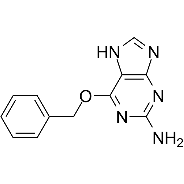
O6-Benzylguanine is an orally active, blood-brain barrier-penetrant inhibitor of O6-methylguanine-DNA methyltransferase (MGMT/AGT). O6-Benzylguanine modulates p53 and its downstream p21 and cyclins to induce cell cycle arrest and apoptosis, and on the other hand activates mTORC1/MAPK and other signaling pathways to induce cellular senescence by inhibiting intracellular MGMT. O6-Benzylguanine is used in research related to various tumors, cellular senescence, and vascular smooth muscle dysfunction.
For research use only. We do not sell to patients.
- Purity : 99.92%
- CAS No.: 19916-73-5
- Formula: C12H11N5O
- Molecular Weight:241.25
-
Storage:
RT, protect from light.
In solvent -80°C, 1 year , -20°C, 6 months

To place orders, for customer services and technical support, please contact: MedChemExpress USA
Tel: 609-228-6898 E-mail: [email protected] [email protected]


-
 Biological Activity
-
Chemical Information
-
Solvent & Solubility
- Protocols
- Purity & Documentation
- References
-
 Help & FAQs


-
HY-13668
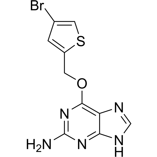
LomeguatribMGMT Inhibitor -
HY-17364
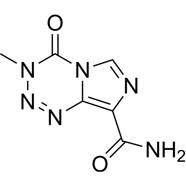
TemozolomideDNA Alkylator -
HY-13585
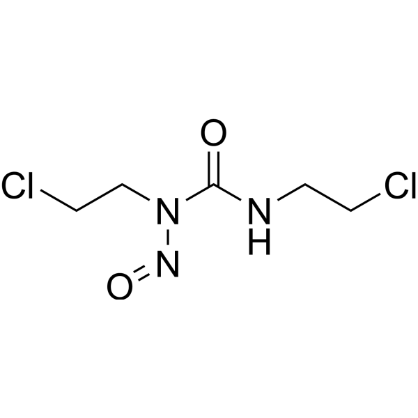
CarmustineDNA/RNA Alkylator -
HY-13765
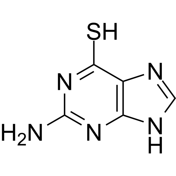
6-ThioguanineImmunosuppressant Agent -
HY-10993
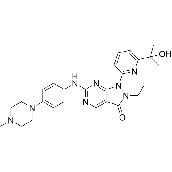
AdavosertibWee1 Inhibitor -

HY-10227
BortezomibProteasome Inhibitor -
HY-15531
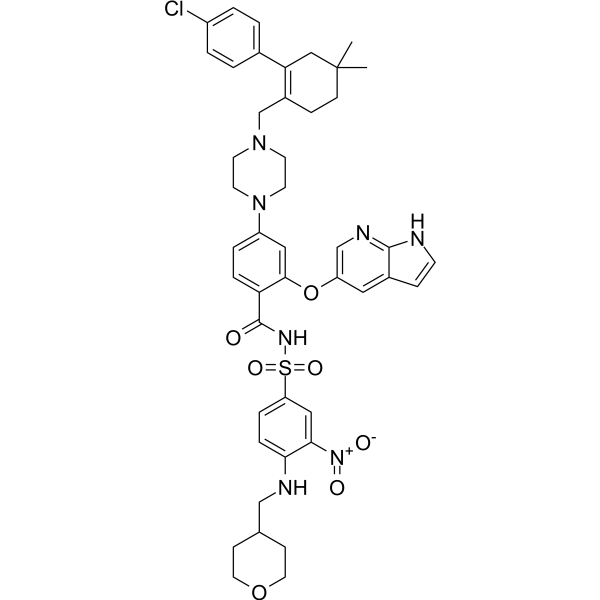
VenetoclaxBCL-2 Inhibitor -
HY-13426
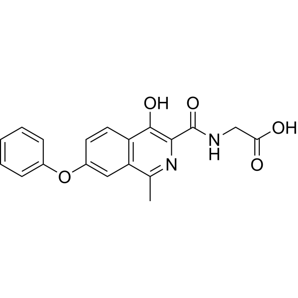
RoxadustatHIF-PH Inhibitor -
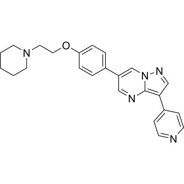
HY-13418A
DorsomorphinAMPK Inhibitor -
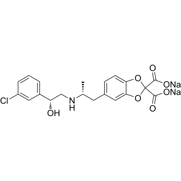
HY-116771A
CL 316243β3-Adrenoceptor Agonist -

HY-W002585R
O6-Benzylguanine (Standard)Reference Standard -
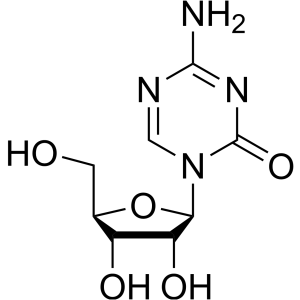
HY-10586
5-AzacytidineDNMT Inhibitor
Publications Citing Use of MedChemExpress (MCE) O6-Benzylguanine
MoreAll DNA Methyltransferase Isoforms
MoreAll Caspase Isoforms
More

Biological Activity
Description
|
MGMT 0.1 μM (ED50) |
CDK1/cyclinB1 |
cdk2/cyclin A |
mTORC1 |
Caspase 9 |
Cellular Effect
|
Cell Line

|
Type | Value | Description | References |
|---|---|---|---|---|
| HeLa | IC50 |
0.62 μM
Compound: 8
|
In vitro inhibition of MGMT using cell free extracts from HeLa S3 cells
In vitro inhibition of MGMT using cell free extracts from HeLa S3 cells
|
[PMID: 11708909] |
| HL-60 | IC50 |
0.039 μM
Compound: O6-BG, BG
|
Inhibition of AGT in human HL60 cells assessed as AGT levels using [3H]AGT after 2 hrs
Inhibition of AGT in human HL60 cells assessed as AGT levels using [3H]AGT after 2 hrs
|
[PMID: 22932317] |
| HT-29 | ED50 |
0.05 μM
Compound: 6-Benzylguanine
|
AGT-inactivating activity was determined by 50% inactivation in HT-29 cells upon incubation for 4 h
AGT-inactivating activity was determined by 50% inactivation in HT-29 cells upon incubation for 4 h
|
[PMID: 7830279] |
| HT-29 | ED50 |
0.05 μM
Compound: 1a
|
Inhibitory activity against alkyl transferase in HT-29 cells
Inhibitory activity against alkyl transferase in HT-29 cells
|
[PMID: 1447749] |
| HT-29 | ED50 |
0.2 μM
Compound: 6-Benzylguanine
|
AGT-inactivating activity was determined by 50% inactivation in HT-29 cell free extract upon incubation for 30 min
AGT-inactivating activity was determined by 50% inactivation in HT-29 cell free extract upon incubation for 30 min
|
[PMID: 7830279] |
| HT-29 | ED50 |
0.2 μM
Compound: 1a
|
In vitro inhibition of human alkyl transferase in HT-29 cell-free extracts.
In vitro inhibition of human alkyl transferase in HT-29 cell-free extracts.
|
[PMID: 1447749] |
| HT-29 | GI50 |
>100 μM
Compound: BG, O6-BG
|
Growth inhibition of human HT-29 cells measured after 24 hrs
Growth inhibition of human HT-29 cells measured after 24 hrs
|
[PMID: 34731767] |
| HT-29 | IC50 |
0.058 μM
Compound: 2
|
concentration required to reduce AGT activity to 50% of control rate in intact HT-29 human colorectal carcinoma cells
concentration required to reduce AGT activity to 50% of control rate in intact HT-29 human colorectal carcinoma cells
|
[PMID: 11063604] |
| HT-29 | IC50 |
0.18 μM
Compound: 2
|
Inhibition of AGT activity to 50% of control rate in HT-29 cell extract
Inhibition of AGT activity to 50% of control rate in HT-29 cell extract
|
[PMID: 11063604] |
| M4Beu cell line | ED50 |
2 μM
Compound: 12
|
In vitro cytotoxicity against M4Beu cells when co-administered with 50 uM cystemustine.
In vitro cytotoxicity against M4Beu cells when co-administered with 50 uM cystemustine.
|
[PMID: 9288172] |
| Raji | IC50 |
0.1 μM
Compound: O6-benzylguanine
|
O6-Alkylguanine-DNA Alkyltransferase-Inactivating Activity in Raji cells (Concentration of inactivator required to produce 50% reduction in ATPase activity)
O6-Alkylguanine-DNA Alkyltransferase-Inactivating Activity in Raji cells (Concentration of inactivator required to produce 50% reduction in ATPase activity)
|
[PMID: 9857094] |
| T98G | GI50 |
91.8 μM
Compound: BG, O6-BG
|
Antiproliferative activity against human T98G cells assessed as cell growth inhibition incubated for 96 hrs by CellTiter-Glo luminescent cell viability assay
Antiproliferative activity against human T98G cells assessed as cell growth inhibition incubated for 96 hrs by CellTiter-Glo luminescent cell viability assay
|
[PMID: 34731767] |
In Vitro
O6-Benzylguanine (100 μM; 1-5 days) sodium inhibits cell growth, induces cellular senescence, and upregulates senescence-related markers such as p27, Bcl2, and MnSOD in rat aortic vascular smooth muscle cells (VSMCs)[3].
O6-Benzylguanine (50 μg/mL; 48 h) sodium downregulates MGMT and cyclin expression, upregulates p21 and pro-apoptotic protein expression, and induces apoptosis in human pancreatic cancer L3.6pl and PANC1 cells[2].
O6-Benzylguanine (up to 1000 μM; 30 min) sodium inactivates AGT in purified Escherichia coli Ada-C mutant protein and human AGT protein (with an ED50 of 0.1 μM when bound to DNA)[4].
MedChemExpress (MCE) has not independently confirmed the accuracy of these methods. They are for reference only. Further protocols information, click here.
-
Cell Line:L3.6pl and PANC1 cells
-
Concentration:50 μg/mL
-
Incubation Time:48 h
-
Result:Decreased the protein expressions of MGMT, cyclin B1, cyclin B2, cyclin A, and ki-67.
Increased the expression of p21, cytochrome C, caspase 9, and the cleavage of PARP1.
-
Cell Line:L3.6pl and PANC1 cells
-
Concentration:50 μg/mL
-
Incubation Time:48 h
-
Result:Reduced the mRNA levels of cyclin B1, cyclin B2, cyclin A, ki-67, and MGMT, and increased the mRNA levels of p21.
-
Cell Line:Rat aortic VSMCs
-
Concentration:100 μM
-
Incubation Time:1, 3, 5 days
-
Result:Increased the protein levels of p27, Bcl2, MnSOD, and pERK1/2, and decreased the levels of PCNA and αSMA.
-
Cell Line:Rat aortic VSMCs
-
Concentration:100 μM
-
Incubation Time:5 days
-
Result:Caused cells to become enlarged in morphology and displayed intense staining signals for γH2A.X.
In Vivo
O6-Benzylguanine (4 mg/kg; p.o.; once every 3 days; 7 weeks) sodium decreases the vasodilatory response of aortic smooth muscle to sodium nitroprusside and enhances its response to K+Cl− in healthy male Wistar rats[3].
O6-Benzylguanine sodium sensitizes human pancreatic tumor xenografts expressing AGT in athymic mice to the antitumor activity of BCNU (HY-13585) and Temozolomide (HY-17364)[1].
O6-Benzylguanine sodium reduces the maximum tolerated dose of BCNU in mice and dogs, indicating that it enhances the toxicity of BCNU to normal tissues[1].
O6-Benzylguanine sodium exhibits superior cerebrospinal fluid penetration compared to its analogs in adult male rhesus monkeys, supporting its use for brain tumor treatment[1].
MedChemExpress (MCE) has not independently confirmed the accuracy of these methods. They are for reference only.
-
Animal Model:NCI-nu (male, 8 to 12 weeks of age, orthotopic injection of human L3.6pl pancreatic cancer cells into the pancreas)[2]
-
Dosage:100 μg
-
Administration:i.p.; once daily Monday-Friday; 5 weeks
-
Result:Reduced median pancreatic tumor volume to 2419 mm3 (vs. 4596 mm3 in control mice).
Reduced median tumor weight to 1.53 g (vs. 2.96 g in control mice).
Decreased expression of MGMT, cyclin B1, cyclin B2, cyclin A, and the proliferation marker ki-67 in tumors.
Increased expression of p21 in tumors.
Increased tumor cell apoptosis (TUNEL-positive staining) compared to control mice.
-
Animal Model:Wistar rats (male, 12-week-old)[3]
-
Dosage:4 mg/kg
-
Administration:p.o.; every third day; 7 weeks
-
Result:Increased response to KCl in aortic rings.
Reduced smooth muscle relaxation response to the NO donor sodium nitrate in aortic rings.
Unchanged responses to phenylephrine and isoprenaline in aortic rings.
Clinical Trial
| NCT Number | Sponsor | Condition | Start Date |
Phase
|
|---|---|---|---|---|
| NCT01329991 | Plexxikon| | 2011-05 | PHASE1 |


Chemical Information
-
CAS No. 19916-73-5
-
Appearance Solid
-
Molecular Weight 241.25
-
Formula C12H11N5O
-
Color White to off-white
-
SMILES
NC(N=C1OCC2=CC=CC=C2)=NC3=C1NC=N3
-
Shipping
Room temperature in continental US; may vary elsewhere.
-
Storage
RT, protect from light
In solvent -80°C 1 year -20°C 6 months


Publications (3)
-
Journal Impact Factor
-
Most Recent
-
Cell Death Dis
Methyltransferase MGMT upregulation drives metastasis by activating epithelial-mesenchymal transition in KRAS mutant colon cancer. [Abstract]2026 May 16;17(1):628. PMID: 42143085 -
J Med Virol
Drug Repurposing: In Vitro Evaluation of Simeprevir as a Novel Antiviral Drug Against Severe Fever With Thrombocytopenia Syndrome Virus. [Abstract]2025 Oct;97(10):e70655. PMID: 41117261 -
Pathol Res Pract
Identification of AGT and CD44 in methotrexate-resistant colorectal cancer and reversal of methotrexate-resistance. [Abstract]2022 Jan:229:153717. PMID: 34952427


Solvent & Solubility
In Vitro:
DMSO : 100 mg/mL (414.51 mM; Need ultrasonic; Hygroscopic DMSO has a significant impact on the solubility of product, please use newly opened DMSO)
Please refer to the solubility information to select the appropriate solvent. Once prepared, please aliquot and store the solution to prevent product inactivation from repeated freeze-thaw cycles.
Storage method and period of stock solution: -80°C, 1 year; -20°C, 6 months. When stored at -80°C, please use it within 1 year. When stored at -20°C, please use it within 6 months.
Please refer to the solubility information to select the appropriate solvent. Once prepared, please aliquot and store the solution to prevent product inactivation from repeated freeze-thaw cycles.
Storage method and period of stock solution: -80°C, 1 year; -20°C, 6 months. When stored at -80°C, please use it within 1 year. When stored at -20°C, please use it within 6 months.
Concentration (start) × Volume (start) = Concentration (final) × Volume (final)
In Vivo:
Select the appropriate dissolution method based on your experimental animal and administration route.
- For the following dissolution methods, please ensure to first prepare a clear stock solution using an In Vitro approach and then sequentially add co-solvents:
- To ensure reliable experimental results, the clarified stock solution can be appropriately stored based on storage conditions. As for the working solution for In Vivo experiments, it is recommended to prepare freshly and use it on the same day.
- The percentages shown for the solvents indicate their volumetric ratio in the final prepared solution. If precipitation or phase separation occurs during preparation, heat and/or sonication can be used to aid dissolution.
Add each solvent one by one: 10% DMSO 40% PEG300 5% Tween-80 45% Saline
Solubility: ≥ 2.75 mg/mL (11.40 mM); Clear solution
This protocol yields a clear solution of ≥ 2.75 mg/mL (saturation unknown).
Taking 1 mL working solution as an example, add 100 μL DMSO stock solution (27.5 mg/mL) to 400 μL PEG300, and mix evenly; then add 50 μL Tween-80 and mix evenly; then add 450 μL Saline to adjust the volume to 1 mL.
Preparation of Saline: Dissolve 0.9 g sodium chloride in ddH₂O and dilute to 100 mL to obtain a clear Saline solution.
In Vivo Dissolution Calculator
Please enter the basic information of animal experiments:
-
-
-
-
Recommended: Prepare an additional quantity of animals to account for potential losses during experiments.
Please enter your animal formula composition:
-
%DMSO +
Recommended: Keep the proportion of DMSO in working solution below 2% if your animal is weak.
-
%+
-
+%Tween-80 + +
-
%Saline +
The co-solvents required include: DMSO, . All of co-solvents are available by MedChemExpress (MCE). , Tween 80. All of co-solvents are available by MedChemExpress (MCE).
Working solution concentration: 0.22 mg/mL
Method for preparing stock solution: mg drug dissolved in μL DMSO. Stock solution concentration: mg/mL.
1. Take μL DMSO stock solution;
2. Add μL .
μL , mix evenly;
3. Then add μL Tween 80, mix evenly;
4. Then add μL
Please ensure that the stock solution in the first step is dissolved to a clear state, and add co-solvents in sequence. You can use ultrasonic heating (ultrasonic cleaner, recommended frequency 20-40 kHz), vortexing, etc. to assist dissolution.

Protocols
-
Apoptosis
Apoptosis, also called programmed cell death, is generally characterized by distinct morphological characteristics.
-
TUNEL staining for apoptotic DNA fragmentation
TUNEL staining detects DNA strand breaks by using terminal deoxynucleotidyl transferase to add labeled nucleotides to exposed 3′-OH DNA termini, generating either microscopic staining in fixed cells or tissue sections, or fluorescence/cytometric signal in cell suspensions. TUNEL positivity reflects DNA fragmentation but should not be interpreted alone as definitive apoptosis, because TUNEL can also label necrotic, autolytic, mechanically damaged, or DNA-repair-associated DNA breaks.
-
Flow cytometric DNA-content cell-cycle staining
Flow cytometric DNA-content cell-cycle staining measures the fluorescence intensity of DNA-bound fluorochromes in single cells or nuclei to estimate DNA content distributions, allowing assignment of populations to G0/G1, S, and G2/M phases by DNA histogram deconvolution. Propidium iodide (PI) intercalates into DNA, and PI fluorescence is proportional to cellular DNA content when staining is performed under conditions that make DNA accessible and minimize non-DNA signal. Cells with G2/M DNA content are expected to show approximately twice the fluorescence intensity of G0/G1 cells, while S-phase cells occupy intermediate fluorescence values. PI-based DNA-content analysis can also detect cells with fractional DNA content, often reported as sub-G1, when DNA fragmentation and extraction during staining reduce retained DNA signal in apoptotic cells. DAPI is an alternative DNA fluorochrome for univariate DNA-content analysis, while bivariate approaches combining DNA content with proliferation
-
Annexin V plus membrane-impermeant dye apoptosis staining
Annexin V-based apoptosis assays rely on the detection of phosphatidylserine (PS) externalization from the inner leaflet of the plasma membrane to the outer leaflet, an early biochemical hallmark of apoptosis. Fluorescently labeled Annexin V binds PS in a calcium-dependent manner, enabling identification of early apoptotic cells by flow cytometry or fluorescence microscopy. When combined with a membrane-impermeant DNA-binding dye (e. g. , propidium iodide), this approach allows discrimination between viable (Annexin V−/dye−), early apoptotic (Annexin V+/dye−), and late apoptotic or necrotic (Annexin V+/dye+) cell populations by assessing membrane integrity and PS exposure.
-
BrdU Incorporation Assay
Bromodeoxyuridine (BrdU) incorporation assay is based on the principle that BrdU, a thymidine analog, is incorporated into newly synthesized DNA during the S phase of the cell cycle, thereby serving as a marker of DNA replication and cellular proliferation. Incorporated BrdU can be detected using anti-BrdU antibodies following DNA denaturation, enabling visualization or quantification of proliferating cells through immunochemical detection methods such as immunofluorescence or immunohistochemistry.
-
Apoptosis Solutions
Apoptosis is a regulated, generally non-lytic cell-death pathway that removes unwanted, damaged, infected, or abnormal cells through coordinated morphological changes, caspase activation, DNA fragmentation, and membrane remodeling. The intrinsic apoptosis pathway is controlled mainly by mitochondrial outer membrane permeabilization, BCL-2 family proteins, cytochrome c release, apoptosome formation, caspase-9 activation, and downstream executioner caspase-3/7 activation. The extrinsic apoptosis pathway is initiated by death receptors such as Fas, TNFR, and TRAIL receptors, which recruit adaptor proteins and activate caspase-8 before engaging executioner caspases or mitochondrial amplification through BID cleavage. Apoptosis is linked to many phenotypes, including cancer cell killing, tissue homeostasis, immune regulation, neurodegeneration, infection response, and treatment-induced cytotoxicity; unresolved questions include how apoptosis interacts with necroptosis, pyroptosis, ferroptos
-
Protocol for Cell Cycle
Cell-cycle analysis by flow cytometry measures DNA content in single cells to estimate the fraction of cells in G0/G1, S, and G2/M phases. Propidium iodide intercalates into DNA, and after RNA removal with RNase, fluorescence intensity reflects cellular DNA content: 2N cells are assigned to G0/G1, cells between 2N and 4N to S phase, and 4N cells to G2/M. DNA-content analysis alone cannot reliably separate G0 from G1 or G2 from M. Ki-67 can distinguish quiescent G0 cells from cycling cells, EdU or BrdU incorporation marks active DNA synthesis in S phase, and phospho-histone H3 staining identifies mitotic cells within the 4N population.
-
Senescence-associated β-galactosidase staining
Senescence-associated β-galactosidase staining detects β-galactosidase activity that is histochemically visible at pH 6. 0 in senescent cells, where X-gal cleavage produces an insoluble blue precipitate observable by bright-field microscopy. This activity reflects increased lysosomal β-galactosidase/lysosomal mass rather than a senescence-essential enzyme, because GLB1 depletion or genetic lysosomal β-galactosidase deficiency can abolish SA-β-gal staining while cells still undergo senescence. SA-β-gal was originally reported in senescent but not presenescent fibroblasts and keratinocytes, absent from quiescent fibroblasts and terminally differentiated keratinocytes, and increased with donor age in human skin samples. Because SA-β-gal can also appear in some non-senescent or tissue-specific contexts, interpretation should be paired with experimental controls and, when possible, independent senescence markers.


Purity & Documentation
-
Data Sheet (295 KB)
-
SDS (393 KB)
- English - EN (393 KB)
- Français - FR (393 KB)
- Deutsch - DE (393 KB)
- Norwegian - NO (393 KB)
- Español - ES (393 KB)
- Swedish - SV (393 KB)
- Italian - IT (393 KB)
- Korean - KR (393 KB)
- Portuguese - PT (393 KB)
-
Handling Instructions (2659 KB)
References
[1]. Rabik CA, et al. Inactivation of O6-alkylguanine DNA alkyltransferase as a means to enhance chemotherapy. Cancer treatment reviews. 2006 Jun;32(4):261-76. [Content Brief]
[2]. Konduri SD, et al. Blockade of MGMT expression by O6 benzyl guanine leads to inhibition of pancreatic cancer growth and induction of apoptosis. Clinical cancer research : an official journal of the American Association for Cancer Research. 2009 Oct 01;15(19):6087-95. [Content Brief]


Complete Stock Solution Preparation Table
Please refer to the solubility information to select the appropriate solvent. Once prepared, please aliquot and store the solution to prevent product inactivation from repeated freeze-thaw cycles.
Storage method and period of stock solution: -80°C, 1 year; -20°C, 6 months. When stored at -80°C, please use it within 1 year. When stored at -20°C, please use it within 6 months.
| Optional Solvent | Concentration Solvent Mass | 1 mg | 5 mg | 10 mg | 25 mg |
|---|---|---|---|---|---|
| DMSO | 1 mM | 4.1451 mL | 20.7254 mL | 41.4508 mL | 103.6269 mL |
| 5 mM | 0.8290 mL | 4.1451 mL | 8.2902 mL | 20.7254 mL | |
| 10 mM | 0.4145 mL | 2.0725 mL | 4.1451 mL | 10.3627 mL | |
| 15 mM | 0.2763 mL | 1.3817 mL | 2.7634 mL | 6.9085 mL | |
| 20 mM | 0.2073 mL | 1.0363 mL | 2.0725 mL | 5.1813 mL | |
| 25 mM | 0.1658 mL | 0.8290 mL | 1.6580 mL | 4.1451 mL | |
| 30 mM | 0.1382 mL | 0.6908 mL | 1.3817 mL | 3.4542 mL | |
| 40 mM | 0.1036 mL | 0.5181 mL | 1.0363 mL | 2.5907 mL | |
| 50 mM | 0.0829 mL | 0.4145 mL | 0.8290 mL | 2.0725 mL | |
| 60 mM | 0.0691 mL | 0.3454 mL | 0.6908 mL | 1.7271 mL | |
| 80 mM | 0.0518 mL | 0.2591 mL | 0.5181 mL | 1.2953 mL | |
| 100 mM | 0.0415 mL | 0.2073 mL | 0.4145 mL | 1.0363 mL |


O6-Benzylguanine Related Classifications
HY-W002585 Related Classifications
 Powered by Bioz