



Tenacissoside G

Based on 1 Customer Validation
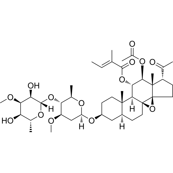
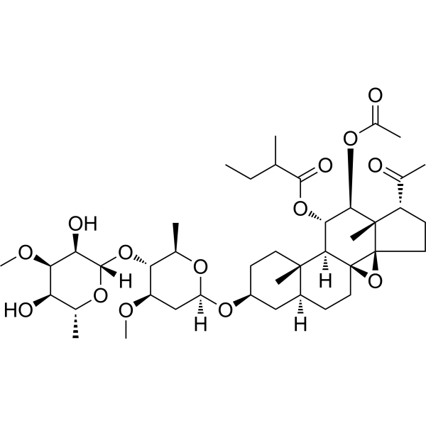
Tenacissoside G is a C21 steroidal glycoside. Tenacissoside G inhibits Src and NF-κB, downregulates the activities of PTN and P-gp, induces DNA damage and apoptosis in cancer cells, inhibits their proliferation and migration, and reverses the resistance of ovarian cancer cells to Paclitaxel (HY-B0015). Tenacissoside G inhibits the expression of iNOS, TNF-α, IL-6, MMP-3, and MMP-13, reduces type II collagen degradation, and alleviates articular cartilage damage. Tenacissoside G can be used in studies related to ovarian cancer, colorectal cancer, and osteoarthritis.
For research use only. We do not sell to patients.
- Purity : 99.64%
- CAS No.: 191729-43-8
- Formula: C42H64O14
- Molecular Weight:792.95
-
Storage:
4°C, protect from light
* In solvent : -80°C, 6 months; -20°C, 1 month (protect from light)

To place orders, for customer services and technical support, please contact: MedChemExpress USA
Tel: 609-228-6898 E-mail: [email protected] [email protected]


-
 Biological Activity
-
Chemical Information
-
Solvent & Solubility
- Protocols
- Purity & Documentation
- References
-
 Help & FAQs


-
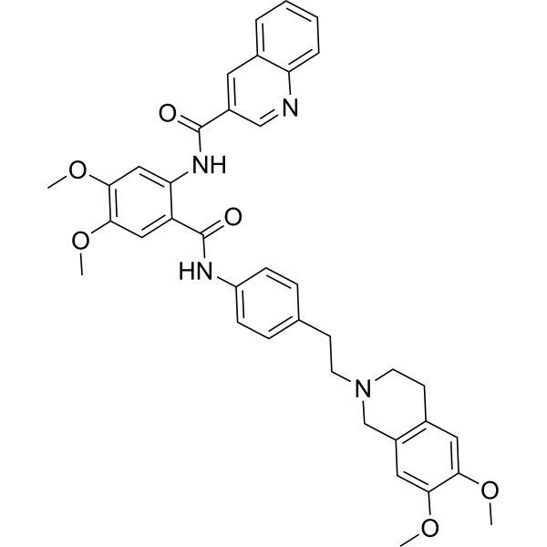
HY-10550
TariquidarP-gp Inhibitor -
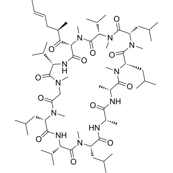
HY-17384
ValspodarP-glycoprotein Inhibitor -
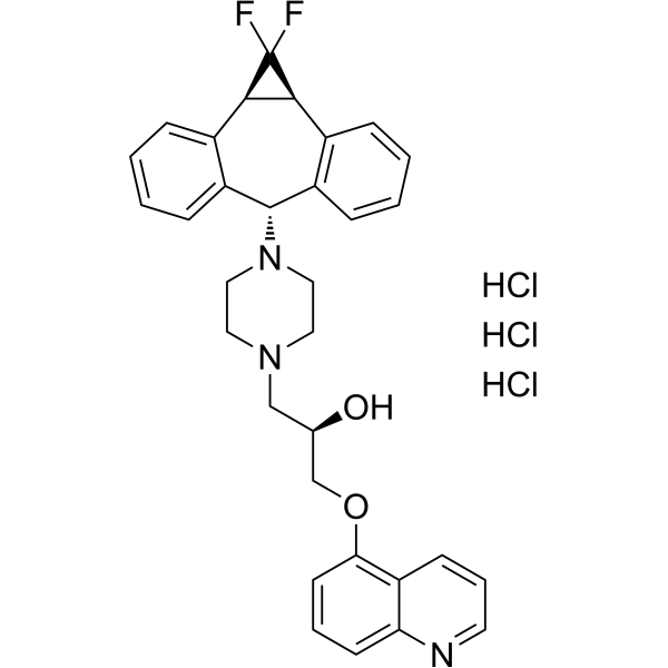
HY-50671
Zosuquidar trihydrochlorideP-gp Inhibitor -
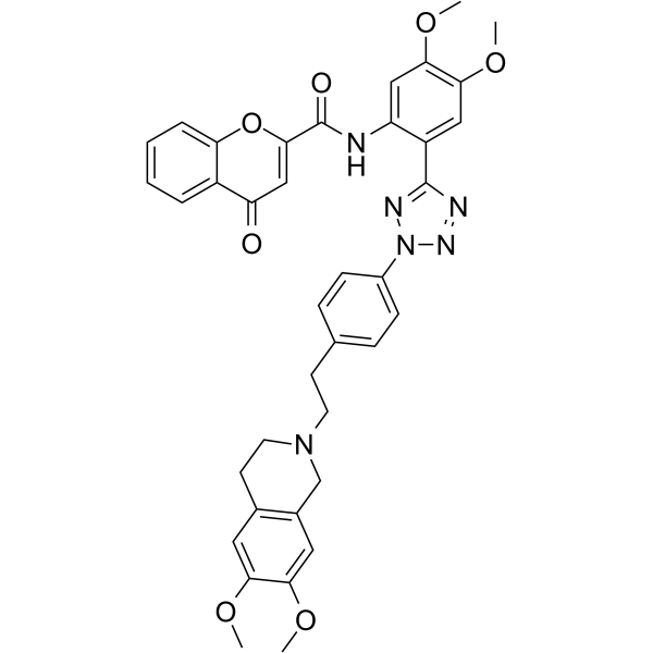
HY-13646
EncequidarP-glycoprotein Inhibitor -
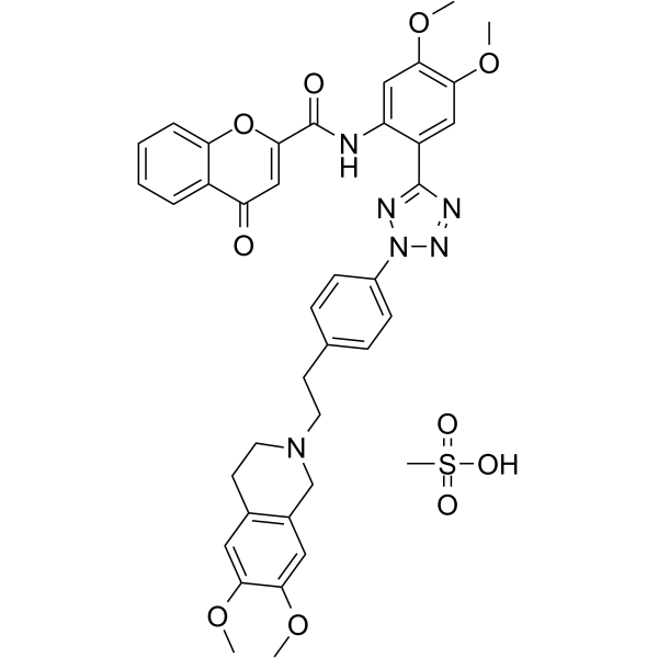
HY-13646A
Encequidar mesylateP-gp Inhibitor -
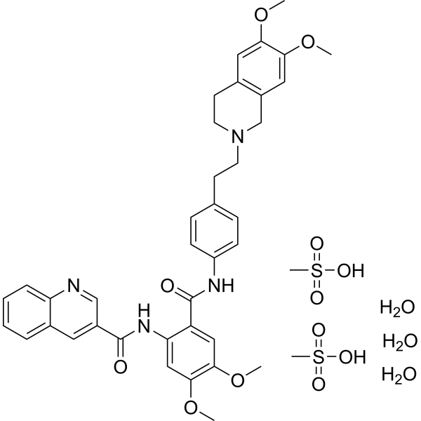
HY-10550A
Tariquidar methanesulfonate, hydrateP-gp Inhibitor -
HY-N0670
Tenacissoside HChinese Medicine Monomer -
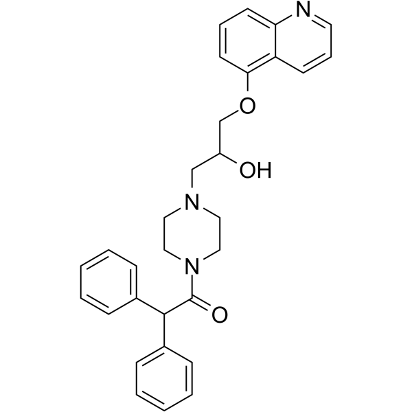
HY-17013
DofequidarP-glycoprotein Inhibitor -
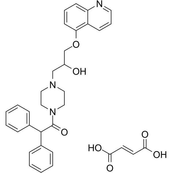
HY-17013A
Dofequidar fumarateP-glycoprotein Inhibitor -
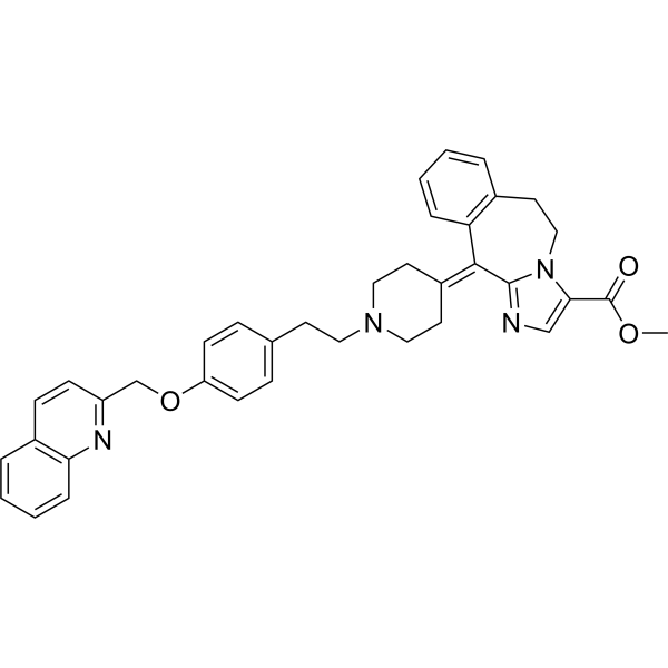
HY-132189
LaniquidarP-glycoprotein Inhibitor -

HY-10550R
Tariquidar (Standard)P-gp Inhibitor -
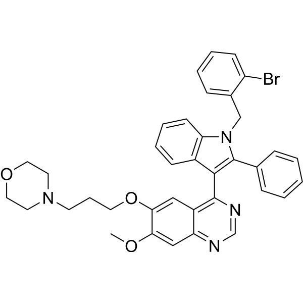
HY-132866
YS-370P-gp Inhibitor
All DNA/RNA Synthesis Isoforms
More

Biological Activity
Description
In Vitro
Tenacissoside G (5-160 μM; 24-48 h) shows no significant cytotoxicity against A2780 and A2780/T cells even at concentrations up to 160 μM, and reverses paclitaxel resistance in A2780/T cells in a concentration-dependent manner, with a maximum reversal fold of 6.96 at 20 μM for 24 h. However, it exerts no significant synergistic cytotoxicity on paclitaxel-sensitive A2780 cells[1].
Tenacissoside G (5-20 μM; 24 h), either used alone or in combination with paclitaxel, downregulates the mRNA and protein expressions of Src, PTN and ABCB1 in A2780/T cells in a dose-dependent manner, and reverses the upregulation of the aforementioned genes and proteins induced by paclitaxel alone[1].
Tenacissoside G (20 μM; 24 h) inhibits the efflux function of P-gp in A2780/T cells and significantly increases the intracellular accumulation of Rhodamine 123 (HY-D0816) and paclitaxel[1].
Tenacissoside G (25-400 μM; 48 h) inhibits the proliferation of human colorectal cancer cell lines RKO, LoVo, HCT-8, HCT116 and HT-29 in a concentration-dependent manner, with its 48 h IC50 values ranging from 55.3 μM (LoVo) to 103.1 μM (HT-29)[3].
Tenacissoside G (50-100 μM; 48 h) reduces DNA replication and inhibits the proliferation of human colorectal cancer cell lines RKO and LoVo[3].
Combined use of Tenacissoside G (5-20 μM; 24 h) and paclitaxel induces apoptosis of A2780/T cells in a dose-dependent manner[1].
Tenacissoside G (50-100 μM; 48 h) induces low-level apoptosis in human colorectal cancer cells RKO and LoVo by activating the caspase cascade[3].
Combination treatment with Tenacissoside G (5-20 μM; 24 h) and paclitaxel induces G2/M cell cycle arrest in A2780/T cells[1].
Tenacissoside G (50-100 μM; 48 h) induces G0/G1 cell cycle arrest in human colorectal cancer cell lines RKO and LoVo by upregulating p53 and p21, and downregulating CDK2, CDK4, CDK6, cyclin D1 and cyclin E[3].
Combined treatment with Tenacissoside G (5-20 μM; 24 h) and paclitaxel inhibits the migration of A2780/T cells in a dose-dependent manner[1].
Tenacissoside G (2.5-160 μM; 24 h) exhibits only extremely low cytotoxicity to primary mouse chondrocytes, with cell viability maintained above 90%[2].
Tenacissoside G (2.5-10 μM; 26 h) dose-dependently protects primary mouse chondrocytes against IL-1β-induced type II collagen degradation and inhibits MMP-13 protein expression[2].
Tenacissoside G (2.5-10 μM; 26 h) dose-dependently inhibits IL-1β-induced activation of the NF-κB pathway in primary mouse chondrocytes by suppressing p65 phosphorylation and maintaining IκBα protein levels[2].
Tenacissoside G (2.5-10 μM; 26 h) dose-dependently maintains the level of type Ⅱ collagen in primary mouse chondrocytes stimulated with IL-1β (detected by immunofluorescence)[2].
Tenacissoside G (2.5-10 μM; 26 h) dose-dependently inhibits the expression of inflammation-related genes (iNOS, TNF-α, IL-6) and matrix degradation-related genes (MMP-3, MMP-13) in IL-1β-induced primary mouse chondrocytes[2].
Tenacissoside G (50-100 μM; 24 h) induces DNA damage in human colorectal cancer cells RKO and LoVo, which is evidenced by an increased percentage of comet tail DNA and upregulated expression of γ-H2AX[3].
MedChemExpress (MCE) has not independently confirmed the accuracy of these methods. They are for reference only. Further protocols information, click here.
-
Cell Line:human paclitaxel-sensitive ovarian cancer A2780 cells, human paclitaxel-resistant ovarian cancer A2780/T cells
-
Concentration:5, 10, 20, 40, 80 and 160 μM (alone); 5, 10 and 20 μM (combined with paclitaxel)
-
Incubation Time:24 h; 48 h
-
Result:Caused no significant decrease in viability of A2780 or A2780/T cells at 160 μM for 24 h or 48 h.
Reduced the IC50 of paclitaxel in A2780/T cells from 28.32 μM to 8.37 μM (3.38-fold reversal) at 10 μM combined with paclitaxel for 24 h.
Reduced the IC50 of paclitaxel in A2780/T cells from 28.32 μM to 4.07 μM (6.96-fold reversal) at 20 μM combined with paclitaxel for 24 h.
Reduced the IC50 of paclitaxel in A2780/T cells from 19.62 μM to 6.80 μM (2.89-fold reversal) at 5 μM combined with paclitaxel for 48 h.
Reduced the IC50 of paclitaxel in A2780/T cells from 19.62 μM to 5.75 μM (3.41-fold reversal) at 10 μM combined with paclitaxel for 48 h.
Reduced the IC50 of paclitaxel in A2780/T cells from 19.62 μM to 2.97 μM (6.60-fold reversal) at 20 μM combined with paclitaxel for 48 h.
Did not significantly reduce cell proliferation in A2780 cells when combined with 5, 10, or 20 μM Tenacissoside G compared to paclitaxel alone.
-
Cell Line:human paclitaxel-resistant ovarian cancer A2780/T cells
-
Concentration:5, 10 and 20 μM (combined with 4 μM paclitaxel)
-
Incubation Time:24 h
-
Result:Increased the apoptosis rate of A2780/T cells to 24% at 5 μM combined with 4 μM paclitaxel compared to 7.5% with paclitaxel alone.
Increased the apoptosis rate of A2780/T cells to 32.1% at 10 μM combined with 4 μM paclitaxel compared to 7.5% with paclitaxel alone.
Increased the apoptosis rate of A2780/T cells to 34.1% at 20 μM combined with 4 μM paclitaxel compared to 7.5% with paclitaxel alone.
-
Cell Line:human paclitaxel-resistant ovarian cancer A2780/T cells
-
Concentration:5, 10 and 20 μM (combined with 4 μM paclitaxel)
-
Incubation Time:24 h
-
Result:Increased the percentage of A2780/T cells in G2/M phase to a maximum of 74.3% (P < 0.001) compared to 43.2% with 4 μM paclitaxel alone.
-
Cell Line:human paclitaxel-resistant ovarian cancer A2780/T cells
-
Concentration:5, 10 and 20 μM (combined with 4 μM paclitaxel)
-
Incubation Time:24 h
-
Result:Significantly reduced the scratch healing rate of A2780/T cells compared to treatment with 4 μM paclitaxel alone.
Decreased the healing rate as Tenacissoside G concentration increased.
-
Cell Line:human paclitaxel-resistant ovarian cancer A2780/T cells
-
Concentration:5, 10 and 20 μM (combined with 4 μM paclitaxel); 20 μM (alone)
-
Incubation Time:24 h
-
Result:Downregulated the mRNA expression of Src, PTN, and ABCB1 at 20 μM alone compared to paclitaxel-induced upregulation.
Downregulated the mRNA expression of Src, PTN, and ABCB1 at 5, 10, or 20 μM combined with paclitaxel compared to paclitaxel-induced upregulation.
Increased the inhibitory effect on mRNA expression with increasing Tenacissoside G concentration.
-
Cell Line:human paclitaxel-resistant ovarian cancer A2780/T cells
-
Concentration:5, 10 and 20 μM (combined with 4 μM paclitaxel); 20 μM (alone)
-
Incubation Time:24 h
-
Result:Downregulated protein levels of Src, phospho-Src, PTN, and P-gp at 20 μM alone compared to paclitaxel-induced upregulation.
Downregulated protein levels of Src, phospho-Src, PTN, and P-gp at 5, 10, or 20 μM combined with paclitaxel compared to paclitaxel-induced upregulation.
Caused a more pronounced reduction in phospho-Src compared to total Src.
Increased the inhibitory effect on protein expression with increasing Tenacissoside G concentration.
-
Cell Line:RKO, LoVo, HCT-8, HCT116, HT-29 human colorectal cancer cell lines
-
Concentration:25, 50, 100, 200, 300 and 400 μM
-
Incubation Time:48 h
-
Result:Inhibited cell proliferation in all five colorectal cancer cell lines in a concentration-dependent manner.
Exhibited 48 h IC50 values of 70.2 μM (RKO), 55.3 μM (LoVo), 100.8 μM (HCT-8), 87.5 μM (HCT116), and 103.1 μM (HT-29).
Rendered RKO and LoVo cells more sensitive than the other three cell lines.
-
Cell Line:RKO, LoVo human colorectal cancer cell lines
-
Concentration:50 and 100 μM
-
Incubation Time:48 h
-
Result:Reduced fluorescence intensity of EdU significantly compared to untreated control cells.
Decreased the fluorescence ratio of EdU to Hoechst 33342 significantly (p < 0.05) in both RKO and LoVo cells.
-
Cell Line:RKO, LoVo human colorectal cancer cell lines
-
Concentration:50 and 100 μM
-
Incubation Time:48 h
-
Result:Increased G0/G1 phase cell accumulation by 1.53% (50 μM) and 1.42% (100 μM) in RKO cells, and by 1.01% (50 μM) and 1.15% (100 μM) in LoVo cells, compared to untreated controls.
Increased p53 expression by 2.5-fold (RKO) and 3.2-fold (LoVo), and p21 expression by 2.1-fold (RKO) and 3.0-fold (LoVo) at 100 μM.
Suppressed expression of CDK2, CDK4, CDK6, cyclin D1, and cyclin E.
-
Cell Line:RKO, LoVo human colorectal cancer cell lines
-
Concentration:50 and 100 μM
-
Incubation Time:48 h
-
Result:Increased apoptotic cell population to 4.06% (50 μM) and 9.73% (100 μM) in RKO cells, and 3.07% (50 μM) and 5.94% (100 μM) in LoVo cells, compared to untreated controls.
Increased expression of cleaved caspase 3, 8, and 9 in a concentration-dependent manner.
In Vivo
Tenacissoside G (50 mg/kg; i.p.; every 2 days; 20 days) inhibits colorectal cancer xenograft growth by 28.4% and reduces tumor weight by 33.7% in BALB/c nude mice without causing significant systemic toxicity[3].
MedChemExpress (MCE) has not independently confirmed the accuracy of these methods. They are for reference only.
-
Animal Model:C57BL/6 (male, 10 weeks old, DMM surgery-induced osteoarthritis)[2]
-
Dosage:4 μg/kg; 8 μg/kg; 16 μg/kg
-
Administration:intra-articular; twice weekly; 8 weeks
-
Result:Increased bone volume/tissue volume (BV/TV) in tibial subchondral bone at 8 weeks post-surgery for 8 μg/kg and 16 μg/kg doses compared to DMM-induction group.
Increased trabecular number (Tb. N) at 8 weeks post-surgery for 8 μg/kg dose compared to DMM-induction group.
Decreased trabecular separation (Tb. Sp) at 8 weeks post-surgery for 16 μg/kg dose compared to DMM-induction group.
Reduced OARSI scores significantly at 4 weeks post-surgery for all doses compared to DMM-induction group, with 16 μg/kg showing greatest reduction.
Lowered OARSI scores significantly at 8 weeks post-surgery for all doses compared to DMM-induction group, with 16 μg/kg showing most significant reduction.
Reduced proteoglycan loss in articular cartilage in a dose-dependent manner, with 16 μg/kg showing least cartilage injury and highest proteoglycan levels at both 4 and 8 weeks.
Increased collagen II protein expression in articular cartilage at both 4 and 8 weeks post-surgery for 8 μg/kg and 16 μg/kg doses compared to DMM-induction group, with 16 μg/kg showing strongest effect.
-
Animal Model:BALB/c nude (female, 4 weeks old, 18-22 g, subcutaneous xenograft model)[3]
-
Dosage:50 mg/kg
-
Administration:i.p.; every 2 days; 20 days
-
Result:Reduced tumor growth by 28.4%.
Reduced tumor weight by 33.7%.
Showed no significant difference in mouse body weight relative to control.
Showed no apparent impairment in heart, liver, spleen, lung, and kidney tissues relative to control.
Increased phosphorylation of p53 at Ser46 in tumor tissue.
Increased cleaved caspase 3 expression in tumor tissue.
Decreased Ki67 expression in tumor tissue.
Induced a small increase in TUNEL-positive apoptotic cells in tumor tissue.


Chemical Information
-
CAS No. 191729-43-8
-
Appearance Solid
-
Molecular Weight 792.95
-
Formula C42H64O14
-
Color White to off-white
-
SMILES
C[C@@]([C@@H]([C@@H](OC(/C(C)=C/C)=O)[C@]1([H])[C@]23C)OC(C)=O)([C@@H]4C(C)=O)[C@]5(CC4)[C@@]1(CC[C@@]2([H])C[C@@H](O[C@@](O[C@H](C)[C@H]6O[C@@](O[C@H](C)[C@@H](O)[C@H]7OC)([H])[C@@H]7O)([H])C[C@H]6OC)CC3)O5
-
Structure Classification
-
Initial Source
-
Shipping
Room temperature in continental US; may vary elsewhere.
-
Storage
4°C, protect from light
* In solvent : -80°C, 6 months; -20°C, 1 month (protect from light)


Solvent & Solubility
In Vitro:
DMSO : 100 mg/mL (126.11 mM; Need ultrasonic; Hygroscopic DMSO has a significant impact on the solubility of product, please use newly opened DMSO)
Please refer to the solubility information to select the appropriate solvent. Once prepared, please aliquot and store the solution to prevent product inactivation from repeated freeze-thaw cycles.
Storage method and period of stock solution: -80°C, 6 months; -20°C, 1 month (protect from light). When stored at -80°C, please use it within 6 months. When stored at -20°C, please use it within 1 month.
Please refer to the solubility information to select the appropriate solvent. Once prepared, please aliquot and store the solution to prevent product inactivation from repeated freeze-thaw cycles.
Storage method and period of stock solution: -80°C, 6 months; -20°C, 1 month (protect from light). When stored at -80°C, please use it within 6 months. When stored at -20°C, please use it within 1 month.
Concentration (start) × Volume (start) = Concentration (final) × Volume (final)
In Vivo:
Select the appropriate dissolution method based on your experimental animal and administration route.
- For the following dissolution methods, please ensure to first prepare a clear stock solution using an In Vitro approach and then sequentially add co-solvents:
- To ensure reliable experimental results, the clarified stock solution can be appropriately stored based on storage conditions. As for the working solution for In Vivo experiments, it is recommended to prepare freshly and use it on the same day.
- The percentages shown for the solvents indicate their volumetric ratio in the final prepared solution. If precipitation or phase separation occurs during preparation, heat and/or sonication can be used to aid dissolution.
Add each solvent one by one: 10% DMSO 40% PEG300 5% Tween-80 45% Saline
Solubility: ≥ 2.5 mg/mL (3.15 mM); Clear solution
This protocol yields a clear solution of ≥ 2.5 mg/mL (saturation unknown).
Taking 1 mL working solution as an example, add 100 μL DMSO stock solution (25.0 mg/mL) to 400 μL PEG300, and mix evenly; then add 50 μL Tween-80 and mix evenly; then add 450 μL Saline to adjust the volume to 1 mL.
Preparation of Saline: Dissolve 0.9 g sodium chloride in ddH₂O and dilute to 100 mL to obtain a clear Saline solution.
Add each solvent one by one: 10% DMSO 90% (20% SBE-β-CD in Saline)
Solubility: ≥ 2.5 mg/mL (3.15 mM); Clear solution
This protocol yields a clear solution of ≥ 2.5 mg/mL (saturation unknown).
Taking 1 mL working solution as an example, add 100 μL DMSO stock solution (25.0 mg/mL) to 900 μL 20% SBE-β-CD in Saline, and mix evenly.
Preparation of 20% SBE-β-CD in Saline (4°C, storage for one week): 2 g SBE-β-CD powder is dissolved in 10 mL Saline, completely dissolve until clear.
In Vivo Dissolution Calculator
Please enter the basic information of animal experiments:
-
-
-
-
Recommended: Prepare an additional quantity of animals to account for potential losses during experiments.
Please enter your animal formula composition:
-
%DMSO +
Recommended: Keep the proportion of DMSO in working solution below 2% if your animal is weak.
-
%+
-
+%Tween-80 + +
-
%Saline +
The co-solvents required include: DMSO, . All of co-solvents are available by MedChemExpress (MCE). , Tween 80. All of co-solvents are available by MedChemExpress (MCE).
Working solution concentration: 0.22 mg/mL
Method for preparing stock solution: mg drug dissolved in μL DMSO. Stock solution concentration: mg/mL. * In solvent : -80°C, 6 months; -20°C, 1 month (protect from light)
1. Take μL DMSO stock solution;
2. Add μL .
μL , mix evenly;
3. Then add μL Tween 80, mix evenly;
4. Then add μL
Please ensure that the stock solution in the first step is dissolved to a clear state, and add co-solvents in sequence. You can use ultrasonic heating (ultrasonic cleaner, recommended frequency 20-40 kHz), vortexing, etc. to assist dissolution.

Protocols
-
RNA extraction experimental
By lysing cells, releasing RNA, and removing impurities such as proteins and DNA, high-purity RNA products are finally obtained. The commonly used traditional method is the guanidine isothiocyanate/phenol/chloroform method (Trizol), which is suitable for a variety of animal materials including animal tissues, microorganisms, cultured cells, etc., and most plant materials.
-
Apoptosis
Apoptosis, also called programmed cell death, is generally characterized by distinct morphological characteristics.
-
TUNEL staining for apoptotic DNA fragmentation
TUNEL staining detects DNA strand breaks by using terminal deoxynucleotidyl transferase to add labeled nucleotides to exposed 3′-OH DNA termini, generating either microscopic staining in fixed cells or tissue sections, or fluorescence/cytometric signal in cell suspensions. TUNEL positivity reflects DNA fragmentation but should not be interpreted alone as definitive apoptosis, because TUNEL can also label necrotic, autolytic, mechanically damaged, or DNA-repair-associated DNA breaks.
-
Mitochondrial membrane-potential fluorescent assay
Mitochondrial membrane potential fluorescent assays estimate ΔΨm in living cells using lipophilic cationic dyes such as TMRM, TMRE, rhodamine 123, and JC-1, which accumulate in mitochondria according to membrane polarization; loss of signal after FCCP or CCCP treatment is interpreted as mitochondrial depolarization. TMRM/TMRE and rhodamine 123 are commonly used for semi-quantitative live-cell microscopy or flow cytometry, while JC-1 can report a shift from red aggregate fluorescence to green monomer fluorescence during depolarization; interpretation requires controls because dye concentration, quenching mode, cell type, dye efflux, and mitochondrial mass can affect fluorescence independently of ΔΨm.
-
Annexin V plus membrane-impermeant dye apoptosis staining
Annexin V-based apoptosis assays rely on the detection of phosphatidylserine (PS) externalization from the inner leaflet of the plasma membrane to the outer leaflet, an early biochemical hallmark of apoptosis. Fluorescently labeled Annexin V binds PS in a calcium-dependent manner, enabling identification of early apoptotic cells by flow cytometry or fluorescence microscopy. When combined with a membrane-impermeant DNA-binding dye (e. g. , propidium iodide), this approach allows discrimination between viable (Annexin V−/dye−), early apoptotic (Annexin V+/dye−), and late apoptotic or necrotic (Annexin V+/dye+) cell populations by assessing membrane integrity and PS exposure.
-
Cell migration
Cell migration is a method that plays an important role in wound healing, cell differentiation, embryonic development, etc.
-
Mitochondrial membrane-potential and mitochondrial mass staining
Mitochondrial membrane potential staining measures the electrochemical polarization across the mitochondrial inner membrane in live cells using lipophilic cationic fluorescent probes; early rhodamine-based work showed that selective mitochondrial dye accumulation is lost when the mitochondrial transmembrane potential is dissipated. JC-1 reports mitochondrial polarization by shifting from green monomer fluorescence to red J-aggregate fluorescence as dye concentration increases within energized mitochondria; therefore, the red/green fluorescence ratio is used as a relative readout of mitochondrial membrane potential. TMRE or TMRM staining provides a single-channel relative readout because these cationic rhodamine esters accumulate in polarized mitochondria, and lower fluorescence indicates reduced mitochondrial polarization when acquisition and dye-loading conditions are controlled. Mitochondrial mass staining is commonly performed with MitoTracker Green FM or related MitoTracker dyes as
-
Apoptosis Solutions
Apoptosis is a regulated, generally non-lytic cell-death pathway that removes unwanted, damaged, infected, or abnormal cells through coordinated morphological changes, caspase activation, DNA fragmentation, and membrane remodeling. The intrinsic apoptosis pathway is controlled mainly by mitochondrial outer membrane permeabilization, BCL-2 family proteins, cytochrome c release, apoptosome formation, caspase-9 activation, and downstream executioner caspase-3/7 activation. The extrinsic apoptosis pathway is initiated by death receptors such as Fas, TNFR, and TRAIL receptors, which recruit adaptor proteins and activate caspase-8 before engaging executioner caspases or mitochondrial amplification through BID cleavage. Apoptosis is linked to many phenotypes, including cancer cell killing, tissue homeostasis, immune regulation, neurodegeneration, infection response, and treatment-induced cytotoxicity; unresolved questions include how apoptosis interacts with necroptosis, pyroptosis, ferroptos
-
Research Protocol for Inflammation-related Diseases
The NLRP3 inflammasome is a cytosolic innate immune signaling platform that integrates priming signals and danger-signal activation to promote caspase-1 activation, maturation of IL-1β and IL-18, and gasdermin D-mediated pyroptotic cell death. The core experimental logic is to determine whether inflammatory disease phenotypes are driven by increased NLRP3 expression, ASC-containing inflammasome assembly, caspase-1 cleavage, GSDMD cleavage, and extracellular release of IL-1β/IL-18 rather than by nonspecific cell injury alone. The pathway is strongly linked to inflammation-related disease phenotypes because monosodium urate crystals activate NALP3/NLRP3 inflammasome signaling in gout-like crystal inflammation, cholesterol crystals activate NLRP3 inflammasomes in atherogenesis models, and DSS-induced intestinal inflammation has been reported to involve NLRP3 inflammasome activity. However, experimental colitis studies also show context-dependent protective effects of NLRP3 inflammasome co
-
Genotoxicity/Mutagenicity Study
The bacterial reverse mutation assay detects point mutations that restore amino-acid prototrophy in auxotrophic Salmonella typhimurium or Escherichia coli tester strains; after exposure to a test article, mutagenic activity is read out as an increased number of revertant colonies on minimal agar compared with the vehicle control. The assay uses tester strains with different mutation targets so that base-substitution and frameshift mutagens can be detected, and testing is performed with and without exogenous mammalian metabolic activation because some chemicals require biotransformation to become mutagenic.
-
Cell Viability Determination by MTT Colorimetric Assay
The following protocol uses the MTT colorimetric assay as a classic literature-established method for assessing cell viability/metabolic activity in cultured mammalian cells. MTT[3-(4,5-dimethylthiazol-2-yl)-2,5-diphenyltetrazolium bromide] is reduced by metabolically active cells to a colored formazan product; the amount of formazan is quantified spectrophotometrically and provides an indirect measure of metabolically active viable cells. Importantly, MTT reduction reflects cellular oxidoreductase/metabolic activity rather than an absolute direct count of living cells, so changes in cellular metabolism can alter the signal independently of cell number.


Purity & Documentation
-
Data Sheet (310 KB)
-
SDS (394 KB)
- English - EN (394 KB)
- Français - FR (394 KB)
- Deutsch - DE (394 KB)
- Norwegian - NO (394 KB)
- Español - ES (394 KB)
- Swedish - SV (394 KB)
- Italian - IT (394 KB)
- Korean - KR (394 KB)
- Portuguese - PT (394 KB)
-
Handling Instructions (2659 KB)
References
[1]. Hu J, et al. Tenacissoside G reverses paclitaxel resistance by inhibiting Src/PTN/P-gp signaling axis activation in ovarian cancer cells. Journal of natural medicines. 2025 May;79(3):621-638. [Content Brief]
[3]. Wang K, et al. Tenacissoside G synergistically potentiates inhibitory effects of 5-fluorouracil to human colorectal cancer. Phytomedicine : international journal of phytotherapy and phytopharmacology. 2021 Jun;86:153553. [Content Brief]


Complete Stock Solution Preparation Table
Please refer to the solubility information to select the appropriate solvent. Once prepared, please aliquot and store the solution to prevent product inactivation from repeated freeze-thaw cycles.
Storage method and period of stock solution: -80°C, 6 months; -20°C, 1 month (protect from light). When stored at -80°C, please use it within 6 months. When stored at -20°C, please use it within 1 month.
| Optional Solvent | Concentration Solvent Mass | 1 mg | 5 mg | 10 mg | 25 mg |
|---|---|---|---|---|---|
| DMSO | 1 mM | 1.2611 mL | 6.3056 mL | 12.6111 mL | 31.5278 mL |
| 5 mM | 0.2522 mL | 1.2611 mL | 2.5222 mL | 6.3056 mL | |
| 10 mM | 0.1261 mL | 0.6306 mL | 1.2611 mL | 3.1528 mL | |
| 15 mM | 0.0841 mL | 0.4204 mL | 0.8407 mL | 2.1019 mL | |
| 20 mM | 0.0631 mL | 0.3153 mL | 0.6306 mL | 1.5764 mL | |
| 25 mM | 0.0504 mL | 0.2522 mL | 0.5044 mL | 1.2611 mL | |
| 30 mM | 0.0420 mL | 0.2102 mL | 0.4204 mL | 1.0509 mL | |
| 40 mM | 0.0315 mL | 0.1576 mL | 0.3153 mL | 0.7882 mL | |
| 50 mM | 0.0252 mL | 0.1261 mL | 0.2522 mL | 0.6306 mL | |
| 60 mM | 0.0210 mL | 0.1051 mL | 0.2102 mL | 0.5255 mL | |
| 80 mM | 0.0158 mL | 0.0788 mL | 0.1576 mL | 0.3941 mL | |
| 100 mM | 0.0126 mL | 0.0631 mL | 0.1261 mL | 0.3153 mL |


Tenacissoside G Related Classifications
HY-N2103 Related Classifications
 Powered by Bioz