



NPS 2390

Based on 1 Customer Validation
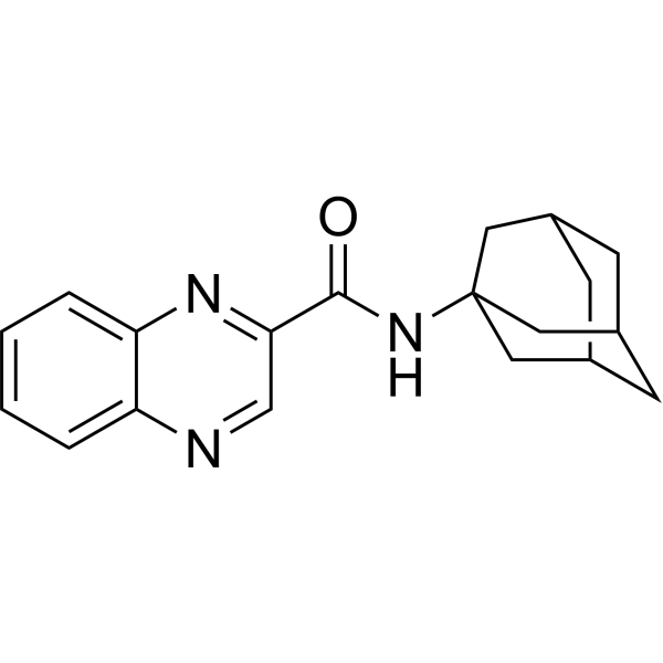
NPS 2390 is an allosteric antagonist of calcium-sensing receptor (CaSR) and mGluR1/5. NPS 2390 inhibits the PI3K/Akt/mTOR signaling pathway, reduces hypoxia-induced intracellular calcium elevation, decreases the expression of autophagy (autophagy) proteins, regulates the expression of phenotypic marker proteins, and inhibits the proliferation of pulmonary artery smooth muscle cells. NPS 2390 attenuates the endogenous apoptosis (apoptosis) pathway, increases the expression level of Bcl-2, downregulates the expression levels of Bax, cytochrome c and caspase-3, alleviates cerebral edema and improves neurological function in rat models. NPS 2390 can be used in studies related to hypoxic pulmonary hypertension, traumatic brain injury, stroke and pain.
For research use only. We do not sell to patients.
- Purity : 99.90%
- CAS No.: 226878-01-9
- Formula: C19H21N3O
- Molecular Weight:307.40
-
Storage:
4°C, sealed storage, away from moisture and light
* In solvent : -80°C, 6 months; -20°C, 1 month (sealed storage, away from moisture and light)

To place orders, for customer services and technical support, please contact: MedChemExpress USA
Tel: 609-228-6898 E-mail: [email protected] [email protected]


-
 Biological Activity
-
Chemical Information
-
Solvent & Solubility
- Protocols
- Purity & Documentation
- References
-
 Help & FAQs


-
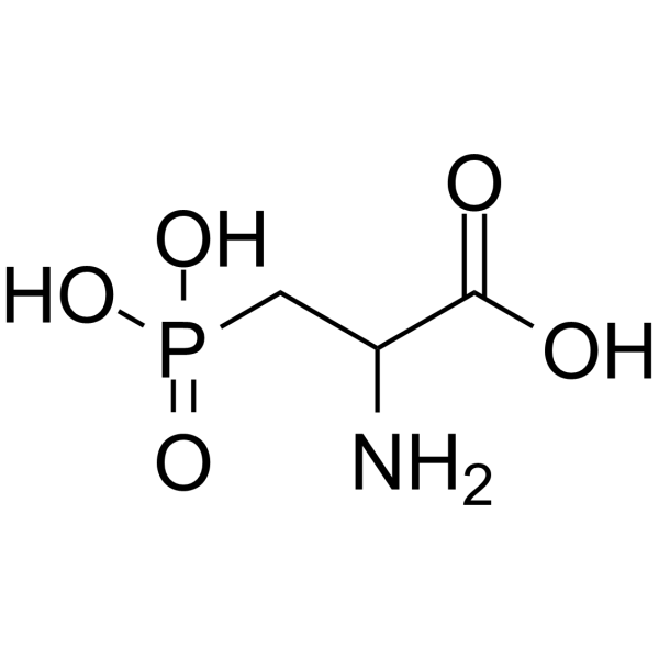
HY-100786
DL-AP3mGluR1 Antagonist -
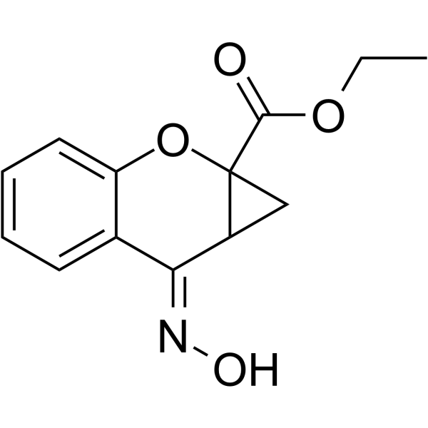
HY-101356
CPCCOEtmGluR1b Antagonist -
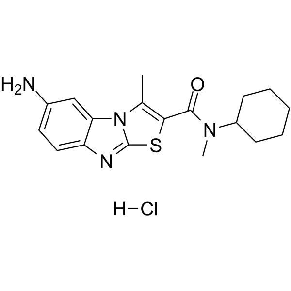
HY-103568
YM-298198 hydrochloridemGluR1 Antagonist -
HY-100405
FTIDCmGluR1 Antagonist -
HY-103566
LY456236mGlu1 Antagonist -
HY-100403
Ro 67-7476mGluR1 PAM -
HY-102095
SIB-1757mGluR5 antagonist -
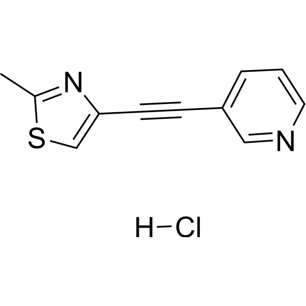
HY-13206
MTEP hydrochloridemGlu5 Antagonist -
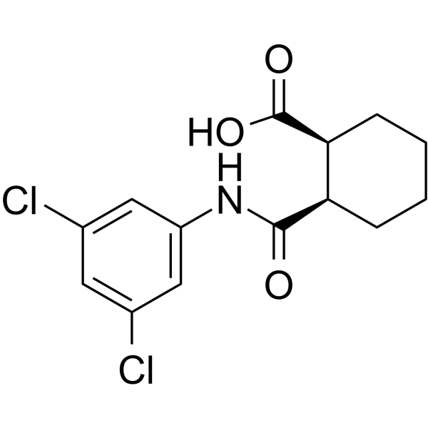
HY-14417
VU0155041mGluR4 Allosteric Agonist -
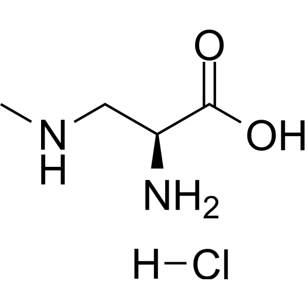
HY-W015546
β-N-methylamino-L-alanine hydrochlorideNeurotoxin -
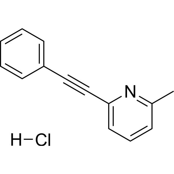
HY-14609
MPEP HydrochloridemGlu5 Receptor Antagonist -
HY-100804
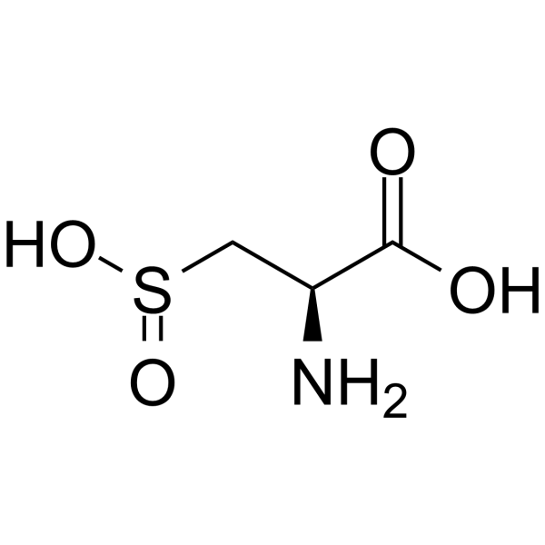
L-Cysteinesulfinic acidmGluR Agonist
All Caspase Isoforms
More

Biological Activity
Description
IC50 & Target
[3]|
mGluR1 5 nM () |
mGluR5 82 nM () |
In Vitro
NPS 2390 (10 µM; 24 h) inhibits hypoxia-induced proliferation and reverses phenotypic modulation in human pulmonary arterial smooth muscle cells by reducing autophagy levels[1].
NPS 2390 (10 µM; 24 h) suppresses hypoxia-induced increases in intracellular calcium concentration in human pulmonary arterial smooth muscle cells[1].
NPS 2390 (10 µM; 24 h) activates the PI3K/Akt/mTOR signaling pathway in hypoxic human pulmonary arterial smooth muscle cells, which mediates its autophagy-inhibiting effects[1].
NPS 2390 (multiple concentrations; 45 min) potently inhibits mGluR1 activity in rat cerebellar granule cells with an IC50 of 0.005 μM[3].
MedChemExpress (MCE) has not independently confirmed the accuracy of these methods. They are for reference only. Further protocols information, click here.
-
Cell Line:human pulmonary arterial smooth muscle cells (HPASMCs)
-
Concentration:10 µM
-
Incubation Time:24 h (under hypoxia)
-
Result:Increased the expression of phosphorylated PI3K, phosphorylated Akt, and phosphorylated mTOR, reversing the hypoxia-induced reduction in their phosphorylation levels.
In Vivo
MedChemExpress (MCE) has not independently confirmed the accuracy of these methods. They are for reference only.
-
Animal Model:Sprague Dawley (male, adult, 300-330 g, traumatic brain injury controlled cortical impact model)[2]
-
Dosage:1.5 mg/kg
-
Administration:s.c.; twice (30 min and 120 min after TBI)
-
Result:Significantly reduced brain water content compared to vehicle-treated TBI rats.
Significantly decreased modified neurological severity scores compared to vehicle-treated TBI rats.
Significantly reduced the number of TUNEL-positive neurons compared to vehicle-treated TBI rats.
Markedly diminished cleaved caspase-3 and Bax protein expression compared to vehicle-treated TBI rats.
Significantly increased Bcl-2 protein expression compared to vehicle-treated TBI rats.
Inhibited the release of cytochrome c into the cytosol compared to vehicle-treated TBI rats.


Chemical Information
-
CAS No. 226878-01-9
-
Appearance Solid
-
Molecular Weight 307.40
-
Formula C19H21N3O
-
Color White to light yellow
-
SMILES
O=C(NC1(C[C@@H]2C3)C[C@H](C2)C[C@H]3C1)C4=NC5=CC=CC=C5N=C4
-
Shipping
Room temperature in continental US; may vary elsewhere.
-
Storage
4°C, sealed storage, away from moisture and light
* In solvent : -80°C, 6 months; -20°C, 1 month (sealed storage, away from moisture and light)


Solvent & Solubility
In Vitro:
DMSO : 6.25 mg/mL (20.33 mM; ultrasonic and warming and heat to 60°C; Hygroscopic DMSO has a significant impact on the solubility of product, please use newly opened DMSO)
Please refer to the solubility information to select the appropriate solvent. Once prepared, please aliquot and store the solution to prevent product inactivation from repeated freeze-thaw cycles.
Storage method and period of stock solution: -80°C, 6 months; -20°C, 1 month (sealed storage, away from moisture and light). When stored at -80°C, please use it within 6 months. When stored at -20°C, please use it within 1 month.
Please refer to the solubility information to select the appropriate solvent. Once prepared, please aliquot and store the solution to prevent product inactivation from repeated freeze-thaw cycles.
Storage method and period of stock solution: -80°C, 6 months; -20°C, 1 month (sealed storage, away from moisture and light). When stored at -80°C, please use it within 6 months. When stored at -20°C, please use it within 1 month.
Concentration (start) × Volume (start) = Concentration (final) × Volume (final)

Protocols
-
RNA extraction experimental
By lysing cells, releasing RNA, and removing impurities such as proteins and DNA, high-purity RNA products are finally obtained. The commonly used traditional method is the guanidine isothiocyanate/phenol/chloroform method (Trizol), which is suitable for a variety of animal materials including animal tissues, microorganisms, cultured cells, etc., and most plant materials.
-
Apoptosis
Apoptosis, also called programmed cell death, is generally characterized by distinct morphological characteristics.
-
TUNEL staining for apoptotic DNA fragmentation
TUNEL staining detects DNA strand breaks by using terminal deoxynucleotidyl transferase to add labeled nucleotides to exposed 3′-OH DNA termini, generating either microscopic staining in fixed cells or tissue sections, or fluorescence/cytometric signal in cell suspensions. TUNEL positivity reflects DNA fragmentation but should not be interpreted alone as definitive apoptosis, because TUNEL can also label necrotic, autolytic, mechanically damaged, or DNA-repair-associated DNA breaks.
-
Annexin V plus membrane-impermeant dye apoptosis staining
Annexin V-based apoptosis assays rely on the detection of phosphatidylserine (PS) externalization from the inner leaflet of the plasma membrane to the outer leaflet, an early biochemical hallmark of apoptosis. Fluorescently labeled Annexin V binds PS in a calcium-dependent manner, enabling identification of early apoptotic cells by flow cytometry or fluorescence microscopy. When combined with a membrane-impermeant DNA-binding dye (e. g. , propidium iodide), this approach allows discrimination between viable (Annexin V−/dye−), early apoptotic (Annexin V+/dye−), and late apoptotic or necrotic (Annexin V+/dye+) cell populations by assessing membrane integrity and PS exposure.
-
Autophagy
Autophagy is a process in which eukaryotic cells use lysosomes to degrade their own cytoplasmic proteins and damaged organelles under the regulation of autophagy related gene (Atg). Microtubule-associated proteins light chain 3 (LC3) is recognized as autophagy marker, which transfers from cytoplasmic LC3 (LC3-I) to membrane type (LC3-II). LC3-II/I ratio could be detected by Western Blot and fluorescence microscopy.
-
Calcium Spark Assay
Calcium sparks are localized, transient increases in intracellular calcium concentration ([Ca2+]i) that occur in cardiac myocytes and represent elementary events underlying excitation-contraction coupling. These events are generated by the coordinated opening of clusters of ryanodine receptors (RyRs) on the sarcoplasmic reticulum membrane, leading to a brief release of Ca2+ into the cytosol. The detection and analysis of calcium sparks provide insights into the mechanisms of calcium handling and signaling in cardiac cells. Imaging techniques using fluorescent calcium indicators such as Fluo-3 are employed to visualize these subcellular calcium transients with high spatial and temporal resolution. The protocol is based on established methodologies described in primary literature for both experimental measurement and automated analysis of calcium sparks.
-
Apoptosis Solutions
Apoptosis is a regulated, generally non-lytic cell-death pathway that removes unwanted, damaged, infected, or abnormal cells through coordinated morphological changes, caspase activation, DNA fragmentation, and membrane remodeling. The intrinsic apoptosis pathway is controlled mainly by mitochondrial outer membrane permeabilization, BCL-2 family proteins, cytochrome c release, apoptosome formation, caspase-9 activation, and downstream executioner caspase-3/7 activation. The extrinsic apoptosis pathway is initiated by death receptors such as Fas, TNFR, and TRAIL receptors, which recruit adaptor proteins and activate caspase-8 before engaging executioner caspases or mitochondrial amplification through BID cleavage. Apoptosis is linked to many phenotypes, including cancer cell killing, tissue homeostasis, immune regulation, neurodegeneration, infection response, and treatment-induced cytotoxicity; unresolved questions include how apoptosis interacts with necroptosis, pyroptosis, ferroptos
-
Research Protocol for Cardiovascular Diseases
Cardiovascular disease can be modeled as maladaptive cardiac remodeling, where ischemic injury or pressure overload activates inflammatory signaling, fibroblast activation, extracellular-matrix deposition, cardiomyocyte hypertrophy, vascular remodeling, and progressive ventricular dysfunction. The TGF-β/SMAD axis is a central profibrotic pathway after myocardial injury and pressure overload, while innate immune and cytokine pathways regulate leukocyte recruitment, scar formation, and adverse remodeling. Key unresolved questions include which inflammatory signals are reparative versus harmful, when fibrosis is protective versus maladaptive, and whether pathway inhibition improves function without weakening necessary infarct healing or compensatory remodeling.
-
Ca2+ Staining Technique
Ca2+ staining is an experimental technique that utilizes specific fluorescent probes (such as Fluo-4 AM, Fura-2, etc.) to qualitatively or quantitatively detect dynamic changes in intracellular Ca2+ concentrations; this is achieved by monitoring the changes in fluorescent signals generated when these probes bind to free intracellular calcium ions. The underlying principle relies primarily on the presence of chelating groups within the probe's molecular structure that possess high affinity for calcium ions.
-
Lysosome and acidic-vesicle live-cell staining
Lysosome and acidic-vesicle live-cell staining detects acidic intracellular compartments by using membrane-permeant acidotropic probes that accumulate in low-pH vesicles, including lysosomes, late endosomes, autolysosomes, and acidic phagosomes. LysoTracker staining is commonly used as an intensity-based readout of acidic lysosomal compartment abundance or enlargement, while acridine orange produces green fluorescence in less concentrated compartments and red fluorescence after concentration-dependent accumulation in acidic vesicular organelles. Loss or reduction of acridine-orange red signal can be used as a readout of lysosomal membrane permeabilization or reduced acidic-vesicle integrity. This protocol is designed for live cultured cells and can be adapted for fluorescence microscopy, high-content imaging, plate-reader readout, or flow cytometry when the selected literature supports the readout. Because these dyes report acidotropic accumulation rather than lysosome identity alone,
-
Macroautophagy Solutions
Macroautophagy is a conserved lysosome-dependent degradation pathway in which cytoplasmic material is sequestered into double-membrane autophagosomes and delivered to lysosomes for degradation and recycling. The pathway supports cellular homeostasis during nutrient limitation, organelle stress, protein-aggregate accumulation, infection, differentiation, and tissue remodeling by coupling cargo sequestration, autophagosome maturation, lysosomal fusion, and degradation of cargo-derived macromolecules. The core molecular sequence includes initiation by nutrient- and stress-regulated autophagy machinery, autophagosome nucleation, LC3/ATG8-family conjugation to autophagosomal membranes, cargo selection through receptors such as SQSTM1/p62, autophagosome-lysosome fusion, and lysosomal degradation. LC3 was identified as a mammalian homolog of yeast Atg8 that localizes to autophagosomal membranes after processing, and p62/SQSTM1 was shown to connect ubiquitinated cargo with autophagic degradati
-
Cell Viability Determination by MTT Colorimetric Assay
The following protocol uses the MTT colorimetric assay as a classic literature-established method for assessing cell viability/metabolic activity in cultured mammalian cells. MTT[3-(4,5-dimethylthiazol-2-yl)-2,5-diphenyltetrazolium bromide] is reduced by metabolically active cells to a colored formazan product; the amount of formazan is quantified spectrophotometrically and provides an indirect measure of metabolically active viable cells. Importantly, MTT reduction reflects cellular oxidoreductase/metabolic activity rather than an absolute direct count of living cells, so changes in cellular metabolism can alter the signal independently of cell number.


Purity & Documentation
-
Data Sheet (277 KB)
-
SDS (394 KB)
- English - EN (394 KB)
- Français - FR (394 KB)
- Deutsch - DE (394 KB)
- Norwegian - NO (394 KB)
- Español - ES (394 KB)
- Swedish - SV (394 KB)
- Italian - IT (394 KB)
- Korean - KR (394 KB)
- Portuguese - PT (394 KB)
-
Handling Instructions (2659 KB)
References
[1]. Peng X, et al. NPS2390, a Selective Calcium-sensing Receptor Antagonist Controls the Phenotypic Modulation of Hypoxic Human Pulmonary Arterial Smooth Muscle Cells by Regulating Autophagy. J Transl Int Med. 2019;7(2):59-68. Published 2019 Jul 11. [Content Brief]
[2]. Xue Z, et al. Calcium-sensing receptor antagonist NPS2390 attenuates neuronal apoptosis though intrinsic pathway following traumatic brain injury in rats. Biochem Biophys Res Commun. 2017;486(2):589-594. [Content Brief]
[3]. Vanejevs M, et al. Positive and negative modulation of group I metabotropic glutamate receptors. J Med Chem. 2008 Feb 14;51(3):634-47. [Content Brief]


Complete Stock Solution Preparation Table
Please refer to the solubility information to select the appropriate solvent. Once prepared, please aliquot and store the solution to prevent product inactivation from repeated freeze-thaw cycles.
Storage method and period of stock solution: -80°C, 6 months; -20°C, 1 month (sealed storage, away from moisture and light). When stored at -80°C, please use it within 6 months. When stored at -20°C, please use it within 1 month.
| Optional Solvent | Concentration Solvent Mass | 1 mg | 5 mg | 10 mg | 25 mg |
|---|---|---|---|---|---|
| DMSO | 1 mM | 3.2531 mL | 16.2655 mL | 32.5309 mL | 81.3273 mL |
| 5 mM | 0.6506 mL | 3.2531 mL | 6.5062 mL | 16.2655 mL | |
| 10 mM | 0.3253 mL | 1.6265 mL | 3.2531 mL | 8.1327 mL | |
| 15 mM | 0.2169 mL | 1.0844 mL | 2.1687 mL | 5.4218 mL | |
| 20 mM | 0.1627 mL | 0.8133 mL | 1.6265 mL | 4.0664 mL |


NPS 2390 Related Classifications
HY-11095 Related Classifications
 Powered by Bioz
Keywords
- NPS 2390
- 226878-01-9
- NPS2390
- NPS-2390
- mGluR
- CaSR
- PI3K
- Akt
- mTOR
- Autophagy
- Apoptosis
- Bcl-2 Family
- Caspase
- metabotropic glutamate receptor 1
- pulmonary arterial smooth muscle cells
- stroke
- rat models
- rat cerebellar granule cells
- traumatic brain injury
- PI3K/Akt/mTOR signaling
- hypoxic pulmonary hypertension
- calcium-sensing receptor
- intrinsic apoptotic pathway
- Inhibitor
- inhibitor
- inhibit