



Solanesol
Based on 2 publication(s) in Google Scholar
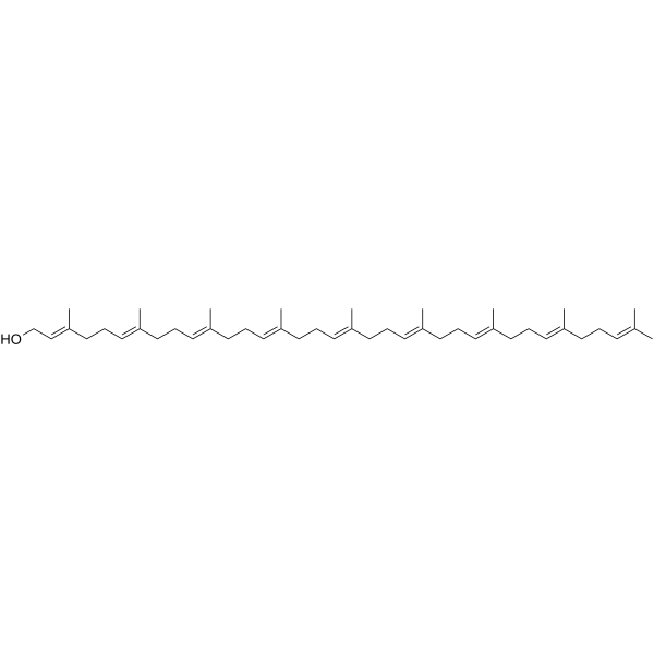
Solanesol is an orally active aliphatic terpene alcohol. Solanesol is mainly found in tobacco and other Solanaceae plants. Solanesol induces HO-1 and Hsp70 expression, activates p38 and Akt signaling pathways, and inhibits Apoptosis (reduces caspase-3 and PARP cleavage). Solanesol has antioxidant, anti-inflammatory, and neuroprotective activities. Solanesol can be used in the research of Huntington's disease, alcoholic liver disease, chronic inflammatory pain, anxiety, Alzheimer's disease, and bipolar disorder.
For research use only. We do not sell to patients.
- Purity : 98.0%
- CAS No.: 13190-97-1
- Formula: C45H74O
- Molecular Weight:631.07
-
Storage:Powder -20°C, 3 years ; In solvent -80°C, 6 months , -20°C, 1 month

To place orders, for customer services and technical support, please contact: MedChemExpress USA
Tel: 609-228-6898 E-mail: [email protected] [email protected]


-
 Biological Activity
-
Chemical Information
-
Solvent & Solubility
- Protocols
- Purity & Documentation
- References
-
 Help & FAQs


-

HY-18739
Phorbol 12-myristate 13-acetatePKC Activator -
HY-13434
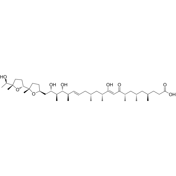
IonomycinCalcium Ionophore -
HY-112251
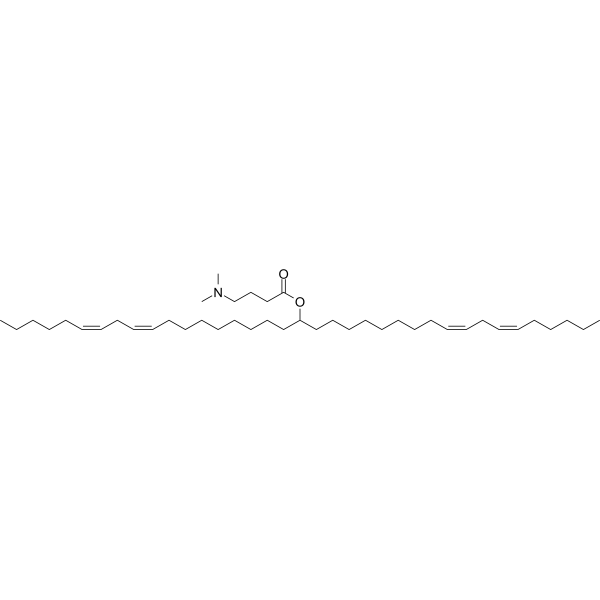
D-Lin-MC3-DMAsiRNA Delivery Vehicle -

HY-N0576R
Solanesol (Standard)Standard Of Solanesol -
HY-15484
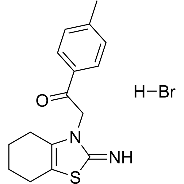
Pifithrin-α hydrobromidep53 Inhibitor -
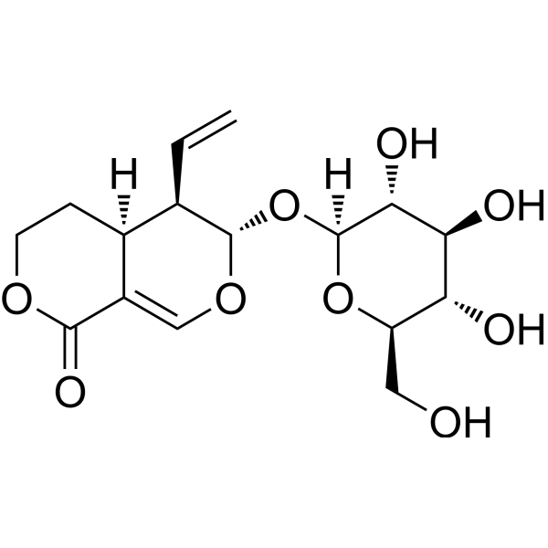
HY-N0806
SwerosideKeap1/Nrf2 Activator -
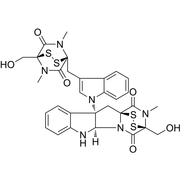
HY-107553
ChetominHsp90/HIF1α Inhibitor -
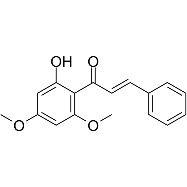
HY-N2132
Flavokawain BAnti-tumor Agent -

HY-N0806R
Sweroside (Standard)Keap1/Nrf2 Activator -

HY-N2132R
Flavokawain B (Standard)Anti-tumor Agent -

HY-15484R
Pifithrin-α hydrobromide (Standard)p53 Inhibitor
Publications Citing Use of MedChemExpress (MCE) Solanesol
More Customer Validation & Images

-
WB

-
IF

All Endogenous Metabolite Isoforms
MoreAll Caspase Isoforms
More

Biological Activity
Description
|
Caspase-3 |
HSP70 |
HO-1 |
In Vitro
Solanesol (40 μM; 0-24 h) induces HO-1 mRNA and protein expression in RAW264.7 cells[2].
Solanesol (10-80 μM; 12–24 h) upregulates HO-1 and Hsp70 expression in human hepatic L02 cells, protecting against ethanol-induced oxidative injury by enhancing Nrf2 and HSF1 nuclear translocation[4].
MedChemExpress (MCE) has not independently confirmed the accuracy of these methods. They are for reference only. Further protocols information, click here.
-
Cell Line:RAW264.7 murine macrophage-like cells
-
Concentration:5 μM, 10 μM, 20 μM, 40 μM
-
Incubation Time:12 h
-
Result:Enhanced phosphorylated p38 and Akt levels.
-
Cell Line:RAW264.7 murine macrophage-like cells
-
Concentration:80 μM
-
Incubation Time:12 h
-
Result:Inhibited apoptosis by decreasing caspase-3 and PARP cleavage.
In Vivo
Solanesol (50 mg/kg, i.p.) alleviates CFA (HY-153808)-induced chronic inflammatory pain in C57BL/6 mice, reducing mechanical and thermal hypersensitivity and suppressing spinal glial cell activation[5].
Solanesol (50 mg/kg; i.p.; 1 week) ameliorates CFA-induced anxiety-like behaviors in mice by downregulating TIA1, inhibiting the expression of pro-inflammatory factors IL-1β and TNF-α, and reducing microglial/astrocytic activation in the anterior cingulate cortex[6].
Solanesol (40-80 mg/kg, p.o.) activates SIRT-1 signaling in Ouabain (HY-B1457)-induced bipolar disorder rats, improving neurobehavioral deficits and restoring Na+/K+-ATPase activity[8].
MedChemExpress (MCE) has not independently confirmed the accuracy of these methods. They are for reference only.
-
Animal Model:Adult C57BL/6 mice (6-8 weeks old, 15-25 g), CFA-induced chronic inflammatory pain model[5]
-
Dosage:50 mg/kg
-
Administration:Intraperitoneal injection
-
Result:Reduced pro-inflammatory cytokines (TNF-α, IL-1β) in spinal cord.
Suppressed microglial and astrocytic activation (Iba-1 and S100β).
Improved mechanical allodynia and thermal hyperalgesia.


Chemical Information
-
CAS No. 13190-97-1
-
Appearance Solid
-
Molecular Weight 631.07
-
Formula C45H74O
-
Color Off-white to light yellow
-
SMILES
C/C(C)=C\CC/C(C)=C/CC/C(C)=C/CC/C(C)=C/CC/C(C)=C/CC/C(C)=C/CC/C(C)=C/CC/C(C)=C/CC/C(C)=C/CO
-
Structure Classification
-
Initial Source
-
Shipping
Room temperature in continental US; may vary elsewhere.
-
Storage
Powder -20°C 3 years In solvent -80°C 6 months -20°C 1 month


Publications (2)
-
Journal Impact Factor
-
Most Recent
-
Vet Microbiol
The Chinese medicine monomer Schisandrin C inhibits PRRSV infection by regulating the OGT-PI3K/AKT/mTOR signaling pathway. [Abstract]2026 May:316:110992. PMID: 41865607 -
Heliyon
Solanesol alleviates CFA-induced chronic inflammatory pain via inhibition of proinflammatory cytokines in spinal glial cells. [Abstract]Heliyon. 2024 Jul 18;10(15):e34870. PMID: 39157324
Solanesol purchased from MedChemExpress. Usage Cited in: Heliyon. Heliyon. 2024 Jul 18;10(15):e34870. [Abstract]
Typical imaging of proinflammatory cytokines TNF-α and IL-1β treated with Solanesol (Sol) (50 mg/kg).
Solanesol purchased from MedChemExpress. Usage Cited in: Heliyon. Heliyon. 2024 Jul 18;10(15):e34870. [Abstract]
Representative images for Iba-1 and S100βin the spinal cord after CFA injection, and Solanesol (Sol) (50 mg/kg) administration suppressed the enhanced activation of Iba-1 and S100β.


Solvent & Solubility
In Vitro:
DMSO : 100 mg/mL (158.46 mM; Need ultrasonic; Hygroscopic DMSO has a significant impact on the solubility of product, please use newly opened DMSO)
Please refer to the solubility information to select the appropriate solvent. Once prepared, please aliquot and store the solution to prevent product inactivation from repeated freeze-thaw cycles.
Storage method and period of stock solution: -80°C, 6 months; -20°C, 1 month. When stored at -80°C, please use it within 6 months. When stored at -20°C, please use it within 1 month.
Please refer to the solubility information to select the appropriate solvent. Once prepared, please aliquot and store the solution to prevent product inactivation from repeated freeze-thaw cycles.
Storage method and period of stock solution: -80°C, 6 months; -20°C, 1 month. When stored at -80°C, please use it within 6 months. When stored at -20°C, please use it within 1 month.
Concentration (start) × Volume (start) = Concentration (final) × Volume (final)
In Vivo:
Select the appropriate dissolution method based on your experimental animal and administration route.
- For the following dissolution methods, please ensure to first prepare a clear stock solution using an In Vitro approach and then sequentially add co-solvents:
- To ensure reliable experimental results, the clarified stock solution can be appropriately stored based on storage conditions. As for the working solution for In Vivo experiments, it is recommended to prepare freshly and use it on the same day.
- The percentages shown for the solvents indicate their volumetric ratio in the final prepared solution. If precipitation or phase separation occurs during preparation, heat and/or sonication can be used to aid dissolution.
Add each solvent one by one: 10% DMSO 40% PEG300 5% Tween-80 45% Saline
Solubility: 2.5 mg/mL (3.96 mM); Suspended solution; Need ultrasonic
This protocol yields a suspended solution of 2.5 mg/mL. Suspended solution can be used for oral and intraperitoneal injection.
Taking 1 mL working solution as an example, add 100 μL DMSO stock solution (25.0 mg/mL) to 400 μL PEG300, and mix evenly; then add 50 μL Tween-80 and mix evenly; then add 450 μL Saline to adjust the volume to 1 mL.
Preparation of Saline: Dissolve 0.9 g sodium chloride in ddH₂O and dilute to 100 mL to obtain a clear Saline solution.
Add each solvent one by one: 10% DMSO 90% (20% SBE-β-CD in Saline)
Solubility: 2.5 mg/mL (3.96 mM); Suspended solution; Need ultrasonic
This protocol yields a suspended solution of 2.5 mg/mL. Suspended solution can be used for oral and intraperitoneal injection.
Taking 1 mL working solution as an example, add 100 μL DMSO stock solution (25.0 mg/mL) to 900 μL 20% SBE-β-CD in Saline, and mix evenly.
Preparation of 20% SBE-β-CD in Saline (4°C, storage for one week): 2 g SBE-β-CD powder is dissolved in 10 mL Saline, completely dissolve until clear.
In Vivo Dissolution Calculator
Please enter the basic information of animal experiments:
-
-
-
-
Recommended: Prepare an additional quantity of animals to account for potential losses during experiments.
Please enter your animal formula composition:
-
%DMSO +
Recommended: Keep the proportion of DMSO in working solution below 2% if your animal is weak.
-
%+
-
+%Tween-80 + +
-
%Saline +
The co-solvents required include: DMSO, . All of co-solvents are available by MedChemExpress (MCE). , Tween 80. All of co-solvents are available by MedChemExpress (MCE).
Working solution concentration: 0.22 mg/mL
Method for preparing stock solution: mg drug dissolved in μL DMSO. Stock solution concentration: mg/mL.
1. Take μL DMSO stock solution;
2. Add μL .
μL , mix evenly;
3. Then add μL Tween 80, mix evenly;
4. Then add μL
Please ensure that the stock solution in the first step is dissolved to a clear state, and add co-solvents in sequence. You can use ultrasonic heating (ultrasonic cleaner, recommended frequency 20-40 kHz), vortexing, etc. to assist dissolution.

Protocols
-
RNA extraction experimental
By lysing cells, releasing RNA, and removing impurities such as proteins and DNA, high-purity RNA products are finally obtained. The commonly used traditional method is the guanidine isothiocyanate/phenol/chloroform method (Trizol), which is suitable for a variety of animal materials including animal tissues, microorganisms, cultured cells, etc., and most plant materials.
-
Apoptosis
Apoptosis, also called programmed cell death, is generally characterized by distinct morphological characteristics.
-
TUNEL staining for apoptotic DNA fragmentation
TUNEL staining detects DNA strand breaks by using terminal deoxynucleotidyl transferase to add labeled nucleotides to exposed 3′-OH DNA termini, generating either microscopic staining in fixed cells or tissue sections, or fluorescence/cytometric signal in cell suspensions. TUNEL positivity reflects DNA fragmentation but should not be interpreted alone as definitive apoptosis, because TUNEL can also label necrotic, autolytic, mechanically damaged, or DNA-repair-associated DNA breaks.
-
Annexin V plus membrane-impermeant dye apoptosis staining
Annexin V-based apoptosis assays rely on the detection of phosphatidylserine (PS) externalization from the inner leaflet of the plasma membrane to the outer leaflet, an early biochemical hallmark of apoptosis. Fluorescently labeled Annexin V binds PS in a calcium-dependent manner, enabling identification of early apoptotic cells by flow cytometry or fluorescence microscopy. When combined with a membrane-impermeant DNA-binding dye (e. g. , propidium iodide), this approach allows discrimination between viable (Annexin V−/dye−), early apoptotic (Annexin V+/dye−), and late apoptotic or necrotic (Annexin V+/dye+) cell populations by assessing membrane integrity and PS exposure.
-
Apoptosis Solutions
Apoptosis is a regulated, generally non-lytic cell-death pathway that removes unwanted, damaged, infected, or abnormal cells through coordinated morphological changes, caspase activation, DNA fragmentation, and membrane remodeling. The intrinsic apoptosis pathway is controlled mainly by mitochondrial outer membrane permeabilization, BCL-2 family proteins, cytochrome c release, apoptosome formation, caspase-9 activation, and downstream executioner caspase-3/7 activation. The extrinsic apoptosis pathway is initiated by death receptors such as Fas, TNFR, and TRAIL receptors, which recruit adaptor proteins and activate caspase-8 before engaging executioner caspases or mitochondrial amplification through BID cleavage. Apoptosis is linked to many phenotypes, including cancer cell killing, tissue homeostasis, immune regulation, neurodegeneration, infection response, and treatment-induced cytotoxicity; unresolved questions include how apoptosis interacts with necroptosis, pyroptosis, ferroptos
-
Formalin-Induced Paw Inflammation/Nociceptive Inflammation
The formalin-induced paw inflammation/nociceptive test is a chemical persistent pain model in rodents in which subcutaneous injection of formalin into the hind paw produces spontaneous nocifensive behaviors such as flinching and licking. The response is classically biphasic, consisting of an early acute phase (Phase I) reflecting direct activation of peripheral nociceptors (particularly C-fiber afferents), followed by a later prolonged phase (Phase II) associated with central sensitization in the spinal dorsal horn driven by sustained afferent input and inflammatory signaling. This model is widely used to evaluate analgesic and anti-inflammatory interventions because it captures both peripheral nociception and central sensitization processes within a single assay system.
-
Research Protocol for Inflammation-related Diseases
The NLRP3 inflammasome is a cytosolic innate immune signaling platform that integrates priming signals and danger-signal activation to promote caspase-1 activation, maturation of IL-1β and IL-18, and gasdermin D-mediated pyroptotic cell death. The core experimental logic is to determine whether inflammatory disease phenotypes are driven by increased NLRP3 expression, ASC-containing inflammasome assembly, caspase-1 cleavage, GSDMD cleavage, and extracellular release of IL-1β/IL-18 rather than by nonspecific cell injury alone. The pathway is strongly linked to inflammation-related disease phenotypes because monosodium urate crystals activate NALP3/NLRP3 inflammasome signaling in gout-like crystal inflammation, cholesterol crystals activate NLRP3 inflammasomes in atherogenesis models, and DSS-induced intestinal inflammation has been reported to involve NLRP3 inflammasome activity. However, experimental colitis studies also show context-dependent protective effects of NLRP3 inflammasome co
-
Alzheimer’s Disease Modeling
Alzheimer’s Disease (AD) is a neurodegenerative disorder characterized by a progressive decline in cognitive functions and loss of specific types of neurons and synapses. Alzheimer's symptoms can be simulated in mice by injecting drugs (such as Aβ) or genetically modified.


Purity & Documentation
-
Data Sheet (274 KB)
-
SDS (394 KB)
- English - EN (394 KB)
- Français - FR (394 KB)
- Deutsch - DE (394 KB)
- Norwegian - NO (394 KB)
- Español - ES (394 KB)
- Swedish - SV (394 KB)
- Italian - IT (394 KB)
- Korean - KR (394 KB)
- Portuguese - PT (394 KB)
-
Handling Instructions (2659 KB)
References
[1]. Yan N, et al. Bioactivities and Medicinal Value of Solanesol and Its Accumulation, Extraction Technology, and Determination Methods. Biomolecules. 2019 Aug 2;9(8):334. [Content Brief]
[2]. Yao X, et al. Solanesol induces the expression of heme oxygenase-1 via p38 and Akt and suppresses the production of proinflammatory cytokines in RAW264.7 cells. Food Funct. 2017 Jan 25;8(1):132-141. [Content Brief]
[3]. Mehan S, et al. Neuroprotective effect of solanesol against 3-nitropropionic acid-induced Huntington's disease-like behavioral, biochemical, and cellular alterations: Restoration of coenzyme-Q10-mediated mitochondrial dysfunction. Indian J Pharmacol. 2018 Nov-Dec;50(6):309-319. [Content Brief]
[4]. Yao X, et al. Solanesol protects human hepatic L02 cells from ethanol-induced oxidative injury via upregulation of HO-1 and Hsp70. Toxicol In Vitro. 2015 Apr;29(3):600-8. [Content Brief]
[5]. Wang YY, et al. Solanesol alleviates CFA-induced chronic inflammatory pain via inhibition of proinflammatory cytokines in spinal glial cells. Heliyon. 2024 Jul 18;10(15):e34870. [Content Brief]
[6]. Ding S, Li Y, et al. Solanesol Ameliorates Anxiety-like Behaviors via the Downregulation of Cingulate T Cell-Restricted Intracellular Antigen-1 in a Complete Freund's Adjuvant-Induced Mouse Model. Int J Mol Sci. 2024 Sep 21;25(18):10165. [Content Brief]
[7]. Wang C, et al. Natural Bioactive Compounds Solanesol and Chlorogenic Acid Assembled Nanomicelles for Alzheimer's Disease Therapy. ACS Appl Mater Interfaces. 2025 Mar 5;17(9):14591-14603. [Content Brief]
[8]. Rajkhowa B, et al. Activating SIRT-1 Signalling with the Mitochondrial-CoQ10 Activator Solanesol Improves Neurobehavioral and Neurochemical Defects in Ouabain-Induced Experimental Model of Bipolar Disorder. Pharmaceuticals (Basel). 2022 Aug 2;15(8):959. [Content Brief]


Complete Stock Solution Preparation Table
Please refer to the solubility information to select the appropriate solvent. Once prepared, please aliquot and store the solution to prevent product inactivation from repeated freeze-thaw cycles.
Storage method and period of stock solution: -80°C, 6 months; -20°C, 1 month. When stored at -80°C, please use it within 6 months. When stored at -20°C, please use it within 1 month.
| Optional Solvent | Concentration Solvent Mass | 1 mg | 5 mg | 10 mg | 25 mg |
|---|---|---|---|---|---|
| DMSO | 1 mM | 1.5846 mL | 7.9231 mL | 15.8461 mL | 39.6153 mL |
| 5 mM | 0.3169 mL | 1.5846 mL | 3.1692 mL | 7.9231 mL | |
| 10 mM | 0.1585 mL | 0.7923 mL | 1.5846 mL | 3.9615 mL | |
| 15 mM | 0.1056 mL | 0.5282 mL | 1.0564 mL | 2.6410 mL | |
| 20 mM | 0.0792 mL | 0.3962 mL | 0.7923 mL | 1.9808 mL | |
| 25 mM | 0.0634 mL | 0.3169 mL | 0.6338 mL | 1.5846 mL | |
| 30 mM | 0.0528 mL | 0.2641 mL | 0.5282 mL | 1.3205 mL | |
| 40 mM | 0.0396 mL | 0.1981 mL | 0.3962 mL | 0.9904 mL | |
| 50 mM | 0.0317 mL | 0.1585 mL | 0.3169 mL | 0.7923 mL | |
| 60 mM | 0.0264 mL | 0.1321 mL | 0.2641 mL | 0.6603 mL | |
| 80 mM | 0.0198 mL | 0.0990 mL | 0.1981 mL | 0.4952 mL | |
| 100 mM | 0.0158 mL | 0.0792 mL | 0.1585 mL | 0.3962 mL |


Solanesol Related Classifications
HY-N0576 Related Classifications
 Powered by Bioz