



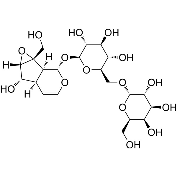
Rehmannioside A
Based on 1 publication(s) in Google Scholar
Rehmannioside A is a compound that can be isolated from Rehmanniae radix. Rehmannioside A is an inhibitor of CYP3A4, 2C9 and 2D6, with IC50 values of 10.08, 12.62 and 16.43 μM, respectively. Rehmannioside A has anti-inflammatory, antioxidant, anti-apoptosis, anti-ferroptosis, cognitive improvement and neuroprotective activities. Rehmannioside A can be used for the research of nervous system and inflammation-related diseases.
For research use only. We do not sell to patients.
- Purity : 99.95%
- CAS No.: 81720-05-0
- Formula: C21H32O15
- Molecular Weight:524.47
-
Storage:Powder -20°C, 3 years , 4°C, 2 years ; In solvent -80°C, 6 months , -20°C, 1 month

To place orders, for customer services and technical support, please contact: MedChemExpress USA
Tel: 609-228-6898 E-mail: [email protected] [email protected]


-
 Biological Activity
-
Chemical Information
-
Solvent & Solubility
- Protocols
- Purity & Documentation
- References
-
 Help & FAQs


-
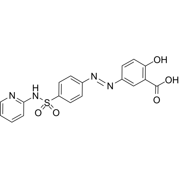
HY-14655
SulfasalazineAnti-rheumatic Agent -
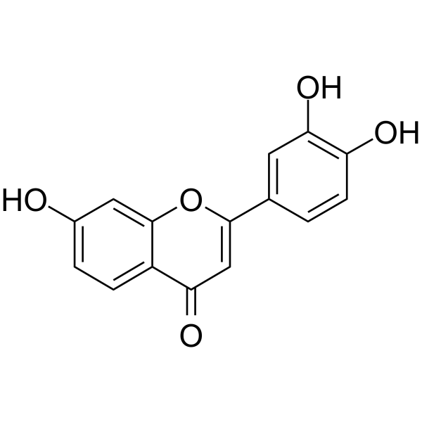
HY-N2736
3′,4′,7-Trihydroxyflavoneflavonoid aglycon compound -
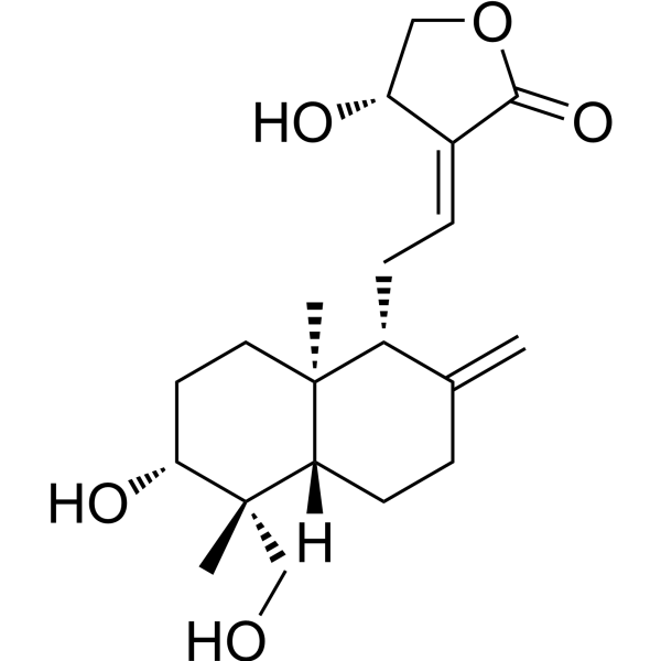
HY-N0191
AndrographolideNF-κB Inhibitor -

HY-14655R
Sulfasalazine (Standard)Anti-rheumatic Agent -
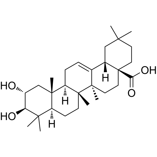
HY-N0629
Maslinic acidBacterial Inhibitor -

HY-N0191R
Andrographolide (Standard)NF-κB Inhibitor -
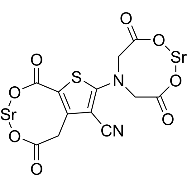
HY-17397
Strontium RanelateAntiosteoporotic Agent -

HY-N0629R
Maslinic acid (Standard)Bacterial Inhibitor -

HY-17397R
Strontium Ranelate (Standard)Antiosteoporotic Agent
Publications Citing Use of MedChemExpress (MCE) Rehmannioside A
MoreAll MEK Isoforms
More

Biological Activity
Description
|
CYP2C9 12.62 μM (IC50) |
CYP3A4 10.08 μM (IC50) |
CYP2D6 16.43 μM (IC50) |
iNOS |
Bcl-2 |
COX-2 |
Cellular Effect
|
Cell Line

|
Type | Value | Description | References |
|---|---|---|---|---|
| A2780 | IC50 |
>10 μM
Compound: 16
|
Cytotoxicity against human A2780 cells by MTT assay
Cytotoxicity against human A2780 cells by MTT assay
|
[PMID: 26859776] |
| A549 | IC50 |
>10 μM
Compound: 16
|
Cytotoxicity against human A549 cells by MTT assay
Cytotoxicity against human A549 cells by MTT assay
|
[PMID: 26859776] |
| Bel-7402 | IC50 |
>10 μM
Compound: 16
|
Cytotoxicity against human Bel7402 cells by MTT assay
Cytotoxicity against human Bel7402 cells by MTT assay
|
[PMID: 26859776] |
| BGC-823 | IC50 |
>10 μM
Compound: 16
|
Cytotoxicity against human BGC823 cells by MTT assay
Cytotoxicity against human BGC823 cells by MTT assay
|
[PMID: 26859776] |
| HT-29 | IC50 |
>10 μM
Compound: 16
|
Cytotoxicity against human HT-29 cells by MTT assay
Cytotoxicity against human HT-29 cells by MTT assay
|
[PMID: 26859776] |
In Vitro
Rehmannioside A (0-100 μM; 24 h) can improve high-glucose-induced decrease in cell viability and inhibit apoptosis and oxidative stress in HK2 cells (increasing the activities of SOD and CAT, reducing the levels of MDA, and decreasing LDH release and ROS production). The mechanism is involved in the inhibition of p38 MAPK and ERK1/2 phosphorylation[1].
Rehmannioside A (0-80 μM; 48 h) inhibits the release of pro-inflammatory mediators and promotes M2 polarization in LPS (HY-D1056)-treated BV2 cells. The mechanism is related to the inhibition of NF-κB and MEK signaling pathways[2].
Rehmannioside A (80 μM; 48 h) reduces neuronal apoptosis and restores the expression of anti-apoptotic protein Bcl-2 in the co-culture system of PC12 and BV2 cells[2].
MedChemExpress (MCE) has not independently confirmed the accuracy of these methods. They are for reference only. Further protocols information, click here.
-
Cell Line:HK2 cells treated high-glucose
-
Concentration:20, 40 and 80 μM
-
Incubation Time:24 h
-
Result:Significantly inhibited the levels of BAX and cleaved PARP proteins and increased Bcl-2 levels.
-
Cell Line:BV2 cells treated LPS (HY-D1056)
-
Concentration:0, 20, 40 and 80 μM
-
Incubation Time:Pretreated with 24 h, then co-incubation for 24 h
-
Result:Inhibited the levels of iNOS and COX-2.
In Vivo
MedChemExpress (MCE) has not independently confirmed the accuracy of these methods. They are for reference only.
-
Animal Model:Female SD rats (200-250 g) with spinal cord injury[2]
-
Dosage:80 mg/kg
-
Administration:Intraperitoneal injection; 28 days
-
Result:Significantly improved the behavioural and histological indices.
Promoted M2 microglial polarization.
Alleviated neuronal apoptosis.
Increased motor function recovery.


Chemical Information
-
CAS No. 81720-05-0
-
Appearance Solid
-
Molecular Weight 524.47
-
Formula C21H32O15
-
Color White to off-white
-
SMILES
OC[C@@]1([C@]([C@@H]2O[C@@H]([C@@H]([C@H]3O)O)O[C@@H]([C@H]3O)CO[C@H]([C@@H]([C@H]4O)O)O[C@@H]([C@@H]4O)CO)([H])[C@](C=CO2)([H])[C@@H]5O)[C@@]5([H])O1
-
Structure Classification
-
Shipping
Room temperature in continental US; may vary elsewhere.
-
Storage
Powder -20°C 3 years 4°C 2 years In solvent -80°C 6 months -20°C 1 month


Publications (1)
-
Journal Impact Factor
-
Most Recent
-
J Ethnopharmacol
Radix Rehmanniae Praeparata promoted zebrafish fin regeneration through aryl hydrocarbon receptor-dependent autophagy. [Abstract]2024 Sep 15:331:118272. PMID: 38710459


Solvent & Solubility
In Vitro:
H2O : ≥ 100 mg/mL (190.67 mM)
DMSO : 50 mg/mL (95.33 mM; Need ultrasonic; Hygroscopic DMSO has a significant impact on the solubility of product, please use newly opened DMSO)
* "≥" means soluble, but saturation unknown.
Please refer to the solubility information to select the appropriate solvent. Once prepared, please aliquot and store the solution to prevent product inactivation from repeated freeze-thaw cycles.
Storage method and period of stock solution: -80°C, 6 months; -20°C, 1 month. When stored at -80°C, please use it within 6 months. When stored at -20°C, please use it within 1 month.
* Note: If you choose water as the stock solution, please dilute it to the working solution, then filter and sterilize it with a 0.22 μm filter before use.
Please refer to the solubility information to select the appropriate solvent. Once prepared, please aliquot and store the solution to prevent product inactivation from repeated freeze-thaw cycles.
Storage method and period of stock solution: -80°C, 6 months; -20°C, 1 month. When stored at -80°C, please use it within 6 months. When stored at -20°C, please use it within 1 month.
* Note: If you choose water as the stock solution, please dilute it to the working solution, then filter and sterilize it with a 0.22 μm filter before use.
Concentration (start) × Volume (start) = Concentration (final) × Volume (final)
In Vivo:
Select the appropriate dissolution method based on your experimental animal and administration route.
- For the following dissolution methods, please ensure to first prepare a clear stock solution using an In Vitro approach and then sequentially add co-solvents:
- To ensure reliable experimental results, the clarified stock solution can be appropriately stored based on storage conditions. As for the working solution for In Vivo experiments, it is recommended to prepare freshly and use it on the same day.
- The percentages shown for the solvents indicate their volumetric ratio in the final prepared solution. If precipitation or phase separation occurs during preparation, heat and/or sonication can be used to aid dissolution.
Add each solvent one by one: 10% DMSO 40% PEG300 5% Tween-80 45% Saline
Solubility: ≥ 1.25 mg/mL (2.38 mM); Clear solution
This protocol yields a clear solution of ≥ 1.25 mg/mL (saturation unknown).
Taking 1 mL working solution as an example, add 100 μL DMSO stock solution (12.5 mg/mL) to 400 μL PEG300, and mix evenly; then add 50 μL Tween-80 and mix evenly; then add 450 μL Saline to adjust the volume to 1 mL.
Preparation of Saline: Dissolve 0.9 g sodium chloride in ddH₂O and dilute to 100 mL to obtain a clear Saline solution.
Add each solvent one by one: 10% DMSO 90% (20% SBE-β-CD in Saline)
Solubility: ≥ 1.25 mg/mL (2.38 mM); Clear solution
This protocol yields a clear solution of ≥ 1.25 mg/mL (saturation unknown).
Taking 1 mL working solution as an example, add 100 μL DMSO stock solution (12.5 mg/mL) to 900 μL 20% SBE-β-CD in Saline, and mix evenly.
Preparation of 20% SBE-β-CD in Saline (4°C, storage for one week): 2 g SBE-β-CD powder is dissolved in 10 mL Saline, completely dissolve until clear.
In Vivo Dissolution Calculator
Please enter the basic information of animal experiments:
-
-
-
-
Recommended: Prepare an additional quantity of animals to account for potential losses during experiments.
Working solution concentration: 0.22 mg/mL
This product has good water solubility, please refer to the measured solubility data in water/PBS/Saline for details.

Protocols
-
Apoptosis
Apoptosis, also called programmed cell death, is generally characterized by distinct morphological characteristics.
-
TUNEL staining for apoptotic DNA fragmentation
TUNEL staining detects DNA strand breaks by using terminal deoxynucleotidyl transferase to add labeled nucleotides to exposed 3′-OH DNA termini, generating either microscopic staining in fixed cells or tissue sections, or fluorescence/cytometric signal in cell suspensions. TUNEL positivity reflects DNA fragmentation but should not be interpreted alone as definitive apoptosis, because TUNEL can also label necrotic, autolytic, mechanically damaged, or DNA-repair-associated DNA breaks.
-
Cell Cytotoxicity Assay
Cytotoxicity assays are usually based on the assessment of cell membrane damage, which can also be indirectly detected by measuring cell viability. Detection methods include MTT assay, CKK-8 assay, LDH assay and ATP assay, etc.
-
Annexin V plus membrane-impermeant dye apoptosis staining
Annexin V-based apoptosis assays rely on the detection of phosphatidylserine (PS) externalization from the inner leaflet of the plasma membrane to the outer leaflet, an early biochemical hallmark of apoptosis. Fluorescently labeled Annexin V binds PS in a calcium-dependent manner, enabling identification of early apoptotic cells by flow cytometry or fluorescence microscopy. When combined with a membrane-impermeant DNA-binding dye (e. g. , propidium iodide), this approach allows discrimination between viable (Annexin V−/dye−), early apoptotic (Annexin V+/dye−), and late apoptotic or necrotic (Annexin V+/dye+) cell populations by assessing membrane integrity and PS exposure.
-
Apoptosis Solutions
Apoptosis is a regulated, generally non-lytic cell-death pathway that removes unwanted, damaged, infected, or abnormal cells through coordinated morphological changes, caspase activation, DNA fragmentation, and membrane remodeling. The intrinsic apoptosis pathway is controlled mainly by mitochondrial outer membrane permeabilization, BCL-2 family proteins, cytochrome c release, apoptosome formation, caspase-9 activation, and downstream executioner caspase-3/7 activation. The extrinsic apoptosis pathway is initiated by death receptors such as Fas, TNFR, and TRAIL receptors, which recruit adaptor proteins and activate caspase-8 before engaging executioner caspases or mitochondrial amplification through BID cleavage. Apoptosis is linked to many phenotypes, including cancer cell killing, tissue homeostasis, immune regulation, neurodegeneration, infection response, and treatment-induced cytotoxicity; unresolved questions include how apoptosis interacts with necroptosis, pyroptosis, ferroptos
-
Research Protocol for Inflammation-related Diseases
The NLRP3 inflammasome is a cytosolic innate immune signaling platform that integrates priming signals and danger-signal activation to promote caspase-1 activation, maturation of IL-1β and IL-18, and gasdermin D-mediated pyroptotic cell death. The core experimental logic is to determine whether inflammatory disease phenotypes are driven by increased NLRP3 expression, ASC-containing inflammasome assembly, caspase-1 cleavage, GSDMD cleavage, and extracellular release of IL-1β/IL-18 rather than by nonspecific cell injury alone. The pathway is strongly linked to inflammation-related disease phenotypes because monosodium urate crystals activate NALP3/NLRP3 inflammasome signaling in gout-like crystal inflammation, cholesterol crystals activate NLRP3 inflammasomes in atherogenesis models, and DSS-induced intestinal inflammation has been reported to involve NLRP3 inflammasome activity. However, experimental colitis studies also show context-dependent protective effects of NLRP3 inflammasome co
-
Ferroptosis Solutions
Ferroptosis is an iron-dependent, non-apoptotic form of regulated cell death characterized by lethal lipid peroxidation and sensitivity to suppression by iron chelators or lipophilic radical-trapping antioxidants. The core pathway links cystine uptake through system Xc−, glutathione availability, GPX4-dependent detoxification of phospholipid hydroperoxides, iron-dependent oxidative reactions, and polyunsaturated-phospholipid metabolism into a cell-death program that is biochemically and morphologically distinct from apoptosis, necrosis, and autophagy. The ferroptosis pathway is experimentally linked to phenotype through chemical and genetic perturbation. Erastin induces ferroptosis by inhibiting cystine uptake through system Xc− and weakening antioxidant defenses, while GPX4 inhibition or depletion causes lipid peroxide accumulation and ferroptotic cancer-cell death. ACSL4 and oxidizable arachidonoyl- or adrenoyl-containing phosphatidylethanolamines shape ferroptosis sensitivity by con


Purity & Documentation
-
Data Sheet (285 KB)
-
SDS (254 KB)
- English - EN (254 KB)
- Français - FR (254 KB)
- Deutsch - DE (254 KB)
- Norwegian - NO (254 KB)
- Español - ES (254 KB)
- Swedish - SV (254 KB)
- Italian - IT (254 KB)
- Korean - KR (254 KB)
- Portuguese - PT (254 KB)
-
Handling Instructions (2659 KB)
References
[2]. Xiao S, et al. Rea regulates microglial polarization and attenuates neuronal apoptosis via inhibition of the NF-κB and MAPK signalings for spinal cord injury repair. J Cell Mol Med. 2021 Feb;25(3):1371-1382. [Content Brief]
[3]. Wang C,et al. Rehmannioside A inhibits the activity of CYP3A4, 2C9 and 2D6 in vitro. Xenobiotica. 2024 Apr;54(4):195-200. [Content Brief]
[4]. Fu C, et al. Rehmannioside A improves cognitive impairment and alleviates ferroptosis via activating PI3K/AKT/Nrf2 and SLC7A11/GPX4 signaling pathway after ischemia. J Ethnopharmacol. 2022 May 10;289:115021. [Content Brief]


Complete Stock Solution Preparation Table
Please refer to the solubility information to select the appropriate solvent. Once prepared, please aliquot and store the solution to prevent product inactivation from repeated freeze-thaw cycles.
Storage method and period of stock solution: -80°C, 6 months; -20°C, 1 month. When stored at -80°C, please use it within 6 months. When stored at -20°C, please use it within 1 month.
| Optional Solvent | Concentration Solvent Mass | 1 mg | 5 mg | 10 mg | 25 mg |
|---|---|---|---|---|---|
| DMSO / H2O | 1 mM | 1.9067 mL | 9.5334 mL | 19.0669 mL | 47.6672 mL |
| 5 mM | 0.3813 mL | 1.9067 mL | 3.8134 mL | 9.5334 mL | |
| 10 mM | 0.1907 mL | 0.9533 mL | 1.9067 mL | 4.7667 mL | |
| 15 mM | 0.1271 mL | 0.6356 mL | 1.2711 mL | 3.1778 mL | |
| 20 mM | 0.0953 mL | 0.4767 mL | 0.9533 mL | 2.3834 mL | |
| 25 mM | 0.0763 mL | 0.3813 mL | 0.7627 mL | 1.9067 mL | |
| 30 mM | 0.0636 mL | 0.3178 mL | 0.6356 mL | 1.5889 mL | |
| 40 mM | 0.0477 mL | 0.2383 mL | 0.4767 mL | 1.1917 mL | |
| 50 mM | 0.0381 mL | 0.1907 mL | 0.3813 mL | 0.9533 mL | |
| 60 mM | 0.0318 mL | 0.1589 mL | 0.3178 mL | 0.7945 mL | |
| 80 mM | 0.0238 mL | 0.1192 mL | 0.2383 mL | 0.5958 mL | |
| H2O | 100 mM | 0.0191 mL | 0.0953 mL | 0.1907 mL | 0.4767 mL |
* Note: If you choose water as the stock solution, please dilute it to the working solution, then filter and sterilize it with a 0.22 μm filter before use.


Rehmannioside A Related Classifications
HY-N0911 Related Classifications
 Powered by Bioz