



3',4'-Dihydroxyflavone

Based on 1 Customer Validation
3',4'-Dihydroxyflavone is an orally active antioxidant. 3',4'-Dihydroxyflavone inhibits the NF-κB, JAK1/STAT1, AP-1, IRF3, and MAPK/MEK/ERK pathways, while activating Nrf2 and reducing Keap1, thereby exerting anti-inflammatory and antioxidant effects. 3',4'-Dihydroxyflavone inhibits NO, PGE2, pro-inflammatory cytokines, and ROS production, upregulates GSH, and activates KATP channels, adenosine A3 receptors, and GABAA receptors. 3',4'-Dihydroxyflavone inhibits PPARγ expression and adipogenic differentiation, induces osteogenic differentiation; it also inhibits 5-lipoxygenase and xanthine oxidase, weakly inhibits PARP1, and scavenges DPPH and superoxide radicals. 3',4'-Dihydroxyflavone can be used for research on peripheral nerve injury, septic shock, obesity, influenza A virus infection, infertility, and diabetic complications.
For research use only. We do not sell to patients.
- Purity : 98.62%
- CAS No.: 4143-64-0
- Formula: C15H10O4
- Molecular Weight:254.24
-
Storage:
4°C, sealed storage, away from moisture
* In solvent : -80°C, 6 months; -20°C, 1 month (sealed storage, away from moisture)

To place orders, for customer services and technical support, please contact: MedChemExpress USA
Tel: 609-228-6898 E-mail: [email protected] [email protected]


-
 Biological Activity
-
Chemical Information
-
Solvent & Solubility
- Protocols
- Purity & Documentation
- References
-
 Help & FAQs


-
HY-111802
3,4'-DihydroxyflavoneAntiviral Agent -
HY-N2609
7,4'-DihydroxyflavoneCCL11/CBR1 Inhibitor -
HY-N0629
Maslinic acidBacterial Inhibitor -
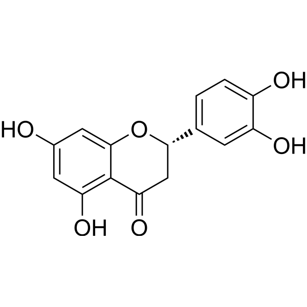
HY-N0637
EriodictyolEndogenous Metabolite -
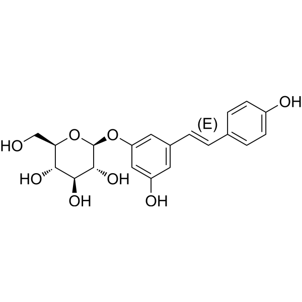
HY-N0120A
PolydatinAutophagy Inducer -
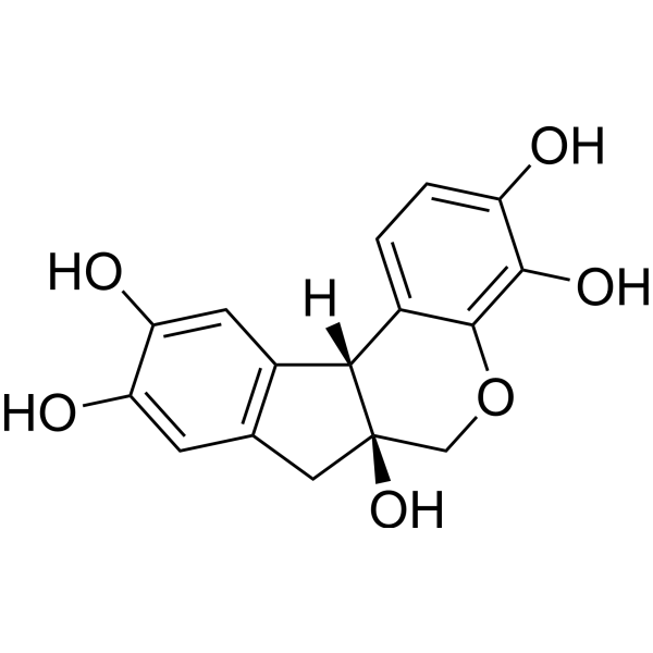
HY-N0116
HematoxylinHistologic Stain -
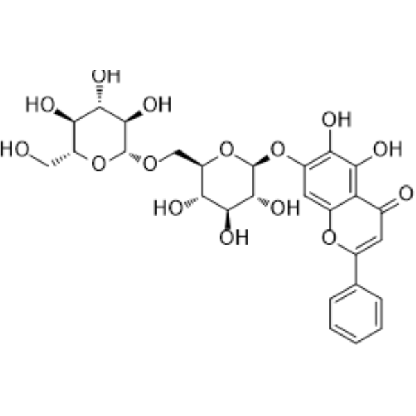
HY-N1435
Oroxin BAntitumor Agent -
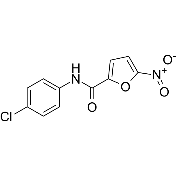
HY-147328
LCS3GSR/TXNRD1 Inhibitor -
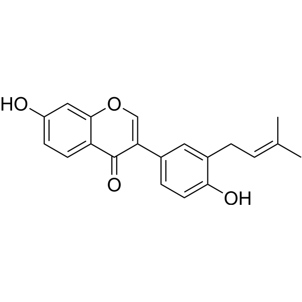
HY-N0720
NeobavaisoflavoneApoptosis Inducer -
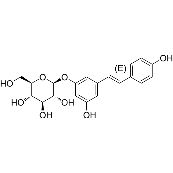
HY-N0120AR
Polydatin (Standard) -
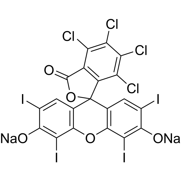
HY-D0214
Acid Red 94 sodiumPhotosensitizer -
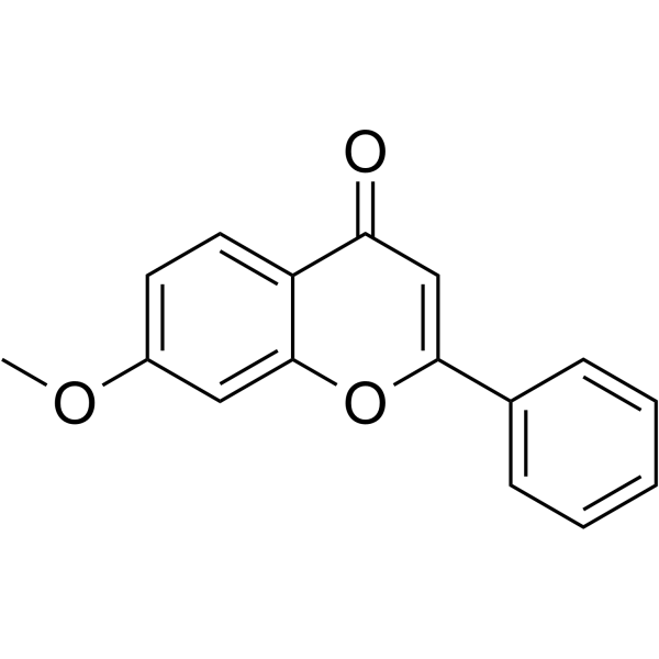
HY-N8487
7-MethoxyflavonePeripheral Analgesic


Biological Activity
Description
|
NF-κB |
JAK1 |
STAT1 |
p38 MAPK |
AP-1 |
MEK |
IRF3 |
ERK |
Nrf2 |
Keap1 |
PGE2 |
GSH |
PARP-1 |
PPARγ |
Cellular Effect
|
Cell Line

|
Type | Value | Description | References |
|---|---|---|---|---|
| MDCK | CC50 |
255 μM
|
Cytotoxicity against Madin-Darby canine kidney MDCK cells assessed as reduction in cell viability incubated for 48 hrs by MTT assay measuring absorbance at 490 nm.
Cytotoxicity against Madin-Darby canine kidney MDCK cells assessed as reduction in cell viability incubated for 48 hrs by MTT assay measuring absorbance at 490 nm.
|
24871979 |
| MDCK | EC50 |
45 μM
|
Antiviral activity against influenza A/PR/8/34 virus infected Madin-Darby canine kidney MDCK cells assessed as increase in cell viability incubated for 48 hrs post-infection by MTT assay measuring absorbance at 490 nm.
Antiviral activity against influenza A/PR/8/34 virus infected Madin-Darby canine kidney MDCK cells assessed as increase in cell viability incubated for 48 hrs post-infection by MTT assay measuring absorbance at 490 nm.
|
24871979 |
| HeLa | EC50 |
62.9 μM
|
Protective activity against MNNG-induced, PARP-mediated parthanatos in human HeLa cells assessed as increase in cell viability by MTT assay.
Protective activity against MNNG-induced, PARP-mediated parthanatos in human HeLa cells assessed as increase in cell viability by MTT assay.
|
LJMU.t.00019422 |
In Vitro
3',4'-Dihydroxyflavone (DHF) (1-5 μM; 24 h) suppresses LPS-induced NO production in RAW 264.7 macrophages[2].
3',4'-Dihydroxyflavone (1-5 μM; 1-4 h) induces Nrf2 nuclear translocation, decreases Keap1 levels, and increases Nrf2 DNA-binding activity in LPS-stimulated RAW 264.7 macrophages[2].
3',4'-Dihydroxyflavone (1-5 μM; 24 h) inhibits LPS-induced PGE2 and pro-inflammatory cytokine production in RAW 264.7 macrophages[2].
3',4'-Dihydroxyflavone (1-5 μM) downregulates the protein levels of iNOS and COX-2 in LPS-stimulated RAW 264.7 macrophages[2].
3',4'-Dihydroxyflavone (1-5 μM) inhibits the phosphorylation of IKKα/β and IκBα and subsequent degradation of IκBα in LPS-stimulated RAW 264.7 macrophages[2].
3',4'-Dihydroxyflavone (1-5 μM) inhibits the LPS-induced phosphorylation of c-Fos, c-Jun, ERK, and p38 in RAW 264.7 macrophages[2].
3',4'-Dihydroxyflavone (1-5 μM) reduces IRF3 phosphorylation and suppresses the LPS-induced phosphorylation of JAK1 and STAT1 in RAW 264.7 macrophages[2].
3',4'-Dihydroxyflavone (1-5 μM) decreases Keap1 levels and upregulates HO-1 in LPS-stimulated RAW 264.7 macrophages[2].
3',4'-Dihydroxyflavone (1-5 μM) downregulates the mRNA levels of iNOS and COX-2 in LPS-stimulated RAW 264.7 macrophages[2].
3',4'-Dihydroxyflavone (1-5 μM; 1 h) inhibits the promoter activities of iNOS and COX-2 in LPS-stimulated RAW 264.7 macrophages[2].
3',4'-Dihydroxyflavone (1-5 μM; 1 h) inhibits the transcriptional activity of NF-κB in LPS-stimulated RAW 264.7 macrophages[2].
3',4'-Dihydroxyflavone (1-5 μM; 1 h) inhibits the transcriptional activity of AP-1 in LPS-stimulated RAW 264.7 macrophages[2].
3',4'-Dihydroxyflavone (1-5 μM; 1 h) inhibits the transcriptional activity of IRF3 in LPS-stimulated RAW 264.7 macrophages[2].
3',4'-Dihydroxyflavone (1-5 μM) inhibits the DNA-binding activities of NF-κB and AP-1 in LPS-stimulated RAW 264.7 macrophages[2].
3',4'-Dihydroxyflavone(1-5 μM) inhibits the nuclear translocation of p65 in LPS-stimulated RAW 264.7 macrophages[2].
3',4'-Dihydroxyflavone (1-5 μM) inhibits the interaction between TRAF3 and TBK1 in LPS-stimulated RAW 264.7 macrophages[2].
3',4'-Dihydroxyflavone (5 μM; 1 h) obstructs the interaction between LPS and MD2 in RAW 264.7 macrophages[2].
3',4'-Dihydroxyflavone (1-5 μM; 1 h) inhibits LPS-induced intracellular ROS production in RAW 264.7 macrophages[2].
3',4'-Dihydroxyflavone (1-5 μM) increases reduced GSH levels in RAW 264.7 macrophages[2].
3',4'-Dihydroxyflavone exhibits significant antioxidant activity with 77.9% DPPH free radical-scavenging activity[7].
3',4'-Dihydroxyflavone scavenges DPPH radicals with an SC50 of 6.5 μM[10].
3',4'-Dihydroxyflavone scavenges superoxide anion radicals with an IC50 of 2.5 μM[10].
3',4'-Dihydroxyflavone inhibits xanthine oxidase activity with an IC50 of 7.9 μM[10].
3',4'-Dihydroxyflavone (10 µM) suppresses lipid accumulation and triglyceride production during adipogenic differentiation of 3T3-L1 preadipocytes[4].
3',4'-Dihydroxyflavone (8 days) suppresses MEK/ERK phosphorylation during adipogenic differentiation of 3T3-L1 preadipocytes[4].
3',4'-Dihydroxyflavone (5-10 μM; 48 h) slightly increases the viability of eADSCs[4].
3',4'-Dihydroxyflavone (10 μM) suppresses lipid accumulation and triglyceride production during adipogenic differentiation of eADSCs[4].
3',4'-Dihydroxyflavone suppresses the expression of adipogenic genes (PPARγ, C/EBPβ, and C/EBPα) in eADSCs[4].
3',4'-Dihydroxyflavone increases SOD2 and catalase expression in eADSCs[4].
3',4'-Dihydroxyflavone elevates the expression of osteogenic differentiation markers (ALP, RUNX2, OCN, and OPN) in eADSCs[4].
3',4'-Dihydroxyflavone suppresses ERK phosphorylation but does not significantly affect AKT phosphorylation during adipogenic differentiation of eADSCs[4].
3',4'-Dihydroxyflavone suppresses the adipogenesis-dependent increase in intracellular ROS levels in eADSCs[4].
3',4'-Dihydroxyflavone treatment increases mitochondrial membrane potential and decreases intracellular ROS in eADSCs[4].
3',4'-Dihydroxyflavone (48 h) increases MDCK cell viability and inhibits A/PR/8/34 virus replication with a CC50 of 255 μM, an EC50 of 45 μM, and a selectivity index of 5.6[5].
3',4'-Dihydroxyflavone (10-100 μM; 48 h) reduces influenza A/PR/8/34 virus titer in MDCK cells at 10 μM and suppresses hemagglutination almost completely at high concentration[5].
3',4'-Dihydroxyflavone (1 h) inhibits influenza A/PR/8/34 viral adsorption onto chicken RBCs by directly interfering with hemagglutinin binding to sialic acid receptors[5].
3',4'-Dihydroxyflavone (10-100 μM; 1 h) inhibits influenza A/PR/8/34 neuraminidase activity[5].
3',4'-Dihydroxyflavone (10-100 μM; 24 h) blocks the synthesis of influenza A/PR/8/34 hemagglutinin and neuraminidase mRNA in MDCK cells at 10, 50, and 100 μM[5].
3',4'-Dihydroxyflavone (DHF) (20 μM) inhibits PARP1 enzyme activity by 24.3% in a cell-free assay[7].
3',4'-Dihydroxyflavone (200 μM; 2 days) inhibits AGEs formation in a BSA-glucose assay with an IC50 of ca. 200 μM[10].
3',4'-Dihydroxyflavonesignificantly decreases hydrogen peroxide-induced intracellular ROS levels in HeLa cells[7].
3',4'-Dihydroxyflavone protects against MNNG (Methylnitronitrosoguanidine) (HY-128612)-induced, PARP-mediated parthanatos in HeLa cells with an EC50 of 62.9 μM[7].
MedChemExpress (MCE) has not independently confirmed the accuracy of these methods. They are for reference only. Further protocols information, click here.
-
Cell Line:RAW 264.7 murine macrophages
-
Concentration:1 μM; 2.5 μM; 5 μM
-
Incubation Time:24 h
-
Result:Reduced LPS-induced PGE2 production in a concentration-dependent manner.
Suppressed the production of pro-inflammatory cytokines such as TNF-α, IL-6, IL-1β, IFN-β, and IP-10 in LPS-stimulated cells.
-
Cell Line:RAW 264.7 murine macrophages
-
Concentration:5 μM
-
Incubation Time:1 h
-
Result:Reduced the co-localization of LPS and MD2.
-
Cell Line:Equine adipose-derived stromal cells (eADSCs)
-
Concentration:5-10 μM
-
Incubation Time:48 h
-
Result:Cell viability slightly increased following treatment with 5 or 10 μM of 3',4'-Dihydroxyflavone.
-
Cell Line:MDCK cells
-
Concentration:10 μM; 50 μM; 100 μM
-
Incubation Time:24 h
-
Result:Blocked the synthesis of influenza hemagglutinin and neuraminidase mRNA.
In Vivo
3',4'-Dihydroxyflavone (50-200 mg/kg; s.c.; 30 min prior to behavioral assessment) exhibits a significant anti-neuropathic effect against Paclitaxel (HY-B0015)-induced peripheral neuropathy in mice[3].
3',4'-Dihydroxyflavone (3,4'-DHF) (1 mg/kg; p.o.; daily; 5 days) decreases lung virus titers to 5.25 log10 EID50/mL, reduces body weight loss, and improves survival in mice infected with influenza A/PR/8/34 (H1N1)[5].
MedChemExpress (MCE) has not independently confirmed the accuracy of these methods. They are for reference only.
-
Animal Model:C57BL/6 (male, 6 weeks old, 18-20 g)[3]
-
Dosage:1 mg/kg; 2.5 mg/kg; 5 mg/kg
-
Administration:i.p.; pretreatment 1 h before LPS injection
-
Result:Increased the survival rate by 40%, 60%, and 80% at doses of 1, 2.5, and 5 mg/kg, respectively.
Suppressed the mRNA expression of iNOS, COX-2, TNF-α, and IL-6 in lung tissue.
Suppressed the LPS-induced inflammatory signaling pathways by inhibiting the phosphorylations of p65, c-Jun, c-Fos, and STAT1 (Y701).
Dose-dependently alleviated oxidative stress-based inflammation pathways by upregulating Nrf2 and HO-1 and downregulating Keap1 expression in lung tissue.
-
Animal Model:Swiss albino mice (either sex, 25-30 g)[4]
-
Dosage:50 mg/kg, 100 mg/kg, 200 mg/kg
-
Administration:s.c.; 30 min prior to behavioral assessment
-
Result:Significantly reduced the paw withdrawal response score in both mechanical and cold allodynia compared to Paclitaxel + vehicle treated animals.
Recorded a significant increase in the reaction time to flick the tail in the hot water tail immersion test in a dose-dependent manner.
-
Animal Model:C57BL/6 (six-week-old female, influenza A virus infection model)[5]
-
Dosage:1 mg/kg
-
Administration:p.o.; daily; 5 days
-
Result:Decreased lung virus titers to 5.25 log10 EID50/mL compared to 7.5 log10 EID50/mL in PBS control.
Reduced body weight loss compared to PBS control.
Improved survival rate to 60-70%.


Chemical Information
-
CAS No. 4143-64-0
-
Appearance Solid
-
Molecular Weight 254.24
-
Formula C15H10O4
-
Color Off-white to light yellow
-
SMILES
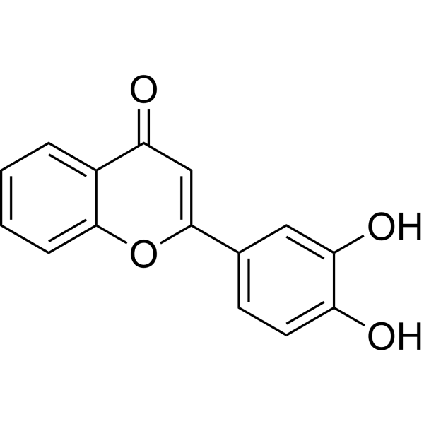
O=C1C=C(C2=CC=C(O)C(O)=C2)OC3=CC=CC=C13
-
Structure Classification
-
Initial Source
-
Shipping
Room temperature in continental US; may vary elsewhere.
-
Storage
4°C, sealed storage, away from moisture
* In solvent : -80°C, 6 months; -20°C, 1 month (sealed storage, away from moisture)


Solvent & Solubility
In Vitro:
DMSO : 125 mg/mL (491.66 mM; ultrasonic and warming and heat to 60°C; Hygroscopic DMSO has a significant impact on the solubility of product, please use newly opened DMSO)
Please refer to the solubility information to select the appropriate solvent. Once prepared, please aliquot and store the solution to prevent product inactivation from repeated freeze-thaw cycles.
Storage method and period of stock solution: -80°C, 6 months; -20°C, 1 month (sealed storage, away from moisture). When stored at -80°C, please use it within 6 months. When stored at -20°C, please use it within 1 month.
Please refer to the solubility information to select the appropriate solvent. Once prepared, please aliquot and store the solution to prevent product inactivation from repeated freeze-thaw cycles.
Storage method and period of stock solution: -80°C, 6 months; -20°C, 1 month (sealed storage, away from moisture). When stored at -80°C, please use it within 6 months. When stored at -20°C, please use it within 1 month.
Concentration (start) × Volume (start) = Concentration (final) × Volume (final)
In Vivo:
Select the appropriate dissolution method based on your experimental animal and administration route.
- For the following dissolution methods, please ensure to first prepare a clear stock solution using an In Vitro approach and then sequentially add co-solvents:
- To ensure reliable experimental results, the clarified stock solution can be appropriately stored based on storage conditions. As for the working solution for In Vivo experiments, it is recommended to prepare freshly and use it on the same day.
- The percentages shown for the solvents indicate their volumetric ratio in the final prepared solution. If precipitation or phase separation occurs during preparation, heat and/or sonication can be used to aid dissolution.
Add each solvent one by one: 10% DMSO 40% PEG300 5% Tween-80 45% Saline
Solubility: ≥ 2.08 mg/mL (8.18 mM); Clear solution
This protocol yields a clear solution of ≥ 2.08 mg/mL (saturation unknown).
Taking 1 mL working solution as an example, add 100 μL DMSO stock solution (20.8 mg/mL) to 400 μL PEG300, and mix evenly; then add 50 μL Tween-80 and mix evenly; then add 450 μL Saline to adjust the volume to 1 mL.
Preparation of Saline: Dissolve 0.9 g sodium chloride in ddH₂O and dilute to 100 mL to obtain a clear Saline solution.
Add each solvent one by one: 10% DMSO 90% (20% SBE-β-CD in Saline)
Solubility: ≥ 2.08 mg/mL (8.18 mM); Clear solution
This protocol yields a clear solution of ≥ 2.08 mg/mL (saturation unknown).
Taking 1 mL working solution as an example, add 100 μL DMSO stock solution (20.8 mg/mL) to 900 μL 20% SBE-β-CD in Saline, and mix evenly.
Preparation of 20% SBE-β-CD in Saline (4°C, storage for one week): 2 g SBE-β-CD powder is dissolved in 10 mL Saline, completely dissolve until clear.
In Vivo Dissolution Calculator
Please enter the basic information of animal experiments:
-
-
-
-
Recommended: Prepare an additional quantity of animals to account for potential losses during experiments.
Please enter your animal formula composition:
-
%DMSO +
Recommended: Keep the proportion of DMSO in working solution below 2% if your animal is weak.
-
%+
-
+%Tween-80 + +
-
%Saline +
The co-solvents required include: DMSO, . All of co-solvents are available by MedChemExpress (MCE). , Tween 80. All of co-solvents are available by MedChemExpress (MCE).
Working solution concentration: 0.22 mg/mL
Method for preparing stock solution: mg drug dissolved in μL DMSO. Stock solution concentration: mg/mL. * In solvent : -80°C, 6 months; -20°C, 1 month (sealed storage, away from moisture)
1. Take μL DMSO stock solution;
2. Add μL .
μL , mix evenly;
3. Then add μL Tween 80, mix evenly;
4. Then add μL
Please ensure that the stock solution in the first step is dissolved to a clear state, and add co-solvents in sequence. You can use ultrasonic heating (ultrasonic cleaner, recommended frequency 20-40 kHz), vortexing, etc. to assist dissolution.

Protocols
-
RNA extraction experimental
By lysing cells, releasing RNA, and removing impurities such as proteins and DNA, high-purity RNA products are finally obtained. The commonly used traditional method is the guanidine isothiocyanate/phenol/chloroform method (Trizol), which is suitable for a variety of animal materials including animal tissues, microorganisms, cultured cells, etc., and most plant materials.
-
Research Protocol for Infectious Diseases
Infectious-disease experiments test how pathogens interact with host barriers, innate immune receptors, inflammatory signaling, pathogen replication, and tissue injury; pattern-recognition receptors such as TLRs, RIG-I-like receptors, NOD-like receptors, and inflammasomes detect microbial molecules and activate NF-κB, interferon, and cytokine responses. The central hypothesis is that infection severity reflects the balance between pathogen burden and host response: protective inflammation restricts pathogen growth, whereas excessive or mislocalized inflammation contributes to tissue damage and disease phenotype. Unresolved questions include which host pathways are protective versus pathogenic, why some infection models fail to translate to human disease, and which combined readouts best predict clinically relevant infection outcomes.
-
LPS-Induced Endotoxemia/Systemic Inflammation
Lipopolysaccharide (LPS)-induced endotoxemia is a widely used in vivo model of acute systemic inflammation in which LPS, a Gram-negative bacterial endotoxin, activates innate immune signaling primarily through TLR4, leading to rapid and transient induction of pro-inflammatory cytokines such as TNF-α, IL-6, and IL-1β in circulation and tissues. This cytokine surge is commonly used as a measurable readout of systemic inflammatory activation and immune dysregulation, and is typically assessed within hours after intraperitoneal LPS administration in mouse models of endotoxemia. The model captures key features of systemic inflammatory response syndrome, including cytokine release, immune cell activation, and downstream tissue responses, and has been used to evaluate anti-inflammatory interventions such as cytokine modulation, lipid mediators, and immune cell-targeting therapies.
-
ROS/oxidative-stress fluorescent staining
ROS/oxidative-stress fluorescent staining uses cell-permeant fluorogenic probes that become fluorescent after oxidation inside cells or tissues; commonly used examples include DCFH-DA/DCFDA for broad cellular oxidant detection, DHE for superoxide-related signal detection, MitoSOX for mitochondrial superoxide-related signal detection, and CellROX probes for oxidative-stress-associated fluorescence readouts. The assay detects probe oxidation rather than a single ROS species unless the probe and analysis method have been chemically validated for that species. DCFH-DA enters cells, is deacetylated by intracellular esterases to DCFH, and produces fluorescent DCF after oxidation, so the readout is used as an operational measure of total cellular oxidative stress rather than a species-specific ROS measurement. DHE and MitoSOX can report superoxide-related oxidation, but red fluorescence alone can include non-specific ethidium-like oxidation products; HPLC or optimized spectral approaches are
-
Mesenchymal stromal/stem cell osteogenic differentiation
Mesenchymal stromal/stem cells can be induced toward an osteoblast-like lineage in vitro by culture in osteogenic medium containing dexamethasone, ascorbic acid or ascorbate-2-phosphate, and β-glycerophosphate; the differentiation process is commonly evaluated by alkaline phosphatase activity, osteogenic marker expression, collagenous matrix formation, and calcium-rich matrix mineralization. The main readouts are alkaline phosphatase activity as an early osteogenic marker and Alizarin Red S staining as a calcium-deposit readout for mineralized extracellular matrix; Alizarin Red S can be inspected microscopically or extracted and measured colorimetrically at 405 nm.
-
Research Protocol for Inflammation-related Diseases
The NLRP3 inflammasome is a cytosolic innate immune signaling platform that integrates priming signals and danger-signal activation to promote caspase-1 activation, maturation of IL-1β and IL-18, and gasdermin D-mediated pyroptotic cell death. The core experimental logic is to determine whether inflammatory disease phenotypes are driven by increased NLRP3 expression, ASC-containing inflammasome assembly, caspase-1 cleavage, GSDMD cleavage, and extracellular release of IL-1β/IL-18 rather than by nonspecific cell injury alone. The pathway is strongly linked to inflammation-related disease phenotypes because monosodium urate crystals activate NALP3/NLRP3 inflammasome signaling in gout-like crystal inflammation, cholesterol crystals activate NLRP3 inflammasomes in atherogenesis models, and DSS-induced intestinal inflammation has been reported to involve NLRP3 inflammasome activity. However, experimental colitis studies also show context-dependent protective effects of NLRP3 inflammasome co
-
Research Protocol for Metabolic Diseases
AMP-activated protein kinase, AMPK, is a conserved cellular energy sensor that responds to reduced cellular energy status and coordinates metabolism by increasing ATP-generating catabolic pathways while suppressing ATP-consuming anabolic processes. In metabolic disease research, the AMPK pathway is experimentally relevant because it regulates hepatic lipid synthesis, fatty acid oxidation, glucose production, skeletal-muscle glucose disposal, mTORC1-linked biosynthesis, autophagy, mitochondrial homeostasis, and whole-body energy balance. The central pathway logic is that energy stress, metformin, exercise-like stimulation, or direct AMPK activators increase AMPKα Thr172 phosphorylation and downstream substrate phosphorylation, including ACC and RAPTOR. Phosphorylation of ACC suppresses lipogenesis and supports fatty acid oxidation, whereas phosphorylation of RAPTOR suppresses mTORC1 signaling and links cellular energy status to growth and protein synthesis control. The pathway is linked


Purity & Documentation
-
Data Sheet (303 KB)
-
SDS (394 KB)
- English - EN (394 KB)
- Français - FR (394 KB)
- Deutsch - DE (394 KB)
- Norwegian - NO (394 KB)
- Español - ES (394 KB)
- Swedish - SV (394 KB)
- Italian - IT (394 KB)
- Korean - KR (394 KB)
- Portuguese - PT (394 KB)
-
Handling Instructions (2659 KB)
References


Complete Stock Solution Preparation Table
Please refer to the solubility information to select the appropriate solvent. Once prepared, please aliquot and store the solution to prevent product inactivation from repeated freeze-thaw cycles.
Storage method and period of stock solution: -80°C, 6 months; -20°C, 1 month (sealed storage, away from moisture). When stored at -80°C, please use it within 6 months. When stored at -20°C, please use it within 1 month.
| Optional Solvent | Concentration Solvent Mass | 1 mg | 5 mg | 10 mg | 25 mg |
|---|---|---|---|---|---|
| DMSO | 1 mM | 3.9333 mL | 19.6665 mL | 39.3329 mL | 98.3323 mL |
| 5 mM | 0.7867 mL | 3.9333 mL | 7.8666 mL | 19.6665 mL | |
| 10 mM | 0.3933 mL | 1.9666 mL | 3.9333 mL | 9.8332 mL | |
| 15 mM | 0.2622 mL | 1.3111 mL | 2.6222 mL | 6.5555 mL | |
| 20 mM | 0.1967 mL | 0.9833 mL | 1.9666 mL | 4.9166 mL | |
| 25 mM | 0.1573 mL | 0.7867 mL | 1.5733 mL | 3.9333 mL | |
| 30 mM | 0.1311 mL | 0.6555 mL | 1.3111 mL | 3.2777 mL | |
| 40 mM | 0.0983 mL | 0.4917 mL | 0.9833 mL | 2.4583 mL | |
| 50 mM | 0.0787 mL | 0.3933 mL | 0.7867 mL | 1.9666 mL | |
| 60 mM | 0.0656 mL | 0.3278 mL | 0.6555 mL | 1.6389 mL | |
| 80 mM | 0.0492 mL | 0.2458 mL | 0.4917 mL | 1.2292 mL | |
| 100 mM | 0.0393 mL | 0.1967 mL | 0.3933 mL | 0.9833 mL |


3',4'-Dihydroxyflavone Related Classifications
HY-127167 Related Classifications
 Powered by Bioz
Keywords
- 3',4'-Dihydroxyflavone
- 4143-64-0
- Free Fatty Acid Receptor
- NF-κB
- JAK
- STAT
- AP-1
- p38 MAPK
- MEK
- ERK
- Keap1-Nrf2
- PGE synthase
- Reactive Oxygen Species (ROS)
- Lipoxygenase
- Xanthine Oxidase
- PARP
- Potassium Channel
- GABA Receptor
- eADSCs
- 3T3-L1 preadipocytes
- Nrf2
- 5-lipoxygenase
- HeLa cells
- influenza A/PR/8/34
- RAW 264.7 macrophages
- MDCK cells
- ERK phosphorylation
- Inhibitor
- inhibitor
- inhibit