


Daphnoretin
Based on 3 publication(s) in Google Scholar
Daphnoretin (Dephnoretin; Thymelol) is a protein kinase C (PKC) activator that inhibits the expression of hepatitis B virus (HBV) surface antigen (HBsAg) and exhibits antiviral activity. Daphnoretin exerts its antitumor effects by inhibiting the activation of the PI3K/AKT signaling pathway and triggers the mitochondrial apoptosis pathway. Daphnoretin alleviates chondrocyte apoptosis and inflammatory responses by inhibiting endoplasmic reticulum stress and activation of the NLRP3 inflammasome. Daphnoretin regulates the differentiation and maturation of dendritic cells, inhibits their immunostimulatory function by downregulating the phosphorylation level of JNK, and thus exerts a protective effect in skin graft rejection.
For research use only. We do not sell to patients.
- Purity: 99.83%
- CAS No.: 2034-69-7
- Formula: C19H12O7
- Molecular Weight:352.29
-
Storage:
4°C, protect from light
* In solvent : -80°C, 6 months; -20°C, 1 month (protect from light)


-
 Biological Activity
-
Chemical Information
-
Solvent & Solubility
- Purity & Documentation
- References
-
 Help & FAQs
-


Anti-Infection Compound Library
HY-L002
-
Apoptosis Compound Library
HY-L003
-
Cell Cycle/DNA Damage Compound Library
HY-L004
-
Epigenetics Compound Library
HY-L005
-
Immunology/Inflammation Compound Library
HY-L007
-
Kinase Inhibitor Library
HY-L009
-
MAPK Compound Library
HY-L010
-
NF-κB Signaling Compound Library
HY-L014
-
PI3K/Akt/mTOR Compound Library
HY-L015
-
Stem Cell Signaling Compound Library
HY-L017
-
TGF-beta/Smad Compound Library
HY-L018
-
Natural Product Library
HY-L021
-
Anti-Cancer Compound Library
HY-L025
-
Antiviral Compound Library
HY-L027
-
Autophagy Compound Library
HY-L029
-
Anti-Aging Compound Library
HY-L034
-
Antioxidant Compound Library
HY-L037
-
Differentiation Inducing Compound Library
HY-L038
-
Reprogramming Compound Library
HY-L039
-
Oxygen Sensing Compound Library
HY-L045
-
Endoplasmic Reticulum Stress Compound Library
HY-L054
-
Phenols Library
HY-L057
-
Glycolysis Compound Library
HY-L058
-
Pyroptosis Compound Library
HY-L059
-
Cytoskeleton Compound Library
HY-L060
-
Glutamine Metabolism Compound Library
HY-L064
-
Traditional Chinese Medicine Active Compound Library
HY-L065
-
Anti-Breast Cancer Compound Library
HY-L074
-
Anti-Lung Cancer Compound Library
HY-L075
-
Anti-Pancreatic Cancer Compound Library
HY-L077
-
Anti-Blood Cancer Compound Library
HY-L079
-
Anti-Cancer Metabolism Compound Library
HY-L083
-
Anti-Parkinson's Disease Compound Library
HY-L085
-
Anti-Obesity Compound Library
HY-L087
-
Angiogenesis-Related Compound Library
HY-L088
-
Mitochondria-Targeted Compound Library
HY-L089
-
Transcription Factor-Targeted Library
HY-L090
-
Glucose Metabolism Compound Library
HY-L092
-
Anti-Liver Cancer Compound Library
HY-L101
-
Anti-Colorectal Cancer Compound Library
HY-L103
-
Anti-Cancer Natural Product Library
HY-L107
-
Antidepressant Compound Library
HY-L108
-
Antiviral Traditional Chinese Medicine Active Compound Library
HY-L113
-
Anti-inflammatory Traditional Chinese Medicine Active Compound Library
HY-L114
-
Plant-Sourced Natural Product Library
HY-L115
-
Human Metabolite Library
HY-L123
-
Anti-Prostate Cancer Compound Library
HY-L124
-
Anti-Pulmonary Fibrosis Compound Library
HY-L125
-
Cancer Stem Cells Compound Library
HY-L135
-
Coagulation and Anti-coagulation Compound Library
HY-L136
-
Pain-Related Compound Library
HY-L139
-
Membrane Protein-targeted Compound Library
HY-L149
-
Membrane Receptor-targeted Compound Library
HY-L150
-
Autoimmune Disease Compound Library
HY-L156
-
Cell Death Library
HY-L162
-
Serine/Threonine Kinase Inhibitor Library
HY-L164
-
Anti-Hematopathy Compound Library
HY-L171
-
Anti-Ovarian Cancer Compound Library
HY-L173
-
Inflammasomes related Compound Library
HY-L175
-
Multi-Target Compound Library
HY-L176
-
Radioprotector Library
HY-L178
-
Mitophagy Compound Library
HY-L180
-
Bioactive Compound Library Max
HY-L181
-
MCE Bioactive Compound Library
HY-L001V
-
Natural Product Library Plus
HY-L021P
-
Natural Product and Natural Product-Like Compound Library
HY-L021M
-
Bioactive Compound Library
HY-L001
-
Anti-Gastric Cancer Compound Library
HY-L184
-
Anti-Fibrosis Compound Library
HY-L185
-
Anti-Brain Cancer Compound Library
HY-L188
-
Miao Ethnicity Medicine Compound Library
HY-L190
-
Tibetan Medicine Compound Library
HY-L191
-
Heat-Clearing and Detoxification Traditional Chinese Medicine Compound Library
HY-L194
-
Protein Kinase Compound Library
HY-L196
-
Non-Alcoholic Fatty Liver Disease (NAFLD) Compound Library
HY-L199
-
RO5 Drug-like Natural Product Library
HY-L200
-
Cell Proliferation Compound Library
HY-L201
-
High-Throughput Bioactive Compound Library
HY-L205
-
High-Throughput Natural Product Library
HY-L206
-
Anti-Rheumatic Arthritis Compound Library
HY-L210
-
Mass Spectrometry Human Metabolite Library
HY-L215
-
Diarrhea-Related Traditional Chinese Medicine Active Compound Library
HY-L224
-
Pattern Recognition Receptors Library
HY-L237
-
Mongolian Medicine Compound Library
HY-L238
-
High-Efficiency Gene Editing Compound Library
HY-L244
-
Natural Product Diversity Scaffold Library
HY-L245
-
Lactylation Compound Library
HY-L249
-
Mass Spectrometry Natural Product Library
HY-L262


Publications Citing Use of MedChemExpress (MCE) Daphnoretin
More Customer Validation & Images

-
WB
-
WB
All Caspase Isoforms
More

Biological Activity
|
PI3K |
Akt |
NLRP3 inflammasome |
Bax |
Bcl-2 |
Caspase-3 |
Caspase-9 |
CDK2/cyclinE |
JNK |
|
Cell Line
|
Type | Value | Description | References |
|---|---|---|---|---|
| MCF7 | IC50 |
2 μM
Compound: Daphnoretin
|
Antiproliferative activity human MCF7 cells assessed as inhibition of cell proliferation incubated for 24 hrs by MTT assay
Antiproliferative activity human MCF7 cells assessed as inhibition of cell proliferation incubated for 24 hrs by MTT assay
|
[PMID: 36154031] |
| MDA-MB-231 | IC50 |
1 μM
Compound: Daphnoretin
|
Antiproliferative activity human MDA-MB-231 cells assessed as inhibition of cell proliferation incubated for 24 hrs by MTT assay
Antiproliferative activity human MDA-MB-231 cells assessed as inhibition of cell proliferation incubated for 24 hrs by MTT assay
|
[PMID: 36154031] |
| P388 | ED50 |
2.2 μg/mL
Compound: Daphnoretin, NSC-291852
|
Cytotoxicity against mouse P388 cells
Cytotoxicity against mouse P388 cells
|
[PMID: 6875580] |
| P388 | ED50 |
3.4 μg/mL
Compound: daphnoretin
|
Cytotoxicity against mouse P388 cells
Cytotoxicity against mouse P388 cells
|
[PMID: 3783168] |
Daphnoretin (0.1-1.0 μM; 48 h) inhibits the expression of hepatitis B surface antigen gene at the mRNA level in human hepatocellular carcinoma Hep3B cells, with an IC50 of 0.1 μM after 48 h of treatment[1].
Daphnoretin (1 μM; 4 h) induces the translocation of protein kinase C (PKC) from the cytosol to the cell membrane, and treatment of human hepatocellular carcinoma Hep3B cells with 800 nM daphnoretin for 24 h downregulates the total intracellular PKC level[1].
Daphnoretin (0.1-8.0 μM; 24-48 h) inhibits the proliferation of human breast cancer MCF-7 and MDA-MB-231 cells in a concentration- and time-dependent manner, with IC50 values of 2.0 μM and 1.0 μM, respectively[2].
Daphnoretin (0.5-4 μM; 24 h) induces S-phase cell cycle arrest in human breast cancer MCF-7 and MDA-MB-231 cells by downregulating the protein levels of cyclin E and CDK2[2].
Daphnoretin (0.5-4 μM; 24 h) induces apoptosis in human breast cancer MCF-7 and MDA-MB-231 cells via a mitochondria-dependent pathway, and reduces BCL-2 levels while increasing the levels of BAX, activated caspase-9 and activated caspase-3[2].
Daphnoretin (0.5-4 μM; 24 h) inhibits the PI3K/AKT pathway in human breast cancer MCF-7 and MDA-MB-231 cells at 24 h post-treatment by reducing the protein levels of p-PI3K and p-AKT[2].
Daphnoretin (0.5-512 μM; 48 h) reduces the viability of ATDC5 chondrocytes, with an IC50 of 72.97 μM after 48 h of treatment[3].
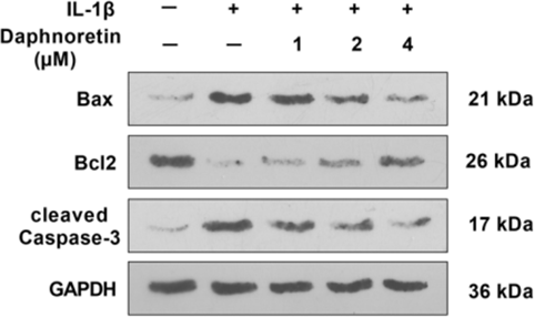
Daphnoretin (1-4 μM; 48 h) increases the viability of IL-1β-induced ATDC5 chondrocytes and reduces their apoptosis rate in a dose-dependent manner[3].
Daphnoretin (1-4 μM) dose-dependently inhibits IL-1β-induced ERS in ATDC5 chondrocytes[3].
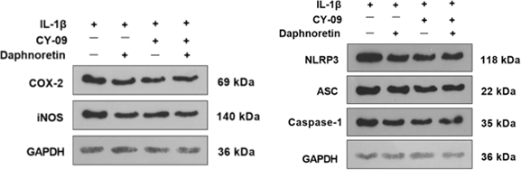
Daphnoretin (1-4 μM; 48 h) dose-dependently inhibits IL-1β-induced production of inflammatory mediators and activation of the NLRP3 inflammasome in ATDC5 chondrocytes[3].
Daphnoretin (1.1-30 μM; 6 days) reduces the viability of human CD14+ monocyte-derived dendritic cells in a dose-dependent manner, with an IC50 of approximately 30 μM; at a concentration of 10 μM, it inhibits dendrite formation without significant cytotoxicity[4].
Daphnoretin (1.1-30 μM; 6 days) dose-dependently downregulates the expression of differentiation and maturation markers, including CD1a, CD40, CD83, DC-SIGN and HLA-DR, on mature dendritic cells derived from human CD14+ monocytes, without inducing their dedifferentiation into macrophages[4].
Daphnoretin (1.1-30 μM; 6 days) dose-dependently impairs the allostimulatory function of mature dendritic cells derived from human CD14+ monocytes and reduces the proliferative capacity of allogeneic naive CD4+CD45RA+ T cells[4].
Daphnoretin (10 μM) specifically downregulates the LPS-induced upregulated expression of phosphorylated JNK in human CD14+ monocyte-derived dendritic cells[4].
MedChemExpress (MCE) has not independently confirmed the accuracy of these methods. They are for reference only.
-
Cell Line:human hepatoma Hep3B cells
-
Concentration:1 μM (PKC translocation); 800 nM (PKC down-regulation)
-
Incubation Time:4 h (PKC translocation); 24 h (PKC down-regulation)
-
Result:Increased membrane-bound PKC by ~20-fold with a concomitant decrease in cytosolic PKC after 4 h of 1 μM treatment. Diminished total extractable cellular PKC to a barely detectable level after 24 h of 800 nM treatment.
-
Cell Line:human breast cancer MCF-7, human breast cancer MDA-MB-231
-
Concentration:1, 2, and 4 μM (MCF-7); 0.5, 1 and 2 μM (MDA-MB-231)
-
Incubation Time:24 h (cell cycle assay; Western blot)
-
Result:Significantly increased the proportion of MCF-7 and MDA-MB-231 cells arrested in the S phase.
Significantly downregulated the protein levels of cyclin E and CDK2 in both cell lines compared to controls.
-
Cell Line:human breast cancer MCF-7, human breast cancer MDA-MB-231
-
Concentration:1, 2, and 4 μM (MCF-7); 0.5, 1 and 2 μM (MDA-MB-231)
-
Incubation Time:24 h (apoptosis assays; Western blot)
-
Result:Revealed typical apoptotic features (chromatin/nuclear condensation, apoptotic bodies) in both daphnoretin-treated cell lines via Hoechst 33258 staining.
Showed significantly increased apoptotic rates in treated MCF-7 and MDA-MB-231 cells compared to controls.
Significantly downregulated BCL-2 protein levels and upregulated BAX, cleaved caspase-9, and cleaved caspase-3 protein levels in both cell lines.
-
Cell Line:human breast cancer MCF-7, human breast cancer MDA-MB-231
-
Concentration:1, 2 and 4 μM (MCF-7, 24 h); 0.5-2 μM (MDA-MB-231, 24 h); 0.5, 1 and 2 μM (0.5, 1, 2 h, both cell lines)
-
Incubation Time:24 h (24 h treatment group); 0.5 h, 1 h, 2 h (short-term treatment group)
-
Result:Significantly downregulated the protein levels of p-PI3K and p-AKT in both MCF-7 and MDA-MB-231 cells after 24 h of treatment.
Showed no significant changes in p-PI3K or p-AKT levels after 0.5, 1, or 2 h of treatment in either cell line.
Caused no significant alterations in total PI3K and AKT levels.
Daphnoretin (0.5-1.0 mg/kg; i.p.; once daily for 7 days) inhibits acute skin allograft rejection in mice without altering body weight or white blood cell counts, and exhibits no obvious dose-dependent effect[4].
MedChemExpress (MCE) has not independently confirmed the accuracy of these methods. They are for reference only.
-
Animal Model:C57BL/6 (8-10-week-old male, DMM-induced OA)[3]
-
Dosage:10 mg/kg
-
Administration:i.p.; daily (first week post-surgery), once every 3 days (subsequent 7 weeks); 8 weeks total
-
Result:Repaired the articular cartilage surface relative to the OA-only group.
Increased the thickness of cartilage and subchondral cortical bone plate relative to the OA-only group.
Reduced the number of TRAP-positive cells relative to the OA-only group.
Significantly improved the thickness of the subchondral cortical bone plate relative to the OA-only group.
Reduced the OARSI score relative to the OA-only group.Downregulated the expression of pro-apoptotic proteins Bax and cleaved-caspase3 in cartilage tissue relative to the OA-only group.
Upregulated the anti-apoptotic protein Bcl2 in cartilage tissue relative to the OA-only group.
Reduced the expression of endoplasmic reticulum stress markers GRP78, CHOP, ATF6, and Caspase-12 relative to the OA-only group.
Reduced the expression of NLRP3, ASC, and Caspase-1 components of the NLRP3 inflammasome relative to the OA-only group.
Abated the mRNA levels of Cox-2, inos, Tnf-α, and Il-6 in cartilage tissues relative to the OA-only group.
-
Animal Model:C57BL/6 (H-2b) (male, 6-8-week-old); BALB/c (H-2d) (male, 6-8-week-old, skin allograft recipient)[4]
-
Dosage:0.5 mg/kg; 1.0 mg/kg
-
Administration:i.p.; once daily for 7 days
-
Result:Increased the survival fraction of skin allografts relative to untreated controls.
Caused no significant changes in recipient body weight or white blood cell count during the observation period.
Showed no evident dose-dependent response between the two tested doses.


Chemical Information
-
CAS No. 2034-69-7
-
Appearance Solid
-
Molecular Weight 352.29
-
Formula C19H12O7
-
Color Off-white to light yellow
-
SMILES
O=C1C(OC2=CC=C(C(O3)=C2)C=CC3=O)=CC4=CC(OC)=C(O)C=C4O1
-
Synonyms
Dephnoretin; Thymelol
-
Structure Classification
-
Initial Source
-
Shipping
Room temperature in continental US; may vary elsewhere.
-
Storage
4°C, protect from light
* In solvent : -80°C, 6 months; -20°C, 1 month (protect from light)


Publications (3)
-
Journal Impact Factor
-
Most Recent
-
J Orthop Surg Res
Daphnoretin relieves IL-1β-mediated chondrocytes apoptosis via repressing endoplasmic reticulum stress and NLRP3 inflammasome. [Abstract]2022 Nov 16;17(1):487. PMID: 36384642
Daphnoretin purchased from MedChemExpress. Usage Cited in: J Orthop Surg Res. 2022 Nov 16;17(1):487. [Abstract]
Daphnoretin (1, 2, 4 µM) reduces the concentrations of TNF-α, IL-6, and PGE2 in mouse chondrocyte culture supernatant in a dose-dependent manner.
Daphnoretin purchased from MedChemExpress. Usage Cited in: J Orthop Surg Res. 2022 Nov 16;17(1):487. [Abstract]
Daphnoretin or CY-09 markedly attenuates the expression of COX-2, iNOS, TNF-α, IL-6, PGE2 and the NLRP3-ASC-Caspase1 infammasome in ATDC5 cells.
-
Vet Microbiol
The Chinese medicine monomer Schisandrin C inhibits PRRSV infection by regulating the OGT-PI3K/AKT/mTOR signaling pathway. [Abstract]2026 May:316:110992. PMID: 41865607 -


Solvent & Solubility
DMSO : 25 mg/mL (70.96 mM; Need ultrasonic; Hygroscopic DMSO has a significant impact on the solubility of product, please use newly opened DMSO)
Please refer to the solubility information to select the appropriate solvent. Once prepared, please aliquot and store the solution to prevent product inactivation from repeated freeze-thaw cycles.
Storage method and period of stock solution: -80°C, 6 months; -20°C, 1 month (protect from light). When stored at -80°C, please use it within 6 months. When stored at -20°C, please use it within 1 month.
Concentration (start) × Volume (start) = Concentration (final) × Volume (final)
Select the appropriate dissolution method based on your experimental animal and administration route.
- For the following dissolution methods, please ensure to first prepare a clear stock solution using an In Vitro approach and then sequentially add co-solvents:
- To ensure reliable experimental results, the clarified stock solution can be appropriately stored based on storage conditions. As for the working solution for In Vivo experiments, it is recommended to prepare freshly and use it on the same day.
- The percentages shown for the solvents indicate their volumetric ratio in the final prepared solution. If precipitation or phase separation occurs during preparation, heat and/or sonication can be used to aid dissolution.
Add each solvent one by one: 10% DMSO 40% PEG300 5% Tween-80 45% Saline
Solubility: 2.08 mg/mL (5.90 mM); Suspended solution; Need ultrasonic
This protocol yields a suspended solution of 2.08 mg/mL. Suspended solution can be used for oral and intraperitoneal injection.
Taking 1 mL working solution as an example, add 100 μL DMSO stock solution (20.8 mg/mL) to 400 μL PEG300, and mix evenly; then add 50 μL Tween-80 and mix evenly; then add 450 μL Saline to adjust the volume to 1 mL.
Preparation of Saline: Dissolve 0.9 g sodium chloride in ddH₂O and dilute to 100 mL to obtain a clear Saline solution.
Add each solvent one by one: 10% DMSO 90% (20% SBE-β-CD in Saline)
Solubility: 2.08 mg/mL (5.90 mM); Suspended solution; Need ultrasonic
This protocol yields a suspended solution of 2.08 mg/mL. Suspended solution can be used for oral and intraperitoneal injection.
Taking 1 mL working solution as an example, add 100 μL DMSO stock solution (20.8 mg/mL) to 900 μL 20% SBE-β-CD in Saline, and mix evenly.
Preparation of 20% SBE-β-CD in Saline (4°C, storage for one week): 2 g SBE-β-CD powder is dissolved in 10 mL Saline, completely dissolve until clear.
Please enter the basic information of animal experiments:
-
-
-
-
Recommended: Prepare an additional quantity of animals to account for potential losses during experiments.
Please enter your animal formula composition:
-
%DMSO +
Recommended: Keep the proportion of DMSO in working solution below 2% if your animal is weak.
-
%+
-
+%Tween-80 + +
-
%Saline +
The co-solvents required include: DMSO, . All of co-solvents are available by MedChemExpress (MCE). , Tween 80. All of co-solvents are available by MedChemExpress (MCE).
Working solution concentration: 0.22 mg/mL
Method for preparing stock solution: mg drug dissolved in μL DMSO. Stock solution concentration: mg/mL. * In solvent : -80°C, 6 months; -20°C, 1 month (protect from light)
1. Take μL DMSO stock solution;
2. Add μL .
μL , mix evenly;
3. Then add μL Tween 80, mix evenly;
4. Then add μL
Please ensure that the stock solution in the first step is dissolved to a clear state, and add co-solvents in sequence. You can use ultrasonic heating (ultrasonic cleaner, recommended frequency 20-40 kHz), vortexing, etc. to assist dissolution.


Purity & Documentation
-
Data Sheet (289 KB)
-
SDS (393 KB)
- English - EN (393 KB)
- Français - FR (393 KB)
- Deutsch - DE (393 KB)
- Norwegian - NO (393 KB)
- Español - ES (393 KB)
- Swedish - SV (393 KB)
- Italian - IT (393 KB)
- Korean - KR (393 KB)
- Portuguese - PT (393 KB)
-
Handling Instructions (2659 KB)
References
[1]. Chen HC, et al. Identification of a protein kinase C (PKC) activator, daphnoretin, that suppresses hepatitis B virus gene expression in human hepatoma cells. Biochem Pharmacol. 1996;52(7):1025-1032. [Content Brief]
[2]. Xie Q, et al. Daphnoretin Arrests the Cell Cycle and Induces Apoptosis in Human Breast Cancer Cells. J Nat Prod. 2022;85(10):2332-2339. [Content Brief]
[3]. Zhou J, et al. Daphnoretin relieves IL-1β-mediated chondrocytes apoptosis via repressing endoplasmic reticulum stress and NLRP3 inflammasome. J Orthop Surg Res. 2022;17(1):487. Published 2022 Nov 16. [Content Brief]
[4]. Chen CA, et al. Daphnoretin modulates differentiation and maturation of human dendritic cells through down-regulation of c-Jun N-terminal kinase. Int Immunopharmacol. 2017;51:25-30. [Content Brief]


Complete Stock Solution Preparation Table
Please refer to the solubility information to select the appropriate solvent. Once prepared, please aliquot and store the solution to prevent product inactivation from repeated freeze-thaw cycles.
Storage method and period of stock solution: -80°C, 6 months; -20°C, 1 month (protect from light). When stored at -80°C, please use it within 6 months. When stored at -20°C, please use it within 1 month.
| Optional Solvent | Concentration Solvent Mass | 1 mg | 5 mg | 10 mg | 25 mg |
|---|---|---|---|---|---|
| DMSO | 1 mM | 2.8386 mL | 14.1929 mL | 28.3857 mL | 70.9643 mL |
| 5 mM | 0.5677 mL | 2.8386 mL | 5.6771 mL | 14.1929 mL | |
| 10 mM | 0.2839 mL | 1.4193 mL | 2.8386 mL | 7.0964 mL | |
| 15 mM | 0.1892 mL | 0.9462 mL | 1.8924 mL | 4.7310 mL | |
| 20 mM | 0.1419 mL | 0.7096 mL | 1.4193 mL | 3.5482 mL | |
| 25 mM | 0.1135 mL | 0.5677 mL | 1.1354 mL | 2.8386 mL | |
| 30 mM | 0.0946 mL | 0.4731 mL | 0.9462 mL | 2.3655 mL | |
| 40 mM | 0.0710 mL | 0.3548 mL | 0.7096 mL | 1.7741 mL | |
| 50 mM | 0.0568 mL | 0.2839 mL | 0.5677 mL | 1.4193 mL | |
| 60 mM | 0.0473 mL | 0.2365 mL | 0.4731 mL | 1.1827 mL |


Daphnoretin Related Classifications
Cat. No.: HY-N0699
 Powered by Bioz
- Daphnoretin
- 2034-69-7
- Dephnoretin
- Thymelol
- PKC
- Influenza Virus
- NOD-like Receptor (NLR)
- Apoptosis
- HBV
- JNK
- PI3K
- Akt
- CDK
- Caspase
- Bcl-2 Family
- CD14? monocyte-derived dendritic cells
- PI3K/AKT pathway
- MCF-7 cells
- ATDC5 chondrocytes
- hepatitis B surface antigen
- NLRP3 inflammasome
- Protein Kinase C
- c-Jun N-terminal kinase
- Hep3B cells
- MDA-MB-231 cells
- Inhibitor
- inhibitor
- inhibit