



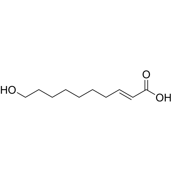
10-Hydroxy-2-decenoic acid
Based on 3 publication(s) in Google Scholar
10-Hydroxy-2-decenoic acid (10-HDA) is an orally active unsaturated medium-chain fatty acid with various physiological activities. 10-Hydroxy-2-decenoic acid induces ROS-mediated apoptosis in A549 cells. 10-Hydroxy-2-decenoic acid inhibits VEGF-induced angiogenesis in human venous endothelial cells. 10-Hydroxy-2-decenoic acid alleviates non-alcoholic fatty liver disease (NAFLD) by activating the AMPK-α signaling pathway. 10-Hydroxy-2-decenoic acid protects against bone loss by inhibiting NF-κB signaling downstream of FFAR4. 10-Hydroxy-2-decenoic acid is an antibiotic against many bacteria and fungi, such as Neurospora sitophila, molds and Staphylococcus aureus. 10-Hydroxy-2-decenoic acid has longevity-promoting effects in C. elegans. 10-Hydroxy-2-decenoic acid prevents osteoarthritis by targeting aspartyl β hydroxylase and inhibiting chondrocyte senescence.
For research use only. We do not sell to patients.
- Purity : 99.93%
- CAS No.: 765-01-5
- Formula: C10H18O3
- Molecular Weight:186.25
-
Storage:Powder -20°C, 3 years , 4°C, 2 years ; In solvent -80°C, 6 months , -20°C, 1 month

To place orders, for customer services and technical support, please contact: MedChemExpress USA
Tel: 609-228-6898 E-mail: [email protected] [email protected]


-
 Biological Activity
-
Chemical Information
-
Solvent & Solubility
- Protocols
- Purity & Documentation
- References
-
 Help & FAQs


-

HY-15141
StaurosporinePKC/PKA Inhibitor -
HY-10218
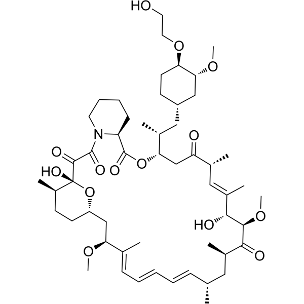
EverolimusmTORC Inhibitor -
HY-122022
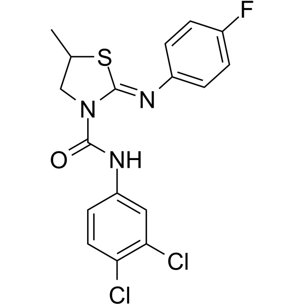
JR-AB2-011mTORC2 Inhibitor -
HY-114384
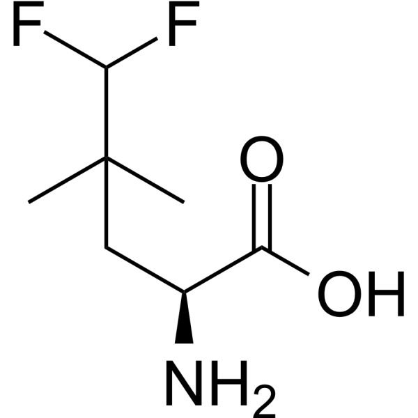
MefluleucinemTORC1 Activator -
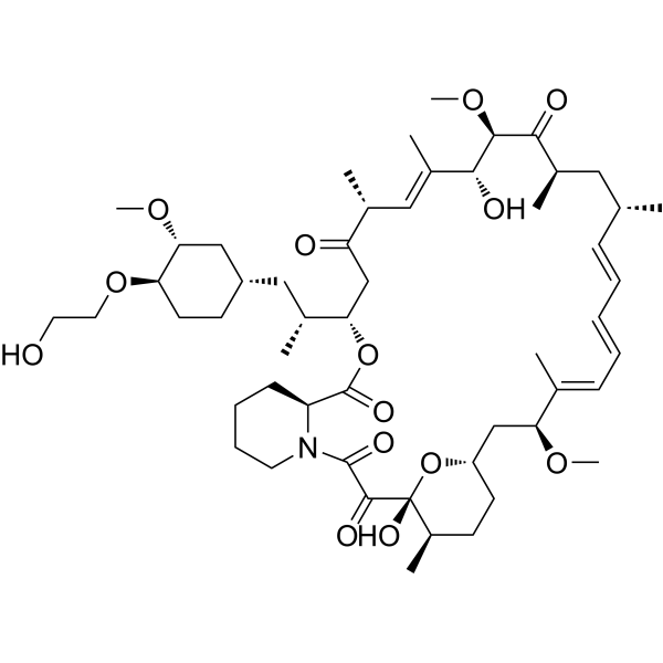
HY-10218R
Everolimus (Standard)mTORC Inhibitor -
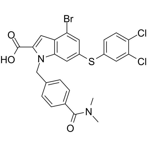
HY-124798
Rheb inhibitor NR1Rheb/mTORC1 Inhibitor -
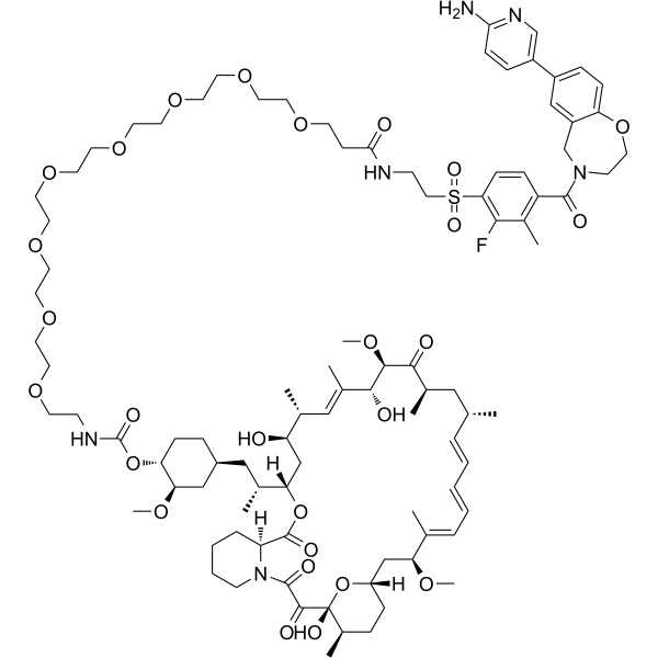
HY-134904
RMC-6272mTORC1-Selective Inhibitor -
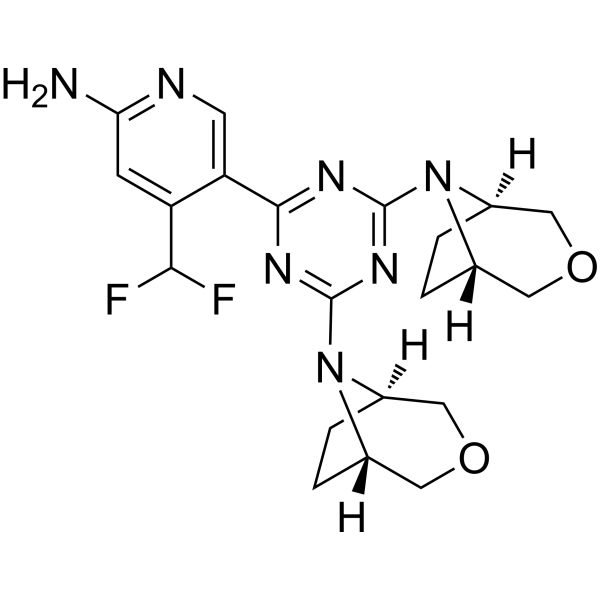
HY-100026
PQR620mTORC1/2 Inhibitor -
HY-141805
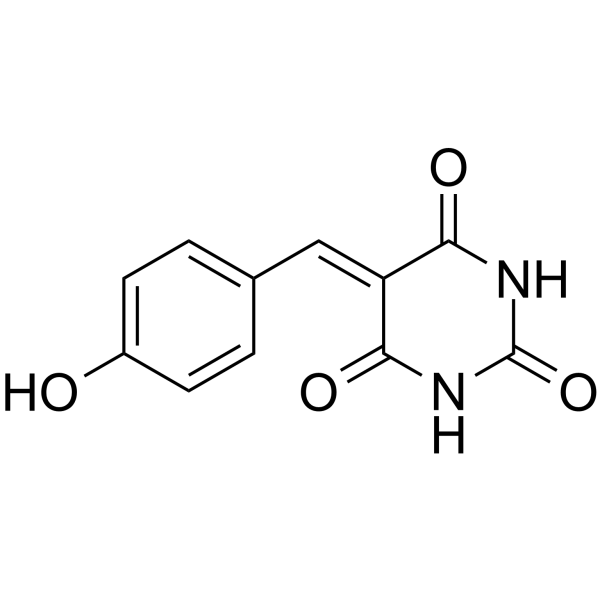
MHY-1685mTOR Inhibitor -

HY-W592871R
10-Hydroxy-2-decenoic acid (Standard)Fatty Acid -

HY-100026R
PQR620 (Standard)mTORC1/2 Inhibitor -
HY-15271A
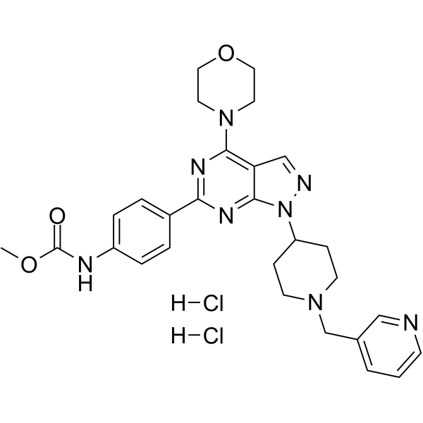
WYE-687 dihydrochloride
Publications Citing Use of MedChemExpress (MCE) 10-Hydroxy-2-decenoic acid
MoreAll AMPK Isoforms
MoreAll Caspase Isoforms
More

Biological Activity
Description
In Vitro
10-Hydroxy-2-decenoic acid (0-40 nM, 0-6 days) boosts proliferation and anabolism, whereas inhibits apoptosis and catabolism in primary chondrocytes and human C28/I2 chondrocytes[1].
10-Hydroxy-2-decenoic acid (0-10 nM, 7 days) enhances anabolism and represses catabolism of chondrocytesIn human osteoarthritis (OA) cartilage explants[1].
10-Hydroxy-2-decenoic acid (0-200 μM, 1-48 h) can bind to ASPH and regulates chondrocyte metabolism dependent on the Asp-Arg-Hydrox domain of ASPH in C28/I2 and HEK-293T cells[1].
10-Hydroxy-2-decenoic acid (0-10 nM, 48 h) reduces senescence-associated β-galactosidase (SA-β-gal) and mRNA levels of cellular senescence marker genes p16 and p21 in C28/I2 cells[1].
10-Hydroxy-2-decenoic acid (0-10 nM, 48 h) inhibits chondrocyte senescence by regulating the ASPH/ERK/p53/p21 and ASPH/GSK3β/p16 signaling pathways in ASPH-KO C28/I2 cells[1].
10-Hydroxy-2-decenoic acid (2 mg/mL) has antitumor activity at low pH values in 6C3HED lympho-sarcoma cells[2].
10-Hydroxy-2-decenoic acid (50-150 μM) prevents FFAs induced lipid accumulation and apoptosis via the activation of AMPK-α in AML12 cells[3].
10-Hydroxy-2-decenoic acid (0.07-1.13 mg/mL, 24 h) reduces the biomass of biofilms and cell viability by destroying the exopolysaccharides (EPS) structure, inhibits the production of hemolysin in S. aureus[5].
10-Hydroxy-2-decenoic acid (20-500 μM,11 days) reduces VEGF-induced tube formation in HUVECs and fibroblasts[6].
10-Hydroxy-2-decenoic acid (20-500 μM, 3 days) inhibits VEGF-induced cell proliferation at 500 µM, however does not inhibit the cell proliferation at 20-100 µM in HUVECs[6].
10-Hydroxy-2-decenoic acid (500 μM, 24 h) inhibits cell migration in HUVECs[6].
10-Hydroxy-2-decenoic acid (1-100 μM, 3-36 h) inhibits the growth of A549, NCI-H460, NCI-H23 cells, with IC50s of 22.68, 44.03, 44.79 μM, respectively, has weak cytotoxicity of IMR90, L-02 cells, and GES-1 cells[8].
10-Hydroxy-2-decenoic acid (30 μM, 3-24 h) increases the levels of ROS and induces apoptosis through regulating the MAPK, NF-κB, and STAT3 signaling pathways in A549 cells[8].
10-Hydroxy-2-decenoic acid (30 μM, 3-24 h) induces cell cycle arrest by regulating cell cycle-associated protein expression in A549 cells[8].
10-Hydroxy-2-decenoic acid (30 μM, 3-24 h) inhibits cell migration by regulating TGF-β1 signaling pathways in A549 Cells[8].
10-Hydroxy-2-decenoic acid (10-100 μM, 50 days) extends mean and maximum lifespan through mechanisms independent of the IIS-DAF-16 pathway in daf-16(mu86) mutants and wild-type C. elegans[9].
10-Hydroxy-2-decenoic acid (0.00004-20mM) activates FFAR4 with EC50 of 1.025 mM[10].
10-Hydroxy-2-decenoic acid (12.5-50 ng/mL, 3 days) suppresses the NF-κB signaling pathway through the FFAR4 receptor so as to inhibit osteoclastogenesis in Primary bone marrow cells[10].
MedChemExpress (MCE) has not independently confirmed the accuracy of these methods. They are for reference only.
-
Cell Line:Primary chondrocytes, Human C28/I2 chondrocytes
-
Concentration:0 nM, 2 nM, 5 nM, 10 nM, 20 nM, 40 nM
-
Incubation Time:0, 2, 4, 6 days
-
Result:Promoted the mRNA levels of CDK1, CDK2, CDK4, CDK6, cyclin A1, cyclin B1, cyclin D1, cyclin E1, proliferating cell nuclear antigen (PCNA), SRY-box transcription factor 9 (SOX9), COL2 and aggrecan (ACAN).
Inhibited the mRNA levels of COL1A1, COL1A2, COL3A1, α-SMA, FMOD, and FNDC1.
Increased the mRNA levels of CDK1, CDK2, CDK4, CDK6, cyclinA1, cyclinB1, cyclinD1, cyclinE1, and PCNA, SOX9, COL2, and ACAN, while inhibiting the expression of MMP13 and IL6 in the presence of IL-1β (10 ng/mL, 48 h).
-
Cell Line:Human OA cartilage explants
-
Concentration:0 nM, 5 nM, 10 nM
-
Incubation Time:7 days
-
Result:Upregulated COL2 and ACAN protein levels and downregulated MMP13 protein levels.
-
Cell Line:C28/I2 cells
-
Concentration:0 nM, 5 nM, 10 nM
-
Incubation Time:7 days
-
Result:Reduced ASPH and COL2 protein levels and increased MMP13 protein levels in knocked down the ASPH expression.
-
Cell Line:HUVECs
-
Concentration:500 µM
-
Incubation Time:24 h
-
Result:Inhibited cell migration.
-
Cell Line:A549 human lung cancer cells
-
Concentration:30 μM
-
Incubation Time:3, 24 h
-
Result:Increased numbers of early and late apoptotic cells, loss of mitochondrial membrane potential (MMP), increased proportion of depolarized cells.
Downregulated Bcl-2, and upregulated BAX, cyto-c, caspase-3, and PARP.
Upregulated p-p38, p-JNK, and I-κB and downregulated p-ERK, p-STAT3, and NF-κB.
-
Cell Line:A549 human lung cancer cells
-
Concentration:30 μM
-
Incubation Time:3, 24 h
-
Result:Increased the percentage of cells in the G0/G1 phase from 62.97% to 80.54%, downregulated AKT, CDK2/4/6, and cyclin D1/E, and upregulated p21 and p27.
-
Cell Line:A549 human lung cancer cells
-
Concentration:30 μM
-
Incubation Time:3, 24 h
-
Result:Inhibited cell migration, reduced the expression levels of TGF-β1, SNAI1, GSK-3β, N-cadherin and vimentin, and increased the expression level of E-cadherin.
-
Cell Line:Primary bone marrow cells
-
Concentration:12.5-50 ng/mL
-
Incubation Time:3 days
-
Result:Reduced protein levels of CtsK, TRAP, V-ATPase D2, and MMP9.
Diminished RANKL-induced phosphorylation and the degradation of IκBα.
-
Cell Line:Primary bone marrow cells
-
Concentration:12.5-50 ng/mL
-
Incubation Time:3 days
-
Result:Had no influence on the expression of Tnfrsf11a (encodes RANK), Csf1r (encodes M-CSFR), Fos, and Mitf. Decreased osteoclastogenesis Nfatc1 and its downstream elements, without affecting the expression of Irf8 and Mafb.
In Vivo
10-Hydroxy-2-decenoic acid (10 mg/kg, IA injection, twice a week for 8 weeks; 100 mg/kg, p.o., three times a week for 8 weeks) relieves chondrocyte senescence and cartilage degeneration in naturally aged mice[1].
10-Hydroxy-2-decenoic acid (0.5-2 mg/mL, s.c., once) has completely prevented the development of tumor in AKR mice and Ascitic tumor connaught mice[2].
10-Hydroxy-2-decenoic acid (10-50 mg/kg, p.o., once a day, 4 weeks) renders strong protective effects against lipid accumulation and liver injury, protects against apoptosis in Methionine (HY-13694)-Choline (HY-B1337) deficient (MCD) diet induced NAFLD mice[3].
10-Hydroxy-2-decenoic acid (basal diet containing 1-5 g/kg, o.p., 21 days) alleviates intestinal damage and growth performance loss through anti-inflammatory, antioxidant and gut microbiota regulatory activities in LPS (HY-D1056)-induced intestinal mucosal injury chickens model[4].
10-Hydroxy-2-decenoic acid (40 mg/kg, p.o., once a day, 4 weeks) inhibits ovariectomy (OVX)-induced osteoclastic bone resorption without affecting bone formation in OVX mice[10].
MedChemExpress (MCE) has not independently confirmed the accuracy of these methods. They are for reference only.
-
Animal Model:DMM mouse (8-week-old male C57BL/6 J) model[1]
-
Dosage:10 mg / kg; 100 mg/kg
-
Administration:IA injection, twice a week for 6 weeks; p.o., three times a week for 7 weeks
-
Result:Increased the number of chondrocytes, improved knee cartilage erosion and superficial cartilage loss, inhibited cartilage degradation, increased the protein and mRNA levels of Col2 and Acan, and reduced the protein and mRNA levels of Mmp13.
Reduced pain-related behaviors associated with OA and DMM surgery.
Was not associated with any significant toxicity in oral administration group.
Inhibited the OA-induced down-regulation of ASPH expression.
Reduced p16 and p21 protein levels.
-
Animal Model:Naturally aged (18 months old, male) C57BL/6 J mice model[1]
-
Dosage:10 mg/kg; 100 mg/kg
-
Administration:IA injection, twice a week for 8 weeks; p.o., three times a week for 8 weeks
-
Result:Reduced knee joint diameter, prevented joint swelling, partially alleviated aging-related cartilage degeneration, and improved OARSI scores.
Reduced p16, p21, and Mmp13 protein levels and increased Col2 protein and ASPH expression.
-
Animal Model:AKR (2-5 million tumor cells) mice (female mice, 5-6 weeks) model[2]
-
Dosage:0.5 mg/mL, 1 mg/mL, 1.5 mg/mL, 2.0 mg/mL, 0.2 mL/mouse
-
Administration:s.c., once
-
Result:Had completely prevented the development of transplantable leukemia at 1.5 mg/mL.
-
Animal Model:Ascitic tumor (the 6CSHED lymphosarcoma, the TAS mammary carcinoma, and the Ehrlich carcinoma, 5-8 million tumor cells) connaught mice (Male mice, 20-22 g) model[2]
-
Dosage:0.6 mg/mL, 1 mg/mL, 2.0 mg/mL
-
Administration:s.c., once
-
Result:Showed slight activity at a level of 0.6 mg/mL, while conferred complete protection at 1.0 mg/mL.
-
Animal Model:MCD diet (5 weeks)-induced NAFLD in mice (Male C57BL/6, 6 weeks of age, weighing 20∼25 g) model[3]
-
Dosage:10 mg/kg; 50 mg/kg
-
Administration:p.o., once a day, 4 weeks
-
Result:Inhibited weight loss, reduced fat droplets, and lowered liver TG by 32% and 60%, respectively.
Reduced hepatocyte vacuoles and ballooning, and attenuated ALT and AST levels.
Reduced TUNEL cell area, reduced BAX expression and increased BCL2 expression.
Was involved in hepatic inflammatory recruitment mainly by regulating the infiltration of macrophages and neutrophils.
Suppressed hepatic lipid accumulation by regulation lipogenesis and fatty acid β-oxidation.
-
Animal Model:LPS-induced (0.5 mg/kg, i.p.) intestinal mucosal injury chickens (one-day-old male) model[4]
-
Dosage:0.1% group, basal diet containing 1 g/kg; 0.5% group, basal diet containing 5 g/kg
-
Administration:p.o., 21 days
-
Result:Reduced feed-to-weight ratios in chickens, but had no significant effect on ADFI and ADG.
Alleviated bleeding in the duodenum, jejunum, and ileum, reduced epithelial villous damage in the jejunum and ileum, reduced serum DAO levels and CD in the jejunum, and increased VCR in the jejunum and ileum.
Reduced serum TNF-α, IL-1β, and IL-6 levels, increased serum IgA and IgG concentrations, had no significant effect on TNF-α and IL-1β levels in the 0.5% group, and increased CAT and GSH-px activities in the 0.5% group.
Downregulated the mRNA expression levels of TLR4, NF-κB, IL-1β, IL-6, TNF-α, and Caspase-3, increased Bcl2 expression, and had no significant effect on the mRNA expression levels of TLR4, IL-1β, IL-6, Bax, and Caspase-3 in the 0.5% group, increased expression levels of CAT, SOD2, ZO-1, and OCLN.
-
Animal Model:OVX mice (Eight-week–old female C57BL6/J) model[10]
-
Dosage:40 mg/kg
-
Administration:p.o., once a day, 4 weeks
-
Result:Inhibited the serum levels of C-terminal telopeptide of type I collagen (CTX-I) that contributes to osteoclast activity, without altering the serum levels of N-terminal propeptide of type I collagen (PINP).
- Detection of Apoptosis
- TUNEL staining for apoptotic DNA fragmentation
- Human pluripotent stem cell endothelial-cell differentiation
- Vascular/Branching Fractal Analysis
- Annexin V plus membrane-impermeant dye apoptosis staining
- ROS/oxidative-stress fluorescent staining
- Endothelial Tube Formation Assay
- Apoptosis Solutions
- Research Protocol for Inflammation-related Diseases
- Research Protocol for Metabolic Diseases
- Endothelial Cell Migration/Angiogenic Sprouting Assay
- Senescence-associated β-galactosidase staining


Chemical Information
-
CAS No. 765-01-5
-
Appearance Solid
-
Molecular Weight 186.25
-
Formula C10H18O3
-
Color White to off-white
-
SMILES
O=C(O)/C=C/CCCCCCCO
-
Synonyms
10-HDA; Queen Bee Acid
-
Structure Classification
-
Initial Source
Royal Jelly
-
Shipping
Room temperature in continental US; may vary elsewhere.
-
Storage
Powder -20°C 3 years 4°C 2 years In solvent -80°C 6 months -20°C 1 month


Publications (3)
-
Journal Impact Factor
-
Most Recent
-
Foods
10-Hydroxy-2-decenoic Acid Suppresses Colorectal Cancer Progression by Inhibiting Wnt/β-Catenin Signaling and Promoting Apoptosis. [Abstract]2026 May 6;15(9):1608. PMID: 42121549 -
-


Solvent & Solubility
In Vitro:
DMSO : ≥ 250 mg/mL (1342.28 mM; Hygroscopic DMSO has a significant impact on the solubility of product, please use newly opened DMSO)
* "≥" means soluble, but saturation unknown.
Please refer to the solubility information to select the appropriate solvent. Once prepared, please aliquot and store the solution to prevent product inactivation from repeated freeze-thaw cycles.
Storage method and period of stock solution: -80°C, 6 months; -20°C, 1 month. When stored at -80°C, please use it within 6 months. When stored at -20°C, please use it within 1 month.
Please refer to the solubility information to select the appropriate solvent. Once prepared, please aliquot and store the solution to prevent product inactivation from repeated freeze-thaw cycles.
Storage method and period of stock solution: -80°C, 6 months; -20°C, 1 month. When stored at -80°C, please use it within 6 months. When stored at -20°C, please use it within 1 month.
Concentration (start) × Volume (start) = Concentration (final) × Volume (final)


Purity & Documentation
-
Data Sheet (295 KB)
-
SDS (254 KB)
- English - EN (254 KB)
- Français - FR (254 KB)
- Deutsch - DE (254 KB)
- Norwegian - NO (254 KB)
- Español - ES (254 KB)
- Swedish - SV (254 KB)
- Italian - IT (254 KB)
- Korean - KR (254 KB)
- Portuguese - PT (254 KB)
-
Handling Instructions (2659 KB)
References
[1]. Geng N, et al.10-hydroxy-2-decenoic acid prevents osteoarthritis by targeting aspartyl β hydroxylase and inhibiting chondrocyte senescence in male mice preclinically. Nat Commun. 2024 Sep 4;15(1):7712. [Content Brief]
[2]. TOWNSEND GF, et al. Studies on the in vitro antitumor activity of fatty acids. I. 10-Hydroxy-2-decenoic acid from royal jelly. Cancer Res. 1960 May;20:503-10 [Content Brief]
[3]. Tang M, et al. 10-Hydroxy-2-decenoic acid attenuates nonalcoholic fatty liver disease by activating AMPK-α signaling pathway. Biochem Pharmacol. 2025 Jan;231:116648. [Content Brief]
[4]. Han L, et al. 10-hydroxy-2-decenoic acid alleviates lipopolysaccharide-induced intestinal mucosal injury through anti-inflammatory, antioxidant, and gut microbiota modulation activities in chickens. Front Microbiol. 2023 Oct 17;14:1285299. [Content Brief]
[5]. Gao K, et al. Anti-Biofilm and Anti-Hemolysis Activities of 10-Hydroxy-2-decenoic Acid against Staphylococcus aureus. Molecules. 2022 Feb 22;27(5):1485. [Content Brief]
[6]. BLUM MS, et al. 10-Hydroxy-delta 2-decenoic acid, an antibiotic found in royal jelly. Science. 1959 Aug 21;130(3373):452-3. [Content Brief]
[7]. Lin XM, et al. 10-HDA Induces ROS-Mediated Apoptosis in A549 Human Lung Cancer Cells by Regulating the MAPK, STAT3, NF-κB, and TGF-β1 Signaling Pathways. Biomed Res Int. 2020 Dec 8;2020:3042636. [Content Brief]
[8]. Honda Y, et al. Lifespan-extending effects of royal jelly and its related substances on the nematode Caenorhabditis elegans. PLoS One. 2011;6(8):e23527. [Content Brief]
[9]. Tsuchiya Y, et al. The key royal jelly component 10-hydroxy-2-decenoic acid protects against bone loss by inhibiting NF-κB signaling downstream of FFAR4. J Biol Chem. 2020 Aug 21;295(34):12224-12232. [Content Brief]


Complete Stock Solution Preparation Table
Please refer to the solubility information to select the appropriate solvent. Once prepared, please aliquot and store the solution to prevent product inactivation from repeated freeze-thaw cycles.
Storage method and period of stock solution: -80°C, 6 months; -20°C, 1 month. When stored at -80°C, please use it within 6 months. When stored at -20°C, please use it within 1 month.
| Optional Solvent | Concentration Solvent Mass | 1 mg | 5 mg | 10 mg | 25 mg |
|---|---|---|---|---|---|
| DMSO | 1 mM | 5.3691 mL | 26.8456 mL | 53.6913 mL | 134.2282 mL |
| 5 mM | 1.0738 mL | 5.3691 mL | 10.7383 mL | 26.8456 mL | |
| 10 mM | 0.5369 mL | 2.6846 mL | 5.3691 mL | 13.4228 mL | |
| 15 mM | 0.3579 mL | 1.7897 mL | 3.5794 mL | 8.9485 mL | |
| 20 mM | 0.2685 mL | 1.3423 mL | 2.6846 mL | 6.7114 mL | |
| 25 mM | 0.2148 mL | 1.0738 mL | 2.1477 mL | 5.3691 mL | |
| 30 mM | 0.1790 mL | 0.8949 mL | 1.7897 mL | 4.4743 mL | |
| 40 mM | 0.1342 mL | 0.6711 mL | 1.3423 mL | 3.3557 mL | |
| 50 mM | 0.1074 mL | 0.5369 mL | 1.0738 mL | 2.6846 mL | |
| 60 mM | 0.0895 mL | 0.4474 mL | 0.8949 mL | 2.2371 mL | |
| 80 mM | 0.0671 mL | 0.3356 mL | 0.6711 mL | 1.6779 mL | |
| 100 mM | 0.0537 mL | 0.2685 mL | 0.5369 mL | 1.3423 mL |


10-Hydroxy-2-decenoic acid Related Classifications
HY-W592871 Related Classifications
 Powered by Bioz
Keywords