



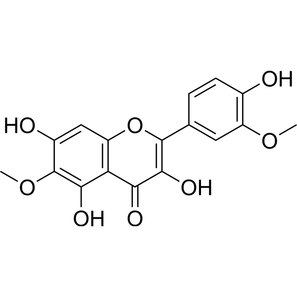
Spinacetin
Based on 1 publication(s) in Google Scholar
Spinacetin is an orally active flavonoid glycoside found in Inula japonica, with antioxidant and anti-inflammatory activities. Spinacetin inhibits major inflammatory signaling pathways such as Syk, MAPK, and NF-κB, and reduces Ca2+-dependent release of allergic mediators. Spinacetin induces protective autophagy and enhances antioxidant defense by upregulating SIRT3/SIRT1/PGC-1α, activating AMPK, and inhibiting mTOR and NF-κB, thereby exerting anti-apoptotic, cardioprotective, bladder-protective, and hepatoprotective effects. Spinacetin alleviates Doxorubicin (HY-15142A)-induced cardiotoxicity and reduces Cyclophosphamide (HY-17420)-induced hemorrhagic cystitis. Spinacetin can be used in research on allergic reactions, cardiotoxicity, cystitis, and cadmium-aggravated hepatic ischemia.
For research use only. We do not sell to patients.
- Purity : 99.98%
- CAS No.: 3153-83-1
- Formula: C17H14O8
- Molecular Weight:346.29
-
Storage:
4°C, sealed storage, away from moisture and light
* In solvent : -80°C, 6 months; -20°C, 1 month (sealed storage, away from moisture and light)

To place orders, for customer services and technical support, please contact: MedChemExpress USA
Tel: 609-228-6898 E-mail: [email protected] [email protected]


-
 Biological Activity
-
Chemical Information
-
Solvent & Solubility
- Protocols
- Purity & Documentation
- References
-
 Help & FAQs


Publications Citing Use of MedChemExpress (MCE) Spinacetin
MoreAll Histamine Receptor Isoforms
MoreAll Phospholipase Isoforms
MoreAll Calcium Channel Isoforms
MoreAll AMPK Isoforms
More

Biological Activity
Description
|
IL-1β |
IL-6 |
SIRT3 |
SIRT1 |
In Vitro
Spinacetin (10 μM; 24 h) protects H9C2 cardiomyoblasts from Doxorubicin-induced cytotoxicity and apoptosis, and this protection involves autophagy[2].
Spinacetin (10 μM) induces autophagy and reduces apoptosis in doxorubicin-treated H9C2 cells, and autophagy inhibition or Atg7 knockdown reverses these protective effects[2].
Spinacetin (10 μM) protects H9C2 cells from doxorubicin-induced cardiotoxicity by activating the SIRT3/AMPK/mTOR pathway to induce protective autophagy and inhibit apoptosis[2].
Spinacetin (1-5 µM; 1 h) suppresses IgE/Ag-induced histamine release and Ca2+ mobilization in BMMCs[1].
Spinacetin (1-5 µM; 1 h) inhibits PLCγ, LAT, and Syk phosphorylation without affecting Lyn or Fyn in IgE/Ag-activated BMMCs[1].
Spinacetin (1-5 µM; 1 h) suppresses IgE/Ag-induced MAPK, cPLA2, Akt/IκB/NF-κB, and PLCγ activation in RBL-2H3 cells[1].
Spinacetin (1-5 µM; 1 h) does not inhibit MAPK, cPLA2, Akt, or IKKα/β phosphorylation in PMA/A23187-stimulated HMC-1 cells[1].
Spinacetin (1-5 µM; 1 h) decreases LTC4 production and blocks cPLA2 activation and MAPK phosphorylation in IgE/Ag-stimulated BMMCs[1].
Spinacetin (1-5 µM; 1 h) suppresses IL-6 and COX-2 expression and Akt/IκBα/NF-κB signaling in IgE/Ag-stimulated BMMCs[1].
Spinacetin (1-5 µM) does not reduce BMMC viability below 5 µM, supporting the use of 1-5 µM for mechanistic assays[1].
MedChemExpress (MCE) has not independently confirmed the accuracy of these methods. They are for reference only. Further protocols information, click here.
-
Cell Line:RBL-2H3 mast cells
-
Concentration:1, 2, 5 µM
-
Incubation Time:1 h (pre-incubation)
-
Result:Stimulation of RBL-2H3 with IgE/Ag increased ERK1/2, JNK1/2, and p38 phosphorylation compared with control cells; this compound reduced activation of the MAPKs.
This compound markedly reduced translocation of phosphorylated cPLA2 from cytosol into the nucleus.
This compound suppressed IκBα phosphorylation and degradation and NF-κB activation, and inhibited Akt.
Phosphorylation of PLCγ was inhibited by this compound.
-
Cell Line:HMC-1 human mast cells
-
Concentration:1, 2, 5 µM
-
Incubation Time:1 h (pre-incubation)
-
Result:However, this compound did not suppress phosphorylation of MAPKs, cPLA2, Akt and IKKα/β in HMC-1 cells.
-
Cell Line:H9C2 cell line
-
Concentration:10 μM
-
Incubation Time:24 h
-
Result:The IC50 of Doxorubicin in H9C2 cells was 4.235 μM.
Significantly inhibited doxorubicin-induced cytotoxicity in H9C2 cells.
Co-administration with CQ significantly diminished the protective effects of this compound.
Annexin V-PI apoptosis analysis verified these results.
-
Cell Line:H9C2 cell line
-
Concentration:10 μM
-
Incubation Time:30 min (MDC staining)
-
Result:Significantly increased the LC3II/LC3I ratio in H9C2 cells.
3-MA reversed the this compound-mediated upregulation of LC3II/LC3I and increased the diminished apoptotic proteins cleaved-caspase3 and cleaved-PARP.
Atg7 knockdown reversed this compound-induced autophagy and protection from apoptosis.
MDC-stained puncta significantly decreased in the doxorubicin-treated group and significantly increased after this compound administration.
-
Cell Line:H9C2 cell line
-
Concentration:10 μM
-
Incubation Time:48 h (SIRT3 siRNA transfection)
-
Result:Activated AMPK (increased p-AMPK), inhibited mTOR (decreased p-mTOR), and increased SIRT3 expression.
SIRT3 knockdown abolished the this compound-mediated upregulation of p-AMPK and inhibition of mTOR.
After SIRT3 knockdown, the LC3II/LC3I ratio did not increase, cleaved-caspase3 and cleaved-PARP remained at high levels, and apoptotic cell rates after doxorubicin exposure were not significantly reduced.
Total AMPK and mTOR levels were not significantly altered.
In Vivo
Spinacetin (20 mg/kg/day; p.o. (gavage); once daily; for 14 consecutive days) attenuates Cyclophosphamide (HY-17420)-induced cystometric dysfunction and biochemical alterations in female Wistar rats[3].
Spinacetin (25-50 mg/kg; p.o.; single dose) attenuates IgE/Ag-mediated passive cutaneous anaphylaxis in mice[1].
Spinacetin (50 mg/kg; p.o.; once daily; for 10 consecutive days) exerts a protective effect against hepatic ischemia/reperfusion injury in male albino rats by restoring antioxidant enzymes, improving liver function markers, reducing apoptosis, and preserving liver histology[4].
Spinacetin (50 mg/kg; p.o.; once daily; for 10 consecutive days) attenuates cadmium-aggravated hepatic ischemia/reperfusion injury in male albino rats by improving antioxidant status, reducing oxidative stress and apoptosis, restoring liver function markers, and protecting liver histology[4].
Spinacetin (20 mg/kg/day; p.o.; once daily; for 14 consecutive days) alone does not significantly alter cystometric parameters in healthy female Wistar rats[3].
MedChemExpress (MCE) has not independently confirmed the accuracy of these methods. They are for reference only.
-
Animal Model:ICR (male, 7 weeks old)[1]
-
Dosage:25 mg/kg; 50 mg/kg
-
Administration:p.o.; once; 1 h before intravenous DNP-HSA challenge
-
Result:This compound markedly decreased the PCA reaction, reflected by the amount of diffused dye and ear thickness, compared with the Ag-challenged group.
Oral administration of this compound dose-dependently attenuated the IgE/Ag-mediated PCA reaction.
There was no variation between the Ag treated group and the this compound treated group.
-
Animal Model:Sprague-Dawley (SD) (male, 250-300 g)[2]
-
Dosage:50 mg/kg; 100 mg/kg
-
Administration:i.g.; once daily; 14 days
-
Result:Interestingly, the application of this compound (lower dose and higher dose) significantly reversed the cytotoxicity of doxorubicin on primary cardiomyocytes.
This compound at the concentration of 50 mg/kg or 100 mg/kg did not present significant cytotoxicity on cardiomyocytes.
Significantly reversed the structure damage induced by doxorubicin in the pathology evaluation of rat hearts.
The management of this compound potently inhibited the doxorubicin-induced over synthesis of those biomarkers.
This compound could also decrease the doxorubicin-mediated upregulation of MDA in a dose-dependent manner.
Single-use of this compound in two doses did not alter the original level of the abovementioned biomarkers or MDA level.
Doxorubicin significantly induced primary myocardial cell apoptosis.
After the administration of this compound, the apoptotic cells in the myocardium significantly decreased.
This compound did not induce myocardial cell apoptosis.
Annexin V/PI flow cytometry of primary myocardial cells printed quadrant values included upper-right quadrant values of 1.54% for control, 30.4% for DOX, 6.23% for DOX+SP, and 3.69% for SP; lower-right quadrant values were 4.28% for control, 3.04% for DOX, 4.83% for DOX+SP, and 4.94% for SP.
The cleaved-caspase3 and cleaved-PARP were potently upregulated in the doxorubicin group.
The administration of this compound significantly decreased the doxorubicin-induced upregulation of cleaved-caspase3 as well as cleaved-PARP.
Doxorubicin significantly decreased the level of LC3II.
The administration of this compound significantly increased the expression level of LC3II in primary myocardial cells.
-
Animal Model:Wistar (female, initially 200-225 g)[3]
-
Dosage:20 mg/kg/day
-
Administration:p.o. (oral gavage); once daily; 14 consecutive days
-
Result:Basal pressure was 2.5 cm H2O.
Threshold pressure was 6.6 cm H2O.
Detrusor overactivity index was 39 cm H2O/mL.
Non-voiding contraction frequency was 0.28 times/filling phase.
Volume threshold to elicit NVC was 55%.
Non-voiding contraction amplitude was 2.3 cm H2O.
Bladder compliance was 0.24 mL/cm H2O.
Micturition voiding pressure was 49 cm H2O.
Intercontraction interval was 994 s.
Voided volume was 0.97 mL.
Post-void residual was 0.074 mL.
Area under the pressure curve was 13 cm H2O/s.
No significant changes in the analyzed urine, detrusor, or urothelial biomarkers were found after this compound alone compared with the saline group.
-
Animal Model:Wistar (female, initially 200-225 g)[3]
-
Dosage:20 mg/kg/day
-
Administration:p.o. (oral gavage); once daily; 14 consecutive days; initiated after Cyclophosphamide
-
Result:Basal pressure was 3.2 cm H2O.
Threshold pressure was 7.8 cm H2O.
Detrusor overactivity index was 213 cm H2O/mL.
Non-voiding contraction frequency was 1.2 times/filling phase.
Volume threshold to elicit NVC was 64%.
Non-voiding contraction amplitude was 3.2 cm H2O.
Bladder compliance was 0.22 mL/cm H2O.
Micturition voiding pressure was 45 cm H2O.
Intercontraction interval was 946 s.
Voided volume was 0.78 mL.
Post-void residual was 0.068 mL.
Area under the pressure curve was 16 cm H2O/s.
This compound prevented the Cyclophosphamide-induced increase in bladder wall thickness.
Bladder edema was attenuated by this compound in the Evans Blue assessment.
This compound lowered Cyclophosphamide-induced c-Fos expression in all three studied central micturition areas: MPA, PMC, and vlPAG.
This compound restored Cyclophosphamide-induced elevations of urine BDNF and NGF, detrusor Rho kinase and VAChT, and urothelial TNF-α, IL-1β, IL-6, CGRP, malondialdehyde, 3-nitrotyrosine, IGFBP-3, occludin, OCT-3, ORM1, PAC1, SNAP23, SNAP25, and SV2A.
This compound restored Cyclophosphamide-induced decreases in HB-EGF, HPX, T-H protein, and ZO1.
-
Animal Model:albino (male, 13-14 weeks old, 225 g)[4]
-
Dosage:50 mg/kg
-
Administration:p.o. (oral gavage); once daily; for 10 consecutive days before the I/R procedure
-
Result:Hepatic oxidative stress markers in the I/R + this compound group were CAT 12.82 U/mg protein; SOD 13.68 U/mg protein; GSR 12.13 nM NADPH oxidized/min/mg tissue; GPx 14.74 U/mg protein; GSH 15.47 μmol/g tissue; GST 17.50 U/mg protein; HO-1 331.45 pmoles bilirubin/mg protein/h; MDA 2.03 nmol/g tissue; ROS 2.07 nmol/g tissue.
Liver function markers in the I/R + this compound group were ALT 46.32 U/L; AST 79.29 U/L; ALP 107.06 U/L; GGT 13.97 IU/mL; total protein 13.89 g/dL; albumin 13.59 g/dL.
Apoptotic biomarkers in the I/R + this compound group were Bax 7.43 pg/mL; Caspase-3 8.79 pg/mL; Caspase-9 9.41 pg/mL; Bcl-2 11.39 ng/mL.
Hepatic histology in the IR + SPI group showed normal histology present; mild damage present; no moderate or severe damage.
mRNA expression changes: I/R + this compound upregulated AMPK, SIRT1, PGC-1α, TFAM, and ERRα and inhibited pro-inflammatory cytokine mRNA expression of NF-κB, IL-6, IL-1β, TNF-α, and COX-2 relative to the I/R group.
-
Animal Model:albino (male, 13-14 weeks old, 225 g)[4]
-
Dosage:50 mg/kg
-
Administration:p.o. (oral gavage); once daily; for 10 consecutive days before the I/R procedure
-
Result:Hepatic oxidative stress markers in the I/R + Cd + this compound group were CAT 9.32 U/mg protein; SOD 9.89 U/mg protein; GSR 9.97 nM NADPH oxidized/min/mg tissue; GPx 11.43 U/mg protein; GSH 12.79 μmol/g tissue; GST 14.88 U/mg protein; HO-1 198.67 pmoles bilirubin/mg protein/h; MDA 2.97 nmol/g tissue; ROS 2.99 nmol/g tissue.
Liver function markers in the I/R + Cd + this compound group were ALT 57.45 U/L; AST 87.34 U/L; ALP 117.41 U/L; GGT 15.89 IU/mL; total protein 10.83 g/dL; albumin 11.32 g/dL.
Apoptotic biomarkers in the I/R + Cd + this compound group were Bax 12.17 pg/mL; Caspase-3 11.76 pg/mL; Caspase-9 12.57 pg/mL; Bcl-2 8.89 ng/mL.
Hepatic histology in the IR + Cd + SPI group showed normal histology present; mild damage present; no moderate or severe damage.
mRNA expression changes: I/R + Cd + this compound suppressed NF-κB, IL-6, IL-1β, TNF-α, and COX-2 and escalated SIRT1, PGC-1α, TFAM, and ERRα relative to the I/R + Cd group.


Chemical Information
-
CAS No. 3153-83-1
-
Appearance Solid
-
Molecular Weight 346.29
-
Formula C17H14O8
-
Color Light yellow to yellow
-
SMILES
COC1=C(O)C=CC(C2=C(O)C(C3=C(C=C(O)C(OC)=C3O)O2)=O)=C1
-
Structure Classification
-
Shipping
Room temperature in continental US; may vary elsewhere.
-
Storage
4°C, sealed storage, away from moisture and light
* In solvent : -80°C, 6 months; -20°C, 1 month (sealed storage, away from moisture and light)


Publications (1)
-
Journal Impact Factor
-
Most Recent
-
Int J Mol Sci
Plant-Derived Spinacetin Mitigates Cyclophosphamide-Induced Hemorrhagic Cystitis in Rats. [Abstract]2026 Mar 27;27(7):3056. PMID: 41977243


Solvent & Solubility
In Vitro:
DMSO : 100 mg/mL (288.78 mM; Need ultrasonic; Hygroscopic DMSO has a significant impact on the solubility of product, please use newly opened DMSO)
Please refer to the solubility information to select the appropriate solvent. Once prepared, please aliquot and store the solution to prevent product inactivation from repeated freeze-thaw cycles.
Storage method and period of stock solution: -80°C, 6 months; -20°C, 1 month (sealed storage, away from moisture and light). When stored at -80°C, please use it within 6 months. When stored at -20°C, please use it within 1 month.
Please refer to the solubility information to select the appropriate solvent. Once prepared, please aliquot and store the solution to prevent product inactivation from repeated freeze-thaw cycles.
Storage method and period of stock solution: -80°C, 6 months; -20°C, 1 month (sealed storage, away from moisture and light). When stored at -80°C, please use it within 6 months. When stored at -20°C, please use it within 1 month.
Concentration (start) × Volume (start) = Concentration (final) × Volume (final)
In Vivo:
Select the appropriate dissolution method based on your experimental animal and administration route.
- For the following dissolution methods, please ensure to first prepare a clear stock solution using an In Vitro approach and then sequentially add co-solvents:
- To ensure reliable experimental results, the clarified stock solution can be appropriately stored based on storage conditions. As for the working solution for In Vivo experiments, it is recommended to prepare freshly and use it on the same day.
- The percentages shown for the solvents indicate their volumetric ratio in the final prepared solution. If precipitation or phase separation occurs during preparation, heat and/or sonication can be used to aid dissolution.
Add each solvent one by one: 10% DMSO 40% PEG300 5% Tween-80 45% Saline
Solubility: ≥ 2.5 mg/mL (7.22 mM); Clear solution
This protocol yields a clear solution of ≥ 2.5 mg/mL (saturation unknown).
Taking 1 mL working solution as an example, add 100 μL DMSO stock solution (25.0 mg/mL) to 400 μL PEG300, and mix evenly; then add 50 μL Tween-80 and mix evenly; then add 450 μL Saline to adjust the volume to 1 mL.
Preparation of Saline: Dissolve 0.9 g sodium chloride in ddH₂O and dilute to 100 mL to obtain a clear Saline solution.
Add each solvent one by one: 10% DMSO 90% (20% SBE-β-CD in Saline)
Solubility: ≥ 2.5 mg/mL (7.22 mM); Clear solution
This protocol yields a clear solution of ≥ 2.5 mg/mL (saturation unknown).
Taking 1 mL working solution as an example, add 100 μL DMSO stock solution (25.0 mg/mL) to 900 μL 20% SBE-β-CD in Saline, and mix evenly.
Preparation of 20% SBE-β-CD in Saline (4°C, storage for one week): 2 g SBE-β-CD powder is dissolved in 10 mL Saline, completely dissolve until clear.
In Vivo Dissolution Calculator
Please enter the basic information of animal experiments:
-
-
-
-
Recommended: Prepare an additional quantity of animals to account for potential losses during experiments.
Please enter your animal formula composition:
-
%DMSO +
Recommended: Keep the proportion of DMSO in working solution below 2% if your animal is weak.
-
%+
-
+%Tween-80 + +
-
%Saline +
The co-solvents required include: DMSO, . All of co-solvents are available by MedChemExpress (MCE). , Tween 80. All of co-solvents are available by MedChemExpress (MCE).
Working solution concentration: 0.22 mg/mL
Method for preparing stock solution: mg drug dissolved in μL DMSO. Stock solution concentration: mg/mL. * In solvent : -80°C, 6 months; -20°C, 1 month (sealed storage, away from moisture and light)
1. Take μL DMSO stock solution;
2. Add μL .
μL , mix evenly;
3. Then add μL Tween 80, mix evenly;
4. Then add μL
Please ensure that the stock solution in the first step is dissolved to a clear state, and add co-solvents in sequence. You can use ultrasonic heating (ultrasonic cleaner, recommended frequency 20-40 kHz), vortexing, etc. to assist dissolution.

Protocols
-
Detection of Apoptosis
Apoptosis, also called programmed cell death, is generally characterized by distinct morphological characteristics.
-
Autophagy
Autophagy is a process in which eukaryotic cells use lysosomes to degrade their own cytoplasmic proteins and damaged organelles under the regulation of autophagy related gene (Atg). Microtubule-associated proteins light chain 3 (LC3) is recognized as autophagy marker, which transfers from cytoplasmic LC3 (LC3-I) to membrane type (LC3-II). LC3-II/I ratio could be detected by Western Blot and fluorescence microscopy.
-
Apoptosis Solutions
Apoptosis is a regulated, generally non-lytic cell-death pathway that removes unwanted, damaged, infected, or abnormal cells through coordinated morphological changes, caspase activation, DNA fragmentation, and membrane remodeling. The intrinsic apoptosis pathway is controlled mainly by mitochondrial outer membrane permeabilization, BCL-2 family proteins, cytochrome c release, apoptosome formation, caspase-9 activation, and downstream executioner caspase-3/7 activation. The extrinsic apoptosis pathway is initiated by death receptors such as Fas, TNFR, and TRAIL receptors, which recruit adaptor proteins and activate caspase-8 before engaging executioner caspases or mitochondrial amplification through BID cleavage. Apoptosis is linked to many phenotypes, including cancer cell killing, tissue homeostasis, immune regulation, neurodegeneration, infection response, and treatment-induced cytotoxicity; unresolved questions include how apoptosis interacts with necroptosis, pyroptosis, ferroptos
-
Research Protocol for Inflammation-related Diseases
The NLRP3 inflammasome is a cytosolic innate immune signaling platform that integrates priming signals and danger-signal activation to promote caspase-1 activation, maturation of IL-1β and IL-18, and gasdermin D-mediated pyroptotic cell death. The core experimental logic is to determine whether inflammatory disease phenotypes are driven by increased NLRP3 expression, ASC-containing inflammasome assembly, caspase-1 cleavage, GSDMD cleavage, and extracellular release of IL-1β/IL-18 rather than by nonspecific cell injury alone. The pathway is strongly linked to inflammation-related disease phenotypes because monosodium urate crystals activate NALP3/NLRP3 inflammasome signaling in gout-like crystal inflammation, cholesterol crystals activate NLRP3 inflammasomes in atherogenesis models, and DSS-induced intestinal inflammation has been reported to involve NLRP3 inflammasome activity. However, experimental colitis studies also show context-dependent protective effects of NLRP3 inflammasome co
-
Ca2+ Staining Technique
Ca2+ staining is an experimental technique that utilizes specific fluorescent probes (such as Fluo-4 AM, Fura-2, etc.) to qualitatively or quantitatively detect dynamic changes in intracellular Ca2+ concentrations; this is achieved by monitoring the changes in fluorescent signals generated when these probes bind to free intracellular calcium ions. The underlying principle relies primarily on the presence of chelating groups within the probe's molecular structure that possess high affinity for calcium ions.
-
Lysosome and acidic-vesicle live-cell staining
Lysosome and acidic-vesicle live-cell staining detects acidic intracellular compartments by using membrane-permeant acidotropic probes that accumulate in low-pH vesicles, including lysosomes, late endosomes, autolysosomes, and acidic phagosomes. LysoTracker staining is commonly used as an intensity-based readout of acidic lysosomal compartment abundance or enlargement, while acridine orange produces green fluorescence in less concentrated compartments and red fluorescence after concentration-dependent accumulation in acidic vesicular organelles. Loss or reduction of acridine-orange red signal can be used as a readout of lysosomal membrane permeabilization or reduced acidic-vesicle integrity. This protocol is designed for live cultured cells and can be adapted for fluorescence microscopy, high-content imaging, plate-reader readout, or flow cytometry when the selected literature supports the readout. Because these dyes report acidotropic accumulation rather than lysosome identity alone,
-
Macroautophagy Solutions
Macroautophagy is a conserved lysosome-dependent degradation pathway in which cytoplasmic material is sequestered into double-membrane autophagosomes and delivered to lysosomes for degradation and recycling. The pathway supports cellular homeostasis during nutrient limitation, organelle stress, protein-aggregate accumulation, infection, differentiation, and tissue remodeling by coupling cargo sequestration, autophagosome maturation, lysosomal fusion, and degradation of cargo-derived macromolecules. The core molecular sequence includes initiation by nutrient- and stress-regulated autophagy machinery, autophagosome nucleation, LC3/ATG8-family conjugation to autophagosomal membranes, cargo selection through receptors such as SQSTM1/p62, autophagosome-lysosome fusion, and lysosomal degradation. LC3 was identified as a mammalian homolog of yeast Atg8 that localizes to autophagosomal membranes after processing, and p62/SQSTM1 was shown to connect ubiquitinated cargo with autophagic degradati


Purity & Documentation
-
Data Sheet (301 KB)
-
SDS (252 KB)
- English - EN (252 KB)
- Français - FR (252 KB)
- Deutsch - DE (252 KB)
- Norwegian - NO (252 KB)
- Español - ES (252 KB)
- Swedish - SV (252 KB)
- Italian - IT (252 KB)
- Korean - KR (252 KB)
- Portuguese - PT (252 KB)
-
Handling Instructions (2659 KB)
References
[1]. Ji N, et al. Spinacetin Suppresses the Mast Cell Activation and Passive Cutaneous Anaphylaxis in Mouse Model. Frontiers in pharmacology. 2018;9:824. [Content Brief]
[3]. Wróbel J, et al. Plant-Derived Spinacetin Mitigates Cyclophosphamide-Induced Hemorrhagic Cystitis in Rats. International journal of molecular sciences. 2026 Mar 27;27(7):3056. [Content Brief]


Complete Stock Solution Preparation Table
Please refer to the solubility information to select the appropriate solvent. Once prepared, please aliquot and store the solution to prevent product inactivation from repeated freeze-thaw cycles.
Storage method and period of stock solution: -80°C, 6 months; -20°C, 1 month (sealed storage, away from moisture and light). When stored at -80°C, please use it within 6 months. When stored at -20°C, please use it within 1 month.
| Optional Solvent | Concentration Solvent Mass | 1 mg | 5 mg | 10 mg | 25 mg |
|---|---|---|---|---|---|
| DMSO | 1 mM | 2.8878 mL | 14.4388 mL | 28.8775 mL | 72.1938 mL |
| 5 mM | 0.5776 mL | 2.8878 mL | 5.7755 mL | 14.4388 mL | |
| 10 mM | 0.2888 mL | 1.4439 mL | 2.8878 mL | 7.2194 mL | |
| 15 mM | 0.1925 mL | 0.9626 mL | 1.9252 mL | 4.8129 mL | |
| 20 mM | 0.1444 mL | 0.7219 mL | 1.4439 mL | 3.6097 mL | |
| 25 mM | 0.1155 mL | 0.5776 mL | 1.1551 mL | 2.8878 mL | |
| 30 mM | 0.0963 mL | 0.4813 mL | 0.9626 mL | 2.4065 mL | |
| 40 mM | 0.0722 mL | 0.3610 mL | 0.7219 mL | 1.8048 mL | |
| 50 mM | 0.0578 mL | 0.2888 mL | 0.5776 mL | 1.4439 mL | |
| 60 mM | 0.0481 mL | 0.2406 mL | 0.4813 mL | 1.2032 mL | |
| 80 mM | 0.0361 mL | 0.1805 mL | 0.3610 mL | 0.9024 mL | |
| 100 mM | 0.0289 mL | 0.1444 mL | 0.2888 mL | 0.7219 mL |


Spinacetin Related Classifications
HY-N10182 Related Classifications
 Powered by Bioz