



Pratol

Based on 1 Customer Validation
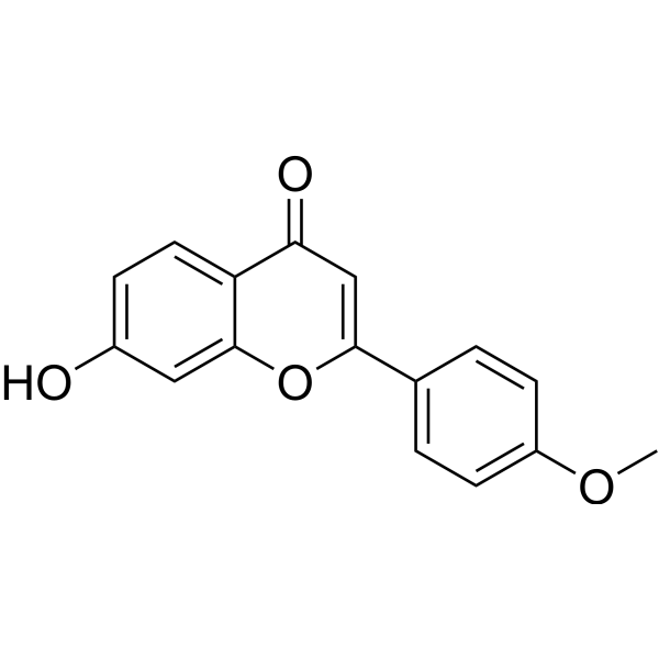
Pratol (7-Hydroxy-4'-methoxyflavone) is an NF-κB inhibitor. Pratol also inhibits NO, PGE2, TNF-α, IL-1β, and IL-6 production. Pratol increases the phosphorylation of p38 MAPK, JNK, and ERK, decreases AKT phosphorylation, and upregulates MITF, tyrosinase, TRP-1, and TRP-2 through a PKA-dependent signaling pathway. Pratol reduces iNOS and COX-2 protein expression, inhibits p65 phosphorylation, and protects IκBα from degradation, thereby inhibiting NF-κB nuclear translocation. Pratol can be used in research on hypopigmentary disorders, inflammatory diseases, cervical cancer, and colon cancer.
Nur für Forschungszwecke. Wir verkaufen nicht an Patienten.
- Reinheit : 99.16%
- CAS. Nr.: 487-24-1
- Formel: C16H12O4
- Molecular Weight:268.26
-
Speicherung:
4°C, stored under nitrogen
* In solvent : -80°C, 6 months; -20°C, 1 month (stored under nitrogen)

To place orders, for customer services and technical support, please contact: MedChemExpress USA
Tel: 609-228-6898 E-mail: [email protected] [email protected]


-
 Biologische Aktivität
-
Chemical Information
-
Lösungsmittel & Löslichkeit
- Protokoll
- Reinheit & Dokumentation
- Verweise
-
 Help & FAQs


Alle TNF Receptor Isoform-spezifische Produkte anzeigen
More

Biologische Aktivität
Beschreibung
|
NF-κB |
IL-1β |
TNF-α |
IL-6 |
p38 MAPK |
JNK |
ERK |
Akt |
PKA |
MITF |
p65 |
TRP-1 |
TRP-2 |
IκBα |
Cellular Effect
|
Cell Line

|
Type | Value | Description | References |
|---|---|---|---|---|
| HeLa | IC50 |
25.73 μg/mL
|
Cytotoxicity against human HeLa cervical cancer cells assessed as reduction in cell viability incubated for 24 hrs by MTT assay.
Cytotoxicity against human HeLa cervical cancer cells assessed as reduction in cell viability incubated for 24 hrs by MTT assay.
|
1.4978148 |
| WiDr | IC50 |
83.75 μg/mL
|
Cytotoxicity against human WiDr colon cancer cells assessed as reduction in cell viability incubated for 24 hrs by MTT assay.
Cytotoxicity against human WiDr colon cancer cells assessed as reduction in cell viability incubated for 24 hrs by MTT assay.
|
1.4978148 |
In Vitro
Pratol (6.25-50 μM; 48 h) significantly increased melanin content in B16F10 mouse melanoma cells in a dose-dependent manner, with no cytotoxicity at concentrations up to 50 μM[1].
Pratol (6.25-50 μM; 72 h) increases tyrosinase activity in B16F10 mouse melanoma cells in a dose-dependent manner[1].
Pratol (6.25-50 μM; 48 h (tyrosinase, TRP-1, TRP-2); 24 h (MITF dose-response); 22-24 h (MITF time-course)) increases the protein expression of tyrosinase, TRP-1, TRP-2, and MITF in B16F10 mouse melanoma cells[1].
Pratol (6.25-50 μM; 4 h) inhibits AKT phosphorylation in B16F10 mouse melanoma cells in a dose-dependent manner[1].
Pratol (6.25-50 μM; 4 h) activates the MAPK pathway by increasing the phosphorylation of p38, ERK, and JNK in B16F10 mouse melanoma cells[1].
Pratol increases tyrosinase activity and melanogenesis gene expression in B16F10 mouse melanoma cells through a PKA-dependent signaling pathway[1].
Pratol (7-Hydroxy-4'-methoxyflavone) (24 h) exhibited active cytotoxic effects against HeLa cervical cancer cells with an IC50 of 25.73 µg/mL, and showed considerable activity against WiDr colon cancer cells with an IC50 of 83.75 µg/mL[3].
Pratol (6.25-50 μM (Pratol); 10 μM (inhibitors))-induced tyrosinase upregulation is associated with the activation of p38 and JNK, but not the ERK pathway, in B16F10 mouse melanoma cells[1].
Pratol (25-100 μM; 24 h) inhibits NO production in LPS-stimulated RAW 264.7 cells, with at least 45% inhibition of production at 100 μM[2].
Pratol (25-100 μM; 24 h) concentration-dependently inhibited PGE2 production in LPS-stimulated RAW 264.7 cells, with at least 85% inhibition of production at 100 μM[2].
Pratol (25-100 μM; 24 h) concentration-dependently inhibited the production of TNF-α, IL-1β, and IL-6 in LPS-stimulated RAW 264.7 cells[2].
Pratol (25-100 μM; 24 h) downregulates iNOS and COX-2 protein expression in LPS-stimulated RAW 264.7 cells[2].
Pratol (25-100 μM; 30 min) protects IκBα from degradation in LPS-stimulated RAW 264.7 cells[2].
Pratol (25-100 μM; 30 min) inhibits p65 phosphorylation in LPS-stimulated RAW 264.7 cells, thereby inhibiting the NF-κB signaling pathway[2].
MedChemExpress (MCE) has not independently confirmed the accuracy of these methods. They are for reference only.
-
Cell Line:B16F10 mouse melanoma cells
-
Concentration:6.25 μM; 12.5 μM; 25 μM; 50 μM
-
Incubation Time:48 h
-
Result:Increased melanin content to 99.2%, 108.9%, 176.2%, and 208.7% at 6.25, 12.5, 25, and 50 μM, respectively.
Maintained cell viability at 101.0%, 99.2%, 92.1%, and 88.4% at 6.25, 12.5, 25, and 50 μM, respectively.
-
Cell Line:B16F10 mouse melanoma cells
-
Concentration:6.25 μM; 12.5 μM; 25 μM; 50 μM
-
Incubation Time:48 h (tyrosinase, TRP-1, TRP-2); 24 h (MITF dose-response); 22-24 h (MITF time-course)
-
Result:Significantly increased the expression of tyrosinase, TRP-1, and TRP-2.
Increased MITF expression in a time- and concentration-dependent manner, reaching a maximum level after 24 h of treatment.
-
Cell Line:B16F10 mouse melanoma cells
-
Concentration:6.25 μM; 12.5 μM; 25 μM; 50 μM
-
Incubation Time:4 h
-
Result:Reduced AKT phosphorylation in a dose-dependent manner, with significant reduction observed at 50 μM.\nSignificantly increased the phosphorylation of p38 MAPK, ERK, and JNK in a dose-dependent manner.
-
Cell Line:RAW 264.7 macrophages
-
Concentration:25, 50, 100 μM
-
Incubation Time:24 h
-
Result:Decreased PGE2 production in a concentration-dependent manner.
Inhibited PGE2 production by at least 85% at 100 μM.\nInhibited the expression of TNF-α, IL-1β, and IL-6 in a concentration-dependent manner relative to LPS alone.
-
Cell Line:RAW 264.7 macrophages
-
Concentration:25, 50, 100 μM
-
Incubation Time:24 h
-
Result:Significantly decreased protein levels of iNOS and COX-2 compared to LPS alone.
-
Cell Line:RAW 264.7 macrophages
-
Concentration:25, 50, 100 μM
-
Incubation Time:30 min
-
Result:Significantly upregulated IκBα expression compared to LPS alone.\nSignificantly decreased p-p65 levels compared to LPS alone.
- Kinase activity and phosphorylation assays
- Western Blot
- RNA extraction experimental
- Cytoplasmic-Nuclear Fractionated Protein Extraction
- Research Protocol for Inflammation-related Diseases
- Protocol for Kinase activity and phosphorylation assays
- Pyroptosis Solutions
- Protocol For Protein Expression And Purification


Chemical Information
-
CAS. Nr. 487-24-1
-
Appearance Solid
-
Molecular Weight 268.26
-
Formel C16H12O4
-
Color White to yellow
-
SMILES
O=C1C=C(C2=CC=C(OC)C=C2)OC3=CC(O)=CC=C13
-
Synonyms
7-Hydroxy-4'-methoxyflavone
-
Structure Classification
-
Initial Source
-
Versand
Room temperature in continental US; may vary elsewhere.
-
Speicherung
4°C, stored under nitrogen
* In solvent : -80°C, 6 months; -20°C, 1 month (stored under nitrogen)


Lösungsmittel & Löslichkeit
In Vitro:
DMSO : 75 mg/mL (279.58 mM; Need ultrasonic; Hygroscopic DMSO has a significant impact on the solubility of product, please use newly opened DMSO)
Please refer to the solubility information to select the appropriate solvent. Once prepared, please aliquot and store the solution to prevent product inactivation from repeated freeze-thaw cycles.
Storage method and period of stock solution: -80°C, 6 months; -20°C, 1 month (stored under nitrogen). When stored at -80°C, please use it within 6 months. When stored at -20°C, please use it within 1 month.
Please refer to the solubility information to select the appropriate solvent. Once prepared, please aliquot and store the solution to prevent product inactivation from repeated freeze-thaw cycles.
Storage method and period of stock solution: -80°C, 6 months; -20°C, 1 month (stored under nitrogen). When stored at -80°C, please use it within 6 months. When stored at -20°C, please use it within 1 month.
Konzentration (Stammlösung) × Volumen (Stammlösung) = Konzentration (Ziellösung) × Volumen (Ziellösung)


Reinheit & Dokumentation
-
Data Sheet (288 KB)
-
SDS (393 KB)
- English - EN (393 KB)
- Français - FR (393 KB)
- Deutsch - DE (393 KB)
- Norwegian - NO (393 KB)
- Español - ES (393 KB)
- Swedish - SV (393 KB)
- Italian - IT (393 KB)
- Korean - KR (393 KB)
- Portuguese - PT (393 KB)
-
Handling Instructions (2659 KB)
Verweise


Complete Stock Solution Preparation Table
Please refer to the solubility information to select the appropriate solvent. Once prepared, please aliquot and store the solution to prevent product inactivation from repeated freeze-thaw cycles.
Storage method and period of stock solution: -80°C, 6 months; -20°C, 1 month (stored under nitrogen). When stored at -80°C, please use it within 6 months. When stored at -20°C, please use it within 1 month.
| Optional Solvent | Concentration Solvent Mass | 1 mg | 5 mg | 10 mg | 25 mg |
|---|---|---|---|---|---|
| DMSO | 1 mM | 3.7277 mL | 18.6386 mL | 37.2773 mL | 93.1932 mL |
| 5 mM | 0.7455 mL | 3.7277 mL | 7.4555 mL | 18.6386 mL | |
| 10 mM | 0.3728 mL | 1.8639 mL | 3.7277 mL | 9.3193 mL | |
| 15 mM | 0.2485 mL | 1.2426 mL | 2.4852 mL | 6.2129 mL | |
| 20 mM | 0.1864 mL | 0.9319 mL | 1.8639 mL | 4.6597 mL | |
| 25 mM | 0.1491 mL | 0.7455 mL | 1.4911 mL | 3.7277 mL | |
| 30 mM | 0.1243 mL | 0.6213 mL | 1.2426 mL | 3.1064 mL | |
| 40 mM | 0.0932 mL | 0.4660 mL | 0.9319 mL | 2.3298 mL | |
| 50 mM | 0.0746 mL | 0.3728 mL | 0.7455 mL | 1.8639 mL | |
| 60 mM | 0.0621 mL | 0.3106 mL | 0.6213 mL | 1.5532 mL | |
| 80 mM | 0.0466 mL | 0.2330 mL | 0.4660 mL | 1.1649 mL | |
| 100 mM | 0.0373 mL | 0.1864 mL | 0.3728 mL | 0.9319 mL |


Pratol Related Classifications
HY-N8719 Related Classifications
 Powered by Bioz
Keywords
- Pratol
- 487-24-1
- 7-Hydroxy-4'-methoxyflavone
- NF-κB
- PGE synthase
- TNF Receptor
- Interleukin Related
- p38 MAPK
- JNK
- ERK
- Akt
- PKA
- Microphthalmia Associated Transcription Factor (MITF)
- TRP Channel
- NO Synthase
- COX
- IKK
- RAW 264.7 cells
- B16F10 mouse melanoma cells
- MITF
- HeLa cervical cancer cells
- melanogenesis inducer
- WiDr colon cancer cells
- Inhibitor
- inhibitor
- inhibit