



Anemonin

Based on 1 Customer Validation
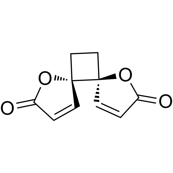
Anemonin (Pulsatilla camphor; Anemonine; trans-Anemonin) is a naturally occurring bislactone small molecule derived from Ranunculaceae with blood-brain barrier permeability, possessing a variety of activities including anti-inflammatory, antioxidant, neuroprotective, melanogenesis-inhibiting, and antiparasitic effects. Anemonin inhibits iNOS to reduce NO release; it inhibits PKC-θ protein expression and downregulates the pro-inflammatory factors TNF-α, IL-1β, and IL-6; it enhances the activities of the antioxidant enzymes SOD, CAT, and GSH-Px, and reduces MDA and ROS levels. Anemonin modulates the Bcl-2 / Bax / caspase-3 pathway to inhibit apoptosis; it downregulates melanogenesis-related molecules including MITF, TYR, TRP1, and TRP2, thereby inhibiting melanin synthesis in human melanocytes. Anemonin inhibits Leishmania and Schistosoma mansoni. Anemonin is used in research related to diseases such as hyperpigmentation, cerebral ischemia/reperfusion injury, inflammation, sepsis-induced acute lung injury, acute ulcerative colitis, leishmaniasis, and schistosomiasis.
For research use only. We do not sell to patients.
- Purity : 99.41%
- CAS No.: 508-44-1
- Formula: C10H8O4
- Molecular Weight:192.17
-
Storage:
4°C, sealed storage, away from moisture and light
* In solvent : -80°C, 6 months; -20°C, 1 month (sealed storage, away from moisture and light)

To place orders, for customer services and technical support, please contact: MedChemExpress USA
Tel: 609-228-6898 E-mail: [email protected] [email protected]


-
 Biological Activity
-
Chemical Information
-
Solvent & Solubility
- Protocols
- Purity & Documentation
- References
-
 Help & FAQs




Biological Activity
Description
|
iNOS |
Leishmania |
Schistosome |
Bax |
Bcl-2 |
Caspase 3 |
IL-1β |
IL-6 |
TNF-α |
Cellular Effect
|
Cell Line

|
Type | Value | Description | References |
|---|---|---|---|---|
| Melanocyte | IC50 |
43.4 μM
|
Inhibition of cellular tyrosinase activity in human melanocytes assessed by measuring L-dopa oxidation at 475 nm after 24 hrs of treatment.
Inhibition of cellular tyrosinase activity in human melanocytes assessed by measuring L-dopa oxidation at 475 nm after 24 hrs of treatment.
|
17766092 |
| RAW264.7 | IC50 |
5.37 μM
|
Inhibition of LPS-induced NO production in murine RAW 264.7 macrophage cells assessed by measuring nitrite concentration in culture medium spectrophotometrically after 24 hrs incubation.
Inhibition of LPS-induced NO production in murine RAW 264.7 macrophage cells assessed by measuring nitrite concentration in culture medium spectrophotometrically after 24 hrs incubation.
|
18281171 |
| Peritoneal macrophage | CC50 |
5.39 μg/mL
|
Cytotoxicity against mouse peritoneal macrophages assessed as reduction in cell viability incubated for 48 hrs by resazurin reduction assay.
Cytotoxicity against mouse peritoneal macrophages assessed as reduction in cell viability incubated for 48 hrs by resazurin reduction assay.
|
molecules26247473 |
In Vitro
Anemonin exhibits low cytotoxicity in human melanocytes, with cell viability remaining above 96% at 50 μM[1].
Anemonin (10-50 μM; 8-48 h) inhibits cellular tyrosinase activity in human melanocytes in a concentration- and time-dependent manner, with an IC50 of 43.4 μM[1].
Anemonin (20-50 μM; 8-48 h) reduces melanin content in human melanocytes; it decreases TYR, TRP1, and TRP2/DCT protein expression in human melanocytes in a dose- and time-dependent manner, particularly affecting TYR and TRP2[1].
Anemonin (20-50 μM; 24 h) downregulates the mRNA expression of TYR, TYRP1, and TYRP2 in human melanocytes in a dose-dependent manner[1].
Anemonin (20-40 μM; 24 h) downregulates MITF mRNA expression in human melanocytes in a dose-dependent manner[1].
Anemonin does not affect the viability of LPS-treated RAW 264.7 macrophages even at 100 μM[3].
Anemonin (2.5-30 μM; 24 h) inhibits LPS-induced NO production in RAW 264.7 macrophages with an IC50 of 5.37 μM[3].
Anemonin (2.5-30 μM; 24 h) inhibits iNOS protein expression in LPS-induced RAW 264.7 macrophages in a concentration-dependent manner[3].
Anemonin (2.5-30 μM; 6 h) inhibits LPS-induced iNOS mRNA expression in RAW 264.7 macrophages in a concentration-dependent manner, without affecting GAPDH mRNA expression[3].
Anemonin (2.5-10 μM; 48 h) shows no cytotoxicity in HT-29 cells and attenuates LPS-induced inflammation in HT-29 cells by downregulating the expression of IL-1β, TNF-α, and IL-6 in a dose-dependent manner[5].
Anemonin (2.5-10 μM; 24 h) is not cytotoxic to LPS-stimulated MH-S and MLE-12 cells[4].
Anemonin (2.5-10 μM; 24 h) dose-dependently inhibits LPS-induced TNF-α, IL-1β, and IL-6 mRNA expression and secretion in MH-S and MLE-12 cells; combination with the NF-κB inhibitor PDTC enhances the inhibition of inflammatory responses; the anti-inflammatory effect is suppressed under Nrf2 knockdown[4].
Anemonin (2.5-10 μM; 24 h) inhibits LPS-induced activation of the NF-κB pathway and enhances activation of the Nrf2/HO-1 pathway in MH-S and MLE-12 cells[4].
Anemonin (2.5-10 μM; 24 h) reverses LPS-induced oxidative stress in MH-S and MLE-12 cells by decreasing MDA content and increasing SOD and CAT activities[4].
Anemonin (2.5-10 μM) inhibits PKC-θ protein expression in a dose-dependent manner in HT-29 cells without affecting PRKCQ gene transcription, significantly reduces TNF-α mRNA levels, and exerts no significant effect on PRKCQ and PTPN2 gene transcription; PRKCQ overexpression reverses the inhibitory effects of Anemonin on cytokine mRNA production and cytokine protein production in HT-29 cells[5].
Anemonin (0.00565-1000 µg/mL; 48 h) exhibits a CC50 of 5.39 µg/mL (28.00 nM) against mouse peritoneal macrophages and shows hemolytic activity against human erythrocytes with an LC50 of 91.00 µg/mL (473.95 nM)[6].
Anemonin (0.000565-100 µg/mL; 72 h) exhibits potent anti-promastigote activity against L. aethiopica and L. donovani with IC50 values of 0.257 µg/mL (1.33 nM) and 0.303 µg/mL (1.58 nM), respectively; and exhibits potent anti-amastigote activity against L. aethiopica and L. donovani with IC50 values of 0.239 µg/mL (1.24 nM) and 0.368 µg/mL (1.91 nM), respectively[6].
Anemonin (1-10 µM; 72 h) exhibits high antischistosomal activity against S. mansoni NTS, with activity of 93.75% at 10 µM and 41.38% at 1 µM; it exhibits moderate antischistosomal activity against adult S. mansoni, with activity of 48.95% at 10 µM[6].
MedChemExpress (MCE) has not independently confirmed the accuracy of these methods. They are for reference only.
In Vivo
Anemonin (150 mg/kg; i.p.; single administration) penetrates the blood-brain barrier in rats and reaches a maximum brain-to-plasma partition coefficient (Ri) of 0.7 at 90 min after administration[2].
Anemonin (5-100 μM; incubation; 6 h for endothelium-denuded rings, 20 min for endothelium-intact rings) reverses LPS-induced hyporeactivity of blood vessels to phenylephrine without affecting eNOS-mediated endothelium-dependent relaxation, which is consistent with selective inhibition of iNOS[3].
Anemonin (10 mg/kg; i.p.; single injection simultaneously with LPS) attenuates sepsis-induced acute lung injury in mice by inhibiting inflammatory responses and oxidative stress[4].
Anemonin (10 mg/kg; i.p.) reduces LPS-induced mortality in a mouse model of sepsis-induced ALI[4].
Anemonin (2-10 mg/kg; i.p.; daily; 7 days) alleviates DSS-induced acute ulcerative colitis in mice by inhibiting colonic tissue inflammation and suppressing PKC-θ protein expression[5].
MedChemExpress (MCE) has not independently confirmed the accuracy of these methods. They are for reference only.
- RNA extraction experimental
- Western Blot
- Annexin V plus membrane-impermeant dye apoptosis staining
- ROS/oxidative-stress fluorescent staining
- Research Protocol for Inflammation-related Diseases
- Protocol For Protein Expression And Purification
- Detection of Apoptosis
- TUNEL staining for apoptotic DNA fragmentation
- LPS-Induced Endotoxemia/Systemic Inflammation
- DSS-Induced Colitis
- TNBS-Induced Colitis
- Transepithelial/transendothelial electrical resistance assay
- Apoptosis Solutions
- Inhalation Toxicity Study
- Pyroptosis Solutions


Chemical Information
-
CAS No. 508-44-1
-
Appearance Solid
-
Molecular Weight 192.17
-
Formula C10H8O4
-
Color Off-white to light yellow
-
SMILES
O=C1C=C[C@@]2([C@]3(CC2)C=CC(O3)=O)O1
-
Synonyms
Pulsatilla camphor; Anemonine; trans-Anemonin
-
Structure Classification
-
Initial Source
-
Shipping
Room temperature in continental US; may vary elsewhere.
-
Storage
4°C, sealed storage, away from moisture and light
* In solvent : -80°C, 6 months; -20°C, 1 month (sealed storage, away from moisture and light)


Solvent & Solubility
In Vitro:
DMSO : 25 mg/mL (130.09 mM; ultrasonic and warming and heat to 60°C; Hygroscopic DMSO has a significant impact on the solubility of product, please use newly opened DMSO)
Please refer to the solubility information to select the appropriate solvent. Once prepared, please aliquot and store the solution to prevent product inactivation from repeated freeze-thaw cycles.
Storage method and period of stock solution: -80°C, 6 months; -20°C, 1 month (sealed storage, away from moisture and light). When stored at -80°C, please use it within 6 months. When stored at -20°C, please use it within 1 month.
Please refer to the solubility information to select the appropriate solvent. Once prepared, please aliquot and store the solution to prevent product inactivation from repeated freeze-thaw cycles.
Storage method and period of stock solution: -80°C, 6 months; -20°C, 1 month (sealed storage, away from moisture and light). When stored at -80°C, please use it within 6 months. When stored at -20°C, please use it within 1 month.
Concentration (start) × Volume (start) = Concentration (final) × Volume (final)


Purity & Documentation
-
Data Sheet (308 KB)
-
SDS (252 KB)
- English - EN (252 KB)
- Français - FR (252 KB)
- Deutsch - DE (252 KB)
- Norwegian - NO (252 KB)
- Español - ES (252 KB)
- Swedish - SV (252 KB)
- Italian - IT (252 KB)
- Korean - KR (252 KB)
- Portuguese - PT (252 KB)
-
Handling Instructions (2659 KB)
References
[3]. Lee TH, et al. Anemonin, from Clematis crassifolia, potent and selective inducible nitric oxide synthase inhibitor. Journal of ethnopharmacology. 2008 Mar 28;116(3):518-27. [Content Brief]
[5]. Jiang L, et al. Anti-inflammatory effects of anemonin on acute ulcerative colitis via targeted regulation of protein kinase C-θ. Chinese medicine. 2022 Mar 28;17(1):39. [Content Brief]


Complete Stock Solution Preparation Table
Please refer to the solubility information to select the appropriate solvent. Once prepared, please aliquot and store the solution to prevent product inactivation from repeated freeze-thaw cycles.
Storage method and period of stock solution: -80°C, 6 months; -20°C, 1 month (sealed storage, away from moisture and light). When stored at -80°C, please use it within 6 months. When stored at -20°C, please use it within 1 month.
| Optional Solvent | Concentration Solvent Mass | 1 mg | 5 mg | 10 mg | 25 mg |
|---|---|---|---|---|---|
| DMSO | 1 mM | 5.2037 mL | 26.0186 mL | 52.0373 mL | 130.0931 mL |
| 5 mM | 1.0407 mL | 5.2037 mL | 10.4075 mL | 26.0186 mL | |
| 10 mM | 0.5204 mL | 2.6019 mL | 5.2037 mL | 13.0093 mL | |
| 15 mM | 0.3469 mL | 1.7346 mL | 3.4692 mL | 8.6729 mL | |
| 20 mM | 0.2602 mL | 1.3009 mL | 2.6019 mL | 6.5047 mL | |
| 25 mM | 0.2081 mL | 1.0407 mL | 2.0815 mL | 5.2037 mL | |
| 30 mM | 0.1735 mL | 0.8673 mL | 1.7346 mL | 4.3364 mL | |
| 40 mM | 0.1301 mL | 0.6505 mL | 1.3009 mL | 3.2523 mL | |
| 50 mM | 0.1041 mL | 0.5204 mL | 1.0407 mL | 2.6019 mL | |
| 60 mM | 0.0867 mL | 0.4336 mL | 0.8673 mL | 2.1682 mL | |
| 80 mM | 0.0650 mL | 0.3252 mL | 0.6505 mL | 1.6262 mL | |
| 100 mM | 0.0520 mL | 0.2602 mL | 0.5204 mL | 1.3009 mL |


Anemonin Related Classifications
HY-N0278 Related Classifications
 Powered by Bioz
Keywords
- Anemonin
- 508-44-1
- Pulsatilla camphor
- Anemonine
- trans-Anemonin
- PKC
- NO Synthase
- Parasite
- Caspase
- Bcl-2 Family
- Reactive Oxygen Species (ROS)
- Tyrosinase
- Interleukin Related
- TNF Receptor
- RAW 264.7 macrophages
- MH-S and MLE-12 cells
- HT-29 cells
- cerebral ischemia/reperfusion injury
- iNOS inhibitor
- tyrosinase inhibitor
- blood-brain barrier-penetrant
- human melanocytes
- Leishmania
- Schistosoma mansoni
- Inhibitor
- inhibitor
- inhibit