



Ginsenoside F2

Based on 1 Customer Validation
Ginsenoside F2 is an orally active bioactive compound that participates in the regulation of metabolism and inflammation. Ginsenoside F2 promotes the phosphorylation of AMPK and ACC, binds to PPARγ, inhibits the phosphorylation of MAPK, activates the PI3K/AKT/GSK-3β pathway, reduces GLRX expression, and regulates lipid metabolism. Ginsenoside F2 reduces ROS production and MDA levels, restores SOD activity in cells, and alleviates oxidative stress. Ginsenoside F2 induces cell apoptosis (Apoptosis) and increases the number of cleaved caspase-3-positive cells. Ginsenoside F2 reduces body weight gain, adipose tissue weight and serum lipid levels in obese mice, and activates the hepatic AMPK signaling pathway and the expression of antioxidant enzymes. Ginsenoside F2 alleviates atopic dermatitis in mice by inhibiting inflammation and reshaping the gut microbiota. Ginsenoside F2 is applicable to research related to insulin resistance, obesity, atopic dermatitis, liver cancer, glioblastoma and glioma.
For research use only. We do not sell to patients.
- Purity : 99.92%
- CAS No.: 62025-49-4
- Formula: C42H72O13
- Molecular Weight:785.01
-
Storage:Powder -20°C, 3 years , 4°C, 2 years ; In solvent -80°C, 6 months , -20°C, 1 month

To place orders, for customer services and technical support, please contact: MedChemExpress USA
Tel: 609-228-6898 E-mail: [email protected] [email protected]


-
 Biological Activity
-
Chemical Information
-
Solvent & Solubility
- Protocols
- Purity & Documentation
- References
-
 Help & FAQs


-
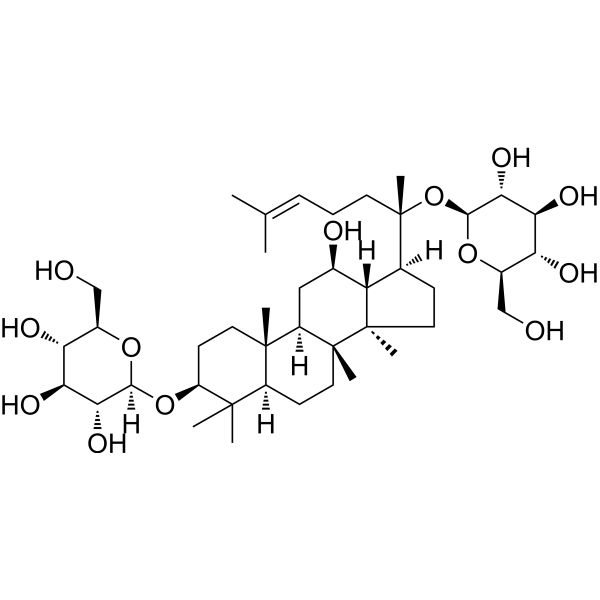
HY-N0605
Ginsenoside Rh2Caspase Activator -
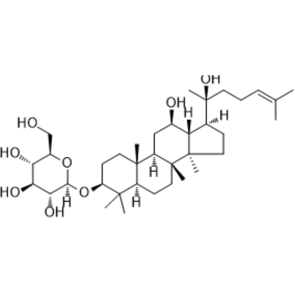
HY-N0043
Ginsenoside RdCytochrome P450 Inhibitor -
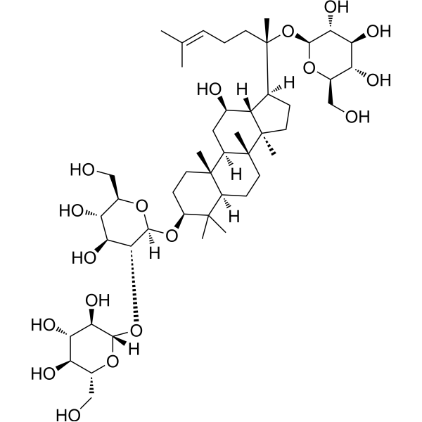
HY-N0039
Ginsenoside Rb1Autophagy Inducer -
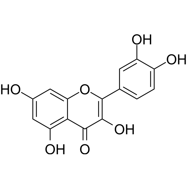
HY-18085
QuercetinFlavonoid Antioxidant -
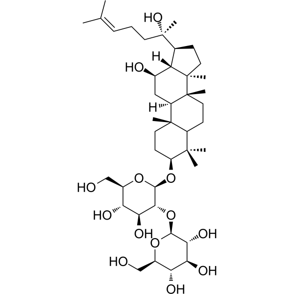
HY-N0603
20(S)-Ginsenoside Rg3Sodium Channel Inhibitor -
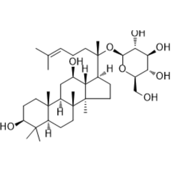
HY-N0904
Ginsenoside C-KNO Synthase/COX Inhibitor -
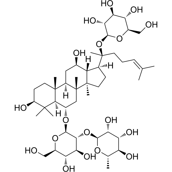
HY-N0044
Ginsenoside ReEndogenous Metabolite -

HY-125848R
Ginsenoside F2 (Standard)Autophagy Inducer -
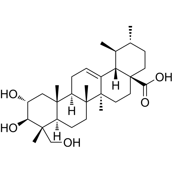
HY-N0194
Asiatic acidApoptosis Inducer -
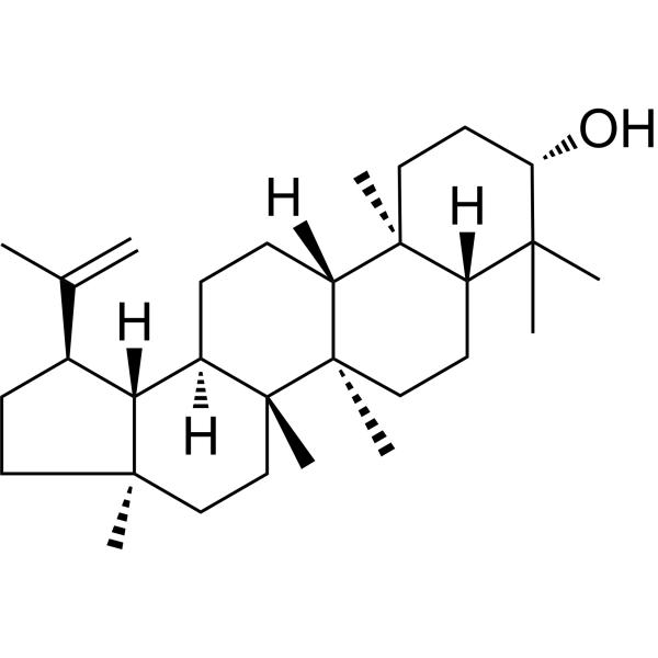
HY-N0790
LupeolAndrogen Receptor Inhibitor -

HY-N0194R
Asiatic acid (Standard)Apoptosis Inducer -
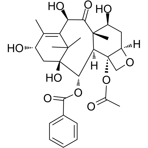
HY-16565
10-Deacetylbaccatin IIIIntermediate


Biological Activity
Description
In Vitro
Ginsenoside F2 (12.5-100 μM; 2-8 days) inhibits the differentiation of 3T3-L1 cells and reduces lipid accumulation[1].
Ginsenoside F2 (100 μM; during differentiation treatment) alters the expression of adipogenesis-related genes in MDI-induced differentiated 3T3-L1 preadipocytes, including upregulating the expression of Grin2d, Pik3cd, MMP9 and Hhip, downregulating the expression of key adipogenic genes such as PPARγ, FASN and ACC, and promoting mitochondrial biogenesis in cells[1].
Ginsenoside F2 (100 μM; 15-180 min) activates the AMPK pathway by increasing the phosphorylation levels of AMPK and ACC in 3T3-L1 preadipocytes[1].
Ginsenoside F2 (100 μM; 24 h) significantly reduces the viability of IR-HepG2 cells[3].
Ginsenoside F2 (12.5-50 μM; 12 h) dose-dependently promotes glucose uptake in IR-HepG2 cells[3].
Ginsenoside F2 (12.5-50 μM; 12 h) dose-dependently upregulates the mRNA expression of GLUT-2 and GLUT-4 in IR-HepG2 cells, and inhibits gluconeogenesis by reducing the mRNA expression of PEPCK and G6Pase[3].
Ginsenoside F2 (12.5-50 μM; 12 h) alleviates oxidative stress in IR-HepG2 cells by reducing ROS production and MDA levels, as well as restoring SOD activity[3].
Ginsenoside F2 (12.5-50 μM; 12 h) promotes glycogen synthesis and increases glycogen content in IR-HepG2 cells, activates the PI3K/AKT/GSK-3β signaling pathway, and elevates the phosphorylation levels of PDK1, AKT and GSK-3β[3].
Ginsenoside F2 (12.5-50 μM; 12 h) dose-dependently inhibits the activation of the MAPK signaling pathway and suppresses the nuclear translocation of NF-κB p65 in high glucose-induced IR-HepG2 cells[3].
Ginsenoside F2 (80 μM; 24 h) induces apoptosis and increases the number of cleaved caspase-3-positive cells in human glioblastoma cell lines U373 and Hs683[4].
Ginsenoside F2 (80 μM; 24 h) induces DNA damage in human glioblastoma cell lines U373 and Hs683, and increases the signal intensity of γH2AX[4].
Ginsenoside F2 (80 μM; 24 h) impairs mitochondrial function in human glioblastoma cell lines U373 and Hs683, which is specifically characterized by a significant decrease in mitochondrial membrane potential[4].
Ginsenoside F2 (20-80 μM; 24 h) upregulates the mRNA expression of p21 and downregulates the mRNA expression of GLRX in a concentration-dependent manner in human glioblastoma cell lines U373 and Hs683[4].
Ginsenoside F2 (20-80 μM; 24 h) inhibits mitochondrial respiration and ATP production in human glioblastoma U373 and Hs683 cells, impairs mitochondrial function, and significantly reduces mitochondrial membrane potential and intracellular NAD+ levels[4].
Ginsenoside F2 (20-80 μM; 24 h) disrupts the intracellular redox balance in human glioblastoma cell lines U373 and Hs683, and reduces the GSH/GSSG ratio[4].
Ginsenoside F2 (20-80 μM; 24 h treatment) induces energy stress in human glioblastoma cell lines U373 and Hs683, which is characterized by a concentration-dependent increase in phosphorylated AMPK levels[4].
MedChemExpress (MCE) has not independently confirmed the accuracy of these methods. They are for reference only. Further protocols information, click here.
-
Cell Line:3T3-L1 preadipocytes
-
Concentration:12.5-100 μM (basic DMEM medium); 12.5-100 μM (MDI medium)
-
Incubation Time:48 h (basic DMEM medium); 48 h (MDI medium)
-
Result:Showed no effect on cell viability in basic DMEM medium at all tested concentrations.
Significantly reduced cell viability in MDI medium at 100 μM compared to the MDI-only group.
-
Cell Line:3T3-L1 preadipocytes
-
Concentration:100 μM
-
Incubation Time:15, 30, 60, 180 min
-
Result:Enhanced the phosphorylation of AMPK and ACC in cells incubated for >30 min. Showed that co-treatment with the AMPK inhibitor compound C did not reduce Ginsenoside F2-induced AMPK phosphorylation.
-
Cell Line:IR-HepG2 cells
-
Concentration:0, 12.5, 25, 50 100 μM
-
Incubation Time:24 h
-
Result:Significantly reduced viability of IR-HepG2 cells at 100 μM.
-
Cell Line:IR-HepG2 cells
-
Concentration:12.5, 25, 50 μM
-
Incubation Time:12 h
-
Result:Enhanced the fluorescent intensity of 2-NBDG.
-
Cell Line:IR-HepG2 cells
-
Concentration:12.5, 25, 50 μM
-
Incubation Time:12 h
-
Result:Upregulated mRNA levels of GLUT-2 and GLUT-4 in a dose-dependent manner. Downregulated mRNA levels of PEPCK and G6Pase in a dose-dependent manner.
-
Cell Line:IR-HepG2 cells
-
Concentration:12.5, 25, 50 μM
-
Incubation Time:12 h
-
Result:Increased phosphorylation of PDK1, AKT, and GSK-3β in a dose-dependent manner. Reversed the high glucose-induced suppression of PDK1, AKT, and GSK-3β phosphorylation.
Resulted in GSK-3β phosphorylation levels exceeding those of the normal control group at 50 μM.、
-
Cell Line:U373 , Hs683 cell
-
Concentration:80 μM
-
Incubation Time:24 h
-
Result:Significantly increased the percentage of cleaved caspase-3-positive cells.\n
Significantly increased relative γH2AX fluorescence signal intensities.\n
Significantly reduced relative TMRE fluorescence signal intensities.
-
Cell Line:U373 , Hs683 cell
-
Concentration:20, 40, 80 μM
-
Incubation Time:24 h
-
Result:Increased relative p21 mRNA expression. Downregulated relative GLRX mRNA expression.
-
Cell Line:U373 , Hs683 cell
-
Concentration:20, 40, 80 μM
-
Incubation Time:24 h
-
Result:Increased relative phosphorylated AMPK band intensities in a concentration-dependent manner in both cell lines.
In Vivo
Ginsenoside F2 (50 mg/kg; p.o.; daily administration; 27 days) significantly alleviates Dermatophagoides farinae extract-induced atopic dermatitis in BALB/c mice by suppressing cutaneous and systemic inflammation, reshaping the gut microbiota to enrich propionate-producing bacteria, and increasing propionate levels in feces and serum[2].
MedChemExpress (MCE) has not independently confirmed the accuracy of these methods. They are for reference only.
-
Animal Model:C57BL/6J (male, 4 weeks old, initial weight 20 g, high-fat diet-induced obesity)[1]
-
Dosage:50 mg/kg; 100 mg/kg
-
Administration:i.g.; daily; 4 weeks
-
Result:Increased body weight and decreased epididymal, perirenal, and mesenteric adipose tissue weight and body fat percentage.
Decreased serum total cholesterol, triglyceride, and aspartate aminotransferase levels in mice at the 100 mg/kg dose.
Decreased adipocyte volume in epididymal adipose tissue in mice at both doses.
Increased mRNA expression of antioxidant enzymes (including SOD1, SOD2, and GSH-Px) in the liver of mice at the 100 mg/kg dose.
Decreased mRNA expression of adipogenesis-related genes (including PPARγ, FASN, and Adipoq) in the liver of mice at the 100 mg/kg dose.
Increased hepatic AMPK and ACC phosphorylation levels in the 100 mg/kg dose group.
-
Animal Model:BALB/c (female, 4 weeks old, SPF conditions)[2]
-
Dosage:50 mg/kg
-
Administration:i.g.; daily; 27 days
-
Result:Improved atopic dermatitis-like skin inflammation and ear tissue thickening, reducing the thickness of the epidermis and dermis in the ear tissue, and decreasing the infiltration of eosinophils and mast cells.
Decreased serum immunoglobulin E (IgE) levels and reduced serum concentrations of IFN-γ, TNF-α, IL-6, IL-31, IL-17A, and CCL2.
Downregulated the mRNA expression of inflammatory cytokines IFN-γ, TNF-α, IL-6, IL-31, IL-17A, and CCL2 in the ear tissue.
Inhibited the expression of NF-κB protein in the ear tissue and increased the expression of IκB-α protein.
Altered the gut microbiota composition (α-diversity indicators showed no significant change, but β-diversity characteristics were significantly altered).
Increased the abundance of Parabacteroides goldsteinii and Lactobacillus plantarum in the gut.
Increases the concentration of propionic acid in feces and serum, and also increases the concentrations of isobutyric acid, monomethylbutyric acid, and isovaleric acid in feces.
Decreases the serum levels of various medium-chain and long-chain fatty acids.


Chemical Information
-
CAS No. 62025-49-4
-
Appearance Solid
-
Molecular Weight 785.01
-
Formula C42H72O13
-
Color White to off-white
-
SMILES
C[C@@]([C@@]12C)(CC[C@@]3([H])C4(C)C)[C@@](C[C@@H](O)[C@]1([H])[C@]([C@@](CC/C=C(C)/C)(C)O[C@@H]([C@@H]([C@@H](O)[C@@H]5O)O)O[C@@H]5CO)([H])CC2)([H])[C@]3(CC[C@@H]4O[C@]([C@@H]([C@@H](O)[C@@H]6O)O)([H])O[C@@H]6CO)C
-
Structure Classification
-
Initial Source
-
Shipping
Room temperature in continental US; may vary elsewhere.
-
Storage
Powder -20°C 3 years 4°C 2 years In solvent -80°C 6 months -20°C 1 month


Solvent & Solubility
In Vitro:
DMSO : 50 mg/mL (63.69 mM; Need ultrasonic; Hygroscopic DMSO has a significant impact on the solubility of product, please use newly opened DMSO)
Please refer to the solubility information to select the appropriate solvent. Once prepared, please aliquot and store the solution to prevent product inactivation from repeated freeze-thaw cycles.
Storage method and period of stock solution: -80°C, 6 months; -20°C, 1 month. When stored at -80°C, please use it within 6 months. When stored at -20°C, please use it within 1 month.
Please refer to the solubility information to select the appropriate solvent. Once prepared, please aliquot and store the solution to prevent product inactivation from repeated freeze-thaw cycles.
Storage method and period of stock solution: -80°C, 6 months; -20°C, 1 month. When stored at -80°C, please use it within 6 months. When stored at -20°C, please use it within 1 month.
Concentration (start) × Volume (start) = Concentration (final) × Volume (final)
In Vivo:
Select the appropriate dissolution method based on your experimental animal and administration route.
- For the following dissolution methods, please ensure to first prepare a clear stock solution using an In Vitro approach and then sequentially add co-solvents:
- To ensure reliable experimental results, the clarified stock solution can be appropriately stored based on storage conditions. As for the working solution for In Vivo experiments, it is recommended to prepare freshly and use it on the same day.
- The percentages shown for the solvents indicate their volumetric ratio in the final prepared solution. If precipitation or phase separation occurs during preparation, heat and/or sonication can be used to aid dissolution.
Add each solvent one by one: 10% DMSO 40% PEG300 5% Tween-80 45% Saline
Solubility: ≥ 2.5 mg/mL (3.18 mM); Clear solution
This protocol yields a clear solution of ≥ 2.5 mg/mL (saturation unknown).
Taking 1 mL working solution as an example, add 100 μL DMSO stock solution (25.0 mg/mL) to 400 μL PEG300, and mix evenly; then add 50 μL Tween-80 and mix evenly; then add 450 μL Saline to adjust the volume to 1 mL.
Preparation of Saline: Dissolve 0.9 g sodium chloride in ddH₂O and dilute to 100 mL to obtain a clear Saline solution.
Add each solvent one by one: 10% DMSO 90% (20% SBE-β-CD in Saline)
Solubility: ≥ 2.5 mg/mL (3.18 mM); Clear solution
This protocol yields a clear solution of ≥ 2.5 mg/mL (saturation unknown).
Taking 1 mL working solution as an example, add 100 μL DMSO stock solution (25.0 mg/mL) to 900 μL 20% SBE-β-CD in Saline, and mix evenly.
Preparation of 20% SBE-β-CD in Saline (4°C, storage for one week): 2 g SBE-β-CD powder is dissolved in 10 mL Saline, completely dissolve until clear.
In Vivo Dissolution Calculator
Please enter the basic information of animal experiments:
-
-
-
-
Recommended: Prepare an additional quantity of animals to account for potential losses during experiments.
Please enter your animal formula composition:
-
%DMSO +
Recommended: Keep the proportion of DMSO in working solution below 2% if your animal is weak.
-
%+
-
+%Tween-80 + +
-
%Saline +
The co-solvents required include: DMSO, . All of co-solvents are available by MedChemExpress (MCE). , Tween 80. All of co-solvents are available by MedChemExpress (MCE).
Working solution concentration: 0.22 mg/mL
Method for preparing stock solution: mg drug dissolved in μL DMSO. Stock solution concentration: mg/mL.
1. Take μL DMSO stock solution;
2. Add μL .
μL , mix evenly;
3. Then add μL Tween 80, mix evenly;
4. Then add μL
Please ensure that the stock solution in the first step is dissolved to a clear state, and add co-solvents in sequence. You can use ultrasonic heating (ultrasonic cleaner, recommended frequency 20-40 kHz), vortexing, etc. to assist dissolution.

Protocols
-
Western Blot
Western blotting (WB) is a commonly used experimental method in molecular biology, biochemistry, and immunogenetics for identifying and quantifying target proteins. It combines gel electrophoresis with immunoassay, enabling researchers to analyze protein expression, post-translational modifications, and molecular weight.
-
RNA extraction experimental
By lysing cells, releasing RNA, and removing impurities such as proteins and DNA, high-purity RNA products are finally obtained. The commonly used traditional method is the guanidine isothiocyanate/phenol/chloroform method (Trizol), which is suitable for a variety of animal materials including animal tissues, microorganisms, cultured cells, etc., and most plant materials.
-
Annexin V plus membrane-impermeant dye apoptosis staining
Annexin V-based apoptosis assays rely on the detection of phosphatidylserine (PS) externalization from the inner leaflet of the plasma membrane to the outer leaflet, an early biochemical hallmark of apoptosis. Fluorescently labeled Annexin V binds PS in a calcium-dependent manner, enabling identification of early apoptotic cells by flow cytometry or fluorescence microscopy. When combined with a membrane-impermeant DNA-binding dye (e. g. , propidium iodide), this approach allows discrimination between viable (Annexin V−/dye−), early apoptotic (Annexin V+/dye−), and late apoptotic or necrotic (Annexin V+/dye+) cell populations by assessing membrane integrity and PS exposure.
-
ROS/oxidative-stress fluorescent staining
ROS/oxidative-stress fluorescent staining uses cell-permeant fluorogenic probes that become fluorescent after oxidation inside cells or tissues; commonly used examples include DCFH-DA/DCFDA for broad cellular oxidant detection, DHE for superoxide-related signal detection, MitoSOX for mitochondrial superoxide-related signal detection, and CellROX probes for oxidative-stress-associated fluorescence readouts. The assay detects probe oxidation rather than a single ROS species unless the probe and analysis method have been chemically validated for that species. DCFH-DA enters cells, is deacetylated by intracellular esterases to DCFH, and produces fluorescent DCF after oxidation, so the readout is used as an operational measure of total cellular oxidative stress rather than a species-specific ROS measurement. DHE and MitoSOX can report superoxide-related oxidation, but red fluorescence alone can include non-specific ethidium-like oxidation products; HPLC or optimized spectral approaches are
-
Research Protocol for Microbiome Analysis
Microbiome analysis characterizes microbial communities in biological or environmental samples by measuring community composition, diversity, taxonomic structure, functional potential, and associations with host or environmental phenotypes. 16S rRNA gene amplicon sequencing is commonly used for bacterial and archaeal taxonomic profiling, while shotgun metagenomics provides higher taxonomic resolution and direct functional information, including microbial genes, pathways, viruses, fungi, and antimicrobial-resistance genes when sequencing depth and host-DNA contamination are adequately controlled. Microbiome results are strongly affected by sample collection, storage, DNA extraction, contamination, sequencing method, reference database, and bioinformatic pipeline; therefore, standardized protocols, negative controls, mock communities, and transparent analysis workflows are required. Unresolved issues include low-biomass contamination, compositional-data bias, inconsistent species-level c
-
Research Protocol for Inflammation-related Diseases
The NLRP3 inflammasome is a cytosolic innate immune signaling platform that integrates priming signals and danger-signal activation to promote caspase-1 activation, maturation of IL-1β and IL-18, and gasdermin D-mediated pyroptotic cell death. The core experimental logic is to determine whether inflammatory disease phenotypes are driven by increased NLRP3 expression, ASC-containing inflammasome assembly, caspase-1 cleavage, GSDMD cleavage, and extracellular release of IL-1β/IL-18 rather than by nonspecific cell injury alone. The pathway is strongly linked to inflammation-related disease phenotypes because monosodium urate crystals activate NALP3/NLRP3 inflammasome signaling in gout-like crystal inflammation, cholesterol crystals activate NLRP3 inflammasomes in atherogenesis models, and DSS-induced intestinal inflammation has been reported to involve NLRP3 inflammasome activity. However, experimental colitis studies also show context-dependent protective effects of NLRP3 inflammasome co
-
Liver Cancer Modeling
Liver cancer can be classified into primary liver cancer and secondary liver cancer. Secondary liver cancer is the metastatic liver cancer. Primary liver cancer includes hepatocellular carcinoma (HCC), intrahepatic cholangiocarcinoma (ICC) and fibrolamellar HCC, of which HCC is the most common form, accounting for approximately 90% of primary liver cancers[1]. HCC mouse models include chemical agent-induced models, transplanted tumor models, and genetic engineered models.
-
Research Protocol for Metabolic Diseases
AMP-activated protein kinase, AMPK, is a conserved cellular energy sensor that responds to reduced cellular energy status and coordinates metabolism by increasing ATP-generating catabolic pathways while suppressing ATP-consuming anabolic processes. In metabolic disease research, the AMPK pathway is experimentally relevant because it regulates hepatic lipid synthesis, fatty acid oxidation, glucose production, skeletal-muscle glucose disposal, mTORC1-linked biosynthesis, autophagy, mitochondrial homeostasis, and whole-body energy balance. The central pathway logic is that energy stress, metformin, exercise-like stimulation, or direct AMPK activators increase AMPKα Thr172 phosphorylation and downstream substrate phosphorylation, including ACC and RAPTOR. Phosphorylation of ACC suppresses lipogenesis and supports fatty acid oxidation, whereas phosphorylation of RAPTOR suppresses mTORC1 signaling and links cellular energy status to growth and protein synthesis control. The pathway is linked
-
Kinase activity and phosphorylation assays
Kinase activity assays measure the ability of kinases to transfer phosphate groups from ATP to specific substrates, while phosphorylation assays detect the presence and levels of phosphorylated proteins. Common methods include radiolabeled ATP incorporation (e. g. ,), ADP release detection via bioluminescence (e. g. ,[3]), enzyme-linked immunosorbent assays (ELISA) for phospho-specific epitopes (e. g. ,[6]), and microtiter-based formats for high-throughput screening (e. g. ,[8]). The ADP-Glo assay quantifies kinase activity by measuring ADP produced during phosphorylation using a luciferase-based system. Radiometric assays involve autoradiography or scintillation counting after incorporation of 32P-labeled ATP into substrate proteins. ELISA-based approaches rely on phospho-specific antibodies to detect activated kinases in cell lysates or purified samples.
-
Apoptosis
Apoptosis, also called programmed cell death, is generally characterized by distinct morphological characteristics.
-
TUNEL staining for apoptotic DNA fragmentation
TUNEL staining detects DNA strand breaks by using terminal deoxynucleotidyl transferase to add labeled nucleotides to exposed 3′-OH DNA termini, generating either microscopic staining in fixed cells or tissue sections, or fluorescence/cytometric signal in cell suspensions. TUNEL positivity reflects DNA fragmentation but should not be interpreted alone as definitive apoptosis, because TUNEL can also label necrotic, autolytic, mechanically damaged, or DNA-repair-associated DNA breaks.
-
Large-size fat particle sorting
Large-size fat particle sorting is widely used to isolate cells up to 200 μm in diameter. Single-cell flow sorting will allow greater insight into adipocyte heterogeneity by identifying gene expression, protein composition, and metabolic signatures at the single-cell level.
-
Research Protocol for Endocrine Diseases
Endocrine diseases often arise from disrupted hormone production, hormone signaling, or target-tissue responsiveness; for diabetes-focused endocrine disease models, insulin signaling regulates glucose uptake, hepatic glucose output, lipid metabolism, and β-cell compensation. Type 2 diabetes develops through interacting defects in insulin resistance, β-cell dysfunction, adipose inflammation, hepatic glucose overproduction, altered incretin signaling, and ectopic lipid metabolism. A major unresolved question is whether endocrine dysfunction is driven primarily by target-tissue insulin resistance, intrinsic β-cell failure, immune/inflammatory stress, or combined multi-organ failure that differs by disease stage.
-
TPA/Croton Oil Ear Edema and Dermatitis
The TPA (12-O-tetradecanoylphorbol-13-acetate) and croton oil-induced mouse ear edema model is a well-established acute cutaneous inflammation system used to evaluate topical anti-inflammatory activity by measuring edema formation, neutrophil infiltration, vascular permeability, and cytokine-mediated skin responses in vivo. The inflammatory response is triggered by topical application of phorbol esters (TPA) or croton oil constituents, leading to rapid activation of protein kinase C signaling, leukocyte recruitment, and increased vascular permeability, which can be quantified by ear thickness, weight, dye extravasation, and biochemical markers such as myeloperoxidase (MPO) activity and pro-inflammatory mediators in ear tissue homogenates. This model is widely used for screening anti-inflammatory agents, where reductions in edema and inflammatory biomarkers reflect suppression of acute dermal inflammation and immune cell infiltration. Histological evaluation typically confirms epidermal
-
Apoptosis Solutions
Apoptosis is a regulated, generally non-lytic cell-death pathway that removes unwanted, damaged, infected, or abnormal cells through coordinated morphological changes, caspase activation, DNA fragmentation, and membrane remodeling. The intrinsic apoptosis pathway is controlled mainly by mitochondrial outer membrane permeabilization, BCL-2 family proteins, cytochrome c release, apoptosome formation, caspase-9 activation, and downstream executioner caspase-3/7 activation. The extrinsic apoptosis pathway is initiated by death receptors such as Fas, TNFR, and TRAIL receptors, which recruit adaptor proteins and activate caspase-8 before engaging executioner caspases or mitochondrial amplification through BID cleavage. Apoptosis is linked to many phenotypes, including cancer cell killing, tissue homeostasis, immune regulation, neurodegeneration, infection response, and treatment-induced cytotoxicity; unresolved questions include how apoptosis interacts with necroptosis, pyroptosis, ferroptos
-
Protocol for Kinase activity and phosphorylation assays
Kinase activity assays measure transfer of phosphate from ATP to a protein or peptide substrate, generating phosphorylated substrate, ADP, or incorporated radiolabeled phosphate as the readout; phosphorylation assays measure site-specific phosphorylation in cells or tissues as a proxy for kinase-pathway activation, inhibition, or substrate regulation. Phosphorylation can be detected by phospho-specific Western blot, immunoprecipitation kinase assay, phospho-immunofluorescence, phospho-flow cytometry, luminescent ADP detection, radiolabeled ATP incorporation, or reporter-based pathway assays, and these readouts can be applied to cancer cells, primary neurons, mouse tumors, organoids, inflammatory macrophages, ferroptosis studies, and mitophagy studies when the kinase target is biologically relevant.


Purity & Documentation
-
Data Sheet (293 KB)
-
SDS (396 KB)
- English - EN (396 KB)
- Français - FR (396 KB)
- Deutsch - DE (396 KB)
- Norwegian - NO (396 KB)
- Español - ES (396 KB)
- Swedish - SV (396 KB)
- Italian - IT (396 KB)
- Korean - KR (396 KB)
- Portuguese - PT (396 KB)
-
Handling Instructions (2659 KB)
References
[1]. Zhou J, et al. Ginsenoside F2 Suppresses Adipogenesis in 3T3-L1 Cells and Obesity in Mice via the AMPK Pathway. J Agric Food Chem. 2021;69(32):9299-9312. [Content Brief]
[2]. Li D, et al. Ginsenoside F2-Mediated Intestinal Microbiota and Its Metabolite Propionic Acid Positively Impact the Gut-Skin Axis in Atopic Dermatitis Mice. J Agric Food Chem. 2024;72(1):339-350. [Content Brief]
[3]. Han S, et al. Ginsenoside F2 enhances glucose metabolism by modulating insulin signal transduction in human hepatocarcinoma cells. J Ginseng Res. 2023;47(3):420-428. [Content Brief]
[4]. Kim TJ, et al. Ginsenoside F2 induces cellular toxicity to glioblastoma through the impairment of mitochondrial function. Phytomedicine. 2021;83:153483. [Content Brief]


Complete Stock Solution Preparation Table
Please refer to the solubility information to select the appropriate solvent. Once prepared, please aliquot and store the solution to prevent product inactivation from repeated freeze-thaw cycles.
Storage method and period of stock solution: -80°C, 6 months; -20°C, 1 month. When stored at -80°C, please use it within 6 months. When stored at -20°C, please use it within 1 month.
| Optional Solvent | Concentration Solvent Mass | 1 mg | 5 mg | 10 mg | 25 mg |
|---|---|---|---|---|---|
| DMSO | 1 mM | 1.2739 mL | 6.3693 mL | 12.7387 mL | 31.8467 mL |
| 5 mM | 0.2548 mL | 1.2739 mL | 2.5477 mL | 6.3693 mL | |
| 10 mM | 0.1274 mL | 0.6369 mL | 1.2739 mL | 3.1847 mL | |
| 15 mM | 0.0849 mL | 0.4246 mL | 0.8492 mL | 2.1231 mL | |
| 20 mM | 0.0637 mL | 0.3185 mL | 0.6369 mL | 1.5923 mL | |
| 25 mM | 0.0510 mL | 0.2548 mL | 0.5095 mL | 1.2739 mL | |
| 30 mM | 0.0425 mL | 0.2123 mL | 0.4246 mL | 1.0616 mL | |
| 40 mM | 0.0318 mL | 0.1592 mL | 0.3185 mL | 0.7962 mL | |
| 50 mM | 0.0255 mL | 0.1274 mL | 0.2548 mL | 0.6369 mL | |
| 60 mM | 0.0212 mL | 0.1062 mL | 0.2123 mL | 0.5308 mL |


Ginsenoside F2 Related Classifications
HY-125848 Related Classifications
 Powered by Bioz
Keywords
- Ginsenoside F2
- 62025-49-4
- Ginsenoside F 2
- Ginsenoside F-2
- Apoptosis
- AMPK
- PPAR
- p38 MAPK
- PI3K
- Akt
- GSK-3
- Reactive Oxygen Species (ROS)
- SOD
- Caspase
- 3T3-L1 preadipocytes
- IR-HepG2 cell
- U373 cell
- Hs683 cell
- C57BL/6J mice
- BALB/c mice
- insulin resistance
- obesity
- atopic dermatitis
- liver cancer
- glioblastoma
- glioma
- Inhibitor
- inhibitor
- inhibit