


Sorafenib tosylate
Based on 292 publication(s) in Google Scholar
Sorafenib (Bay 43-9006) tosylate is a potent oral active multikinase inhibitor. Sorafenib blocks autophosphorylation and activity of receptor tyrosine kinases (VEGFR-2, VEGFR-3) and RAF family kinases, thereby suppressing the RAF/MEK/ERK and PI3K/Akt pathways, inhibiting STAT3 phosphorylation, and selectively inhibiting the MAPK pathway in cancer cells. Sorafenib tosylate induces cell cycle arrest, autophagy, apoptosis, and PARP cleavage, reduces Bcl-2, Bcl-XL, cyclin D1 levels, and activates Bak and Bax. Sorafenib tosylate inhibits tumor growth and metastasis in mouse and rat models. Sorafenib tosylate can be used for cancer research, such as colon, breast, non-small-cell lung cancer (NSCLC), ovarian, pancreatic, melanoma, colorectal and hepatocellular carcinoma.
For research use only. We do not sell to patients.
- Purity: 99.98%
- CAS No.: 475207-59-1
- Formula: C28H24ClF3N4O6S
- Molecular Weight:637.03
-
Storage:
4°C, sealed storage, away from moisture
* In solvent : -80°C, 1 year; -20°C, 6 months (sealed storage, away from moisture)


-
 Biological Activity
-
Chemical Information
-
Solvent & Solubility
- Purity & Documentation
- References
-
 Help & FAQs
-


Anti-Infection Compound Library
HY-L002
-
Apoptosis Compound Library
HY-L003
-
Cell Cycle/DNA Damage Compound Library
HY-L004
-
Epigenetics Compound Library
HY-L005
-
Immunology/Inflammation Compound Library
HY-L007
-
JAK/STAT Compound Library
HY-L008
-
Kinase Inhibitor Library
HY-L009
-
MAPK Compound Library
HY-L010
-
Metabolism/Protease Compound Library
HY-L012
-
PI3K/Akt/mTOR Compound Library
HY-L015
-
Protein Tyrosine Kinase Compound Library
HY-L016
-
Stem Cell Signaling Compound Library
HY-L017
-
FDA-Approved Drug Library
HY-L022
-
Anti-Cancer Compound Library
HY-L025
-
CNS-Penetrant Compound Library
HY-L028
-
Autophagy Compound Library
HY-L029
-
Small Molecule Immuno-Oncology Compound Library
HY-L031
-
Anti-Aging Compound Library
HY-L034
-
Drug Repurposing Compound Library
HY-L035
-
Differentiation Inducing Compound Library
HY-L038
-
Reprogramming Compound Library
HY-L039
-
Oxygen Sensing Compound Library
HY-L045
-
Ferroptosis Compound Library
HY-L051
-
Anti-COVID-19 Compound Library
HY-L052
-
NMPA-Approved Drug Library
HY-L053
-
Glycolysis Compound Library
HY-L058
-
Pyroptosis Compound Library
HY-L059
-
Cytoskeleton Compound Library
HY-L060
-
Orally Active Compound Library
HY-L061
-
Glutamine Metabolism Compound Library
HY-L064
-
FDA Approved & Pharmacopeial Drug Library
HY-L066
-
Neuroprotective Compound Library
HY-L070
-
Anti-Breast Cancer Compound Library
HY-L074
-
Anti-Lung Cancer Compound Library
HY-L075
-
Drug-Induced Liver Injury (DILI) Compound Library
HY-L076
-
Anti-Pancreatic Cancer Compound Library
HY-L077
-
Anti-Blood Cancer Compound Library
HY-L079
-
Targeted Therapy Drug Library
HY-L080
-
Anti-Cancer Metabolism Compound Library
HY-L083
-
Neurodegenerative Disease-related Compound Library
HY-L086
-
Anti-Obesity Compound Library
HY-L087
-
Angiogenesis-Related Compound Library
HY-L088
-
Mitochondria-Targeted Compound Library
HY-L089
-
Transcription Factor-Targeted Library
HY-L090
-
Glucose Metabolism Compound Library
HY-L092
-
Food-Sourced Compound Library
HY-L094
-
Anti-Liver Cancer Compound Library
HY-L101
-
Rare Diseases Drug Library
HY-L102
-
Anti-Colorectal Cancer Compound Library
HY-L103
-
Antidepressant Compound Library
HY-L108
-
EMA-Approved Drug Library
HY-L116
-
FDA-Approved Anticancer Drug Library
HY-L122
-
Human Metabolite Library
HY-L123
-
Anti-Prostate Cancer Compound Library
HY-L124
-
Anti-Pulmonary Fibrosis Compound Library
HY-L125
-
Cancer Stem Cells Compound Library
HY-L135
-
Heterocyclic Compound Library
HY-L138
-
Pain-Related Compound Library
HY-L139
-
Off-patent Drug Library
HY-L141
-
Mitochondrial Protection Compound Library
HY-L144
-
Membrane Protein-targeted Compound Library
HY-L149
-
Membrane Receptor-targeted Compound Library
HY-L150
-
Highly Selective Inhibitors Library
HY-L158
-
Highly Selective Activators Library
HY-L159
-
Cytokine Inhibitors Library
HY-L161
-
Cell Death Library
HY-L162
-
Serine/Threonine Kinase Inhibitor Library
HY-L164
-
Anti-Hematopathy Compound Library
HY-L171
-
Anti-Ovarian Cancer Compound Library
HY-L173
-
Multi-Target Compound Library
HY-L176
-
Mitophagy Compound Library
HY-L180
-
Bioactive Compound Library Max
HY-L181
-
MCE Bioactive Compound Library
HY-L001V
-
Drug Repurposing Compound Library Plus
HY-L035P
-
FDA-Approved Drug Library Plus
HY-L022P
-
FDA-Approved Drug Library Mini
HY-L022M
-
Bioactive Compound Library
HY-L001
-
Anti-Gastric Cancer Compound Library
HY-L184
-
Anti-Fibrosis Compound Library
HY-L185
-
Anti-Brain Cancer Compound Library
HY-L188
-
Protein Kinase Compound Library
HY-L196
-
Non-Alcoholic Fatty Liver Disease (NAFLD) Compound Library
HY-L199
-
Cell Proliferation Compound Library
HY-L201
-
High-Throughput Bioactive Compound Library
HY-L205
-
Anti-Rheumatic Arthritis Compound Library
HY-L210
-
Mass Spectrometry Human Metabolite Library
HY-L215
-
Posttranslational Modification Library
HY-L226
-
FDA Kinase Inhibitor Library
HY-L230
-
RNA Binding Bioactive Compound Library
HY-L248
-
Lactylation Compound Library
HY-L249


Publications Citing Use of MedChemExpress (MCE) Sorafenib tosylate
More- Signal Transduct Target Ther. 2025 Aug 1;10(1):244. [Abstract]
- Signal Transduct Target Ther. 2023 Sep 14;8(1):348. [Abstract]
- Cancer Discov. 2021 Oct;11(10):2544-2563. [Abstract]
- Cancer Discov. 2019 Dec;9(12):1686-1695. [Abstract]
- Cell Metab. 2021 Oct 5;33(10):2021-2039.e8. [Abstract]
- Nat Cancer. 2024 Jul;5(7):1082-1101. [Abstract]
- Cancer Res. 2025 Aug 21. [Abstract]
- Mol Cell. 2023 Dec 7;83(23):4334-4351.e7. [Abstract]
- ACS Nano. 2023 Nov 28;17(22):22240-22258. [Abstract]
- Nat Commun. 2025 Nov 18;16(1):10096. [Abstract]
- Nat Commun. 2025 Jan 2;16(1):87. [Abstract]
- Nat Commun. 2024 Sep 10;15(1):7923. [Abstract]
- Nat Commun. 2022 Mar 18;13(1):1478. [Abstract]
- Cell Death Differ. 2021 Apr;28(4):1222-1236. [Abstract]
- Bone Res. 2024 Apr 10;12(1):24. [Abstract]
- Acta Pharm Sin B. 2025 Nov;15(11):5817-5831. [Abstract]
- Acta Pharm Sin B. 2023 Oct;13(10):4253-4272. [Abstract]
- Acta Pharm Sin B. 2023 Jun;13(6):2645-2662. [Abstract]
- Sci Transl Med. 2018 Jul 18;10(450):eaaq1093. [Abstract]
- J Extracell Vesicles. 2024 Jul;13(7):e12468. [Abstract]
- Autophagy. 2026 Feb 15:1-19. [Abstract]
- Autophagy. 2022 Jan;18(1):191-203. [Abstract]
- Adv Sci (Weinh). 2025 Aug 18:e08613. [Abstract]
- Adv Sci (Weinh). 2025 Jul 13:e02501. [Abstract]
- Adv Sci (Weinh). 2025 May 8:e2501730. [Abstract]
- Adv Sci (Weinh). 2024 Jul 9:e2402327. [Abstract]
- Adv Sci (Weinh). 2023 Sep;10(27):e2302640. [Abstract]
- Adv Sci (Weinh). 2023 Aug;10(22):e2206798. [Abstract]
- Adv Sci (Weinh). 2023 Jan;10(3):e2203718. [Abstract]
- Adv Sci (Weinh). 2022 May;9(15):e2105894. [Abstract]
- Adv Sci (Weinh). 2021 Jan 29;8(6):2002831. [Abstract]
- Exp Hematol Oncol. 2016 Jul 29:5:22. [Abstract]
- Theranostics. 2020 Aug 21;10(23):10498-10512. [Abstract]
- Biomaterials. 2022 Oct:289:121800. [Abstract]
- J Exp Clin Cancer Res. 2023 Jan 6;42(1):6. [Abstract]
- J Exp Clin Cancer Res. 2022 Mar 30;41(1):116. [Abstract]
- J Exp Clin Cancer Res. 2018 Nov 29;37(1):295. [Abstract]
- Cell Discov. 2022 May 3;8(1):40. [Abstract]
- Redox Biol. 2026 May:92:104086. [Abstract]
- Redox Biol. 2025 Sep:85:103786. [Abstract]
- Redox Biol. 2024 Nov 5:78:103419. [Abstract]
- Redox Biol. 2024 Jun:72:103131. [Abstract]
- Redox Biol. 2023 Feb:59:102564. [Abstract]
- J Hazard Mater. 2025 Oct 19:499:140193. [Abstract]
- Gut Microbes. 2024 Jan-Dec;16(1):2410474. [Abstract]
- MedComm (2020). 2025 Nov 23;6(12):e70488. [Abstract]
- Cell Rep Med. 2025 Apr 2:102053. [Abstract]
- J Immunother Cancer. 2022 Aug;10(8):e004006. [Abstract]
- Pharmacol Res. 2022 Jun;180:106244. [Abstract]
- Pharmacol Res. 2022 Mar:177:106050. [Abstract]
- Clin Cancer Res. 2024 Sep 13;30(18):4179-4189. [Abstract]
- Cancer Lett. 2022 Jan 1:524:161-171. [Abstract]
- Cancer Lett. 2021 Nov 1:520:243-254. [Abstract]
- Cancer Lett. 2019 May 28:450:132-143. [Abstract]
- Int J Biol Sci. 2018 Apr 25;14(5):577-585. [Abstract]
- Cell Death Dis. 2026 Jan 3. [Abstract]
- Cell Death Dis. 2025 Nov 17;16(1):839. [Abstract]
- Cell Death Dis. 2025 Aug 28;16(1):654. [Abstract]
- Cell Death Dis. 2025 Jul 11;16(1):512. [Abstract]
- Cell Death Dis. 2025 Apr 6;16(1):257. [Abstract]
- Cell Death Dis. 2025 Jan 26;16(1):42. [Abstract]
- Cell Death Dis. 2024 Jun 5;15(6):395. [Abstract]
- Cell Death Dis. 2024 Jan 18;15(1):66. [Abstract]
- Adv Healthc Mater. 2023 Jul;12(19):e2203120. [Abstract]
- Cell Death Dis. 2023 Jan 12;14(1):22. [Abstract]
- Cell Death Dis. 2022 Feb 4;13(2):118. [Abstract]
- Cell Death Dis. 2021 Jul 9;12(7):691. [Abstract]
- Cell Death Dis. 2019 Apr 15;10(5):331. [Abstract]
- Genes Dis. 2023 Sep 16;11(4):101114. [Abstract]
- Cell Commun Signal. 2024 Aug 7;22(1):391. [Abstract]
- Int J Biol Macromol. 2026 Mar:351:150982. [Abstract]
- Int J Biol Macromol. 2025 Oct 9;330(Pt 3):148223. [Abstract]
- Acta Pharmacol Sin. 2021 Jan;42(1):108-114. [Abstract]
- Phytomedicine. 2026 Jun:155:158086. [Abstract]
- Phytomedicine. 2025 Oct 20:148:157423. [Abstract]
- Phytomedicine. 2025 May 10:143:156840. [Abstract]
- Phytomedicine. 2025 Apr 1:141:156692. [Abstract]
- Phytomedicine. 2024 Jul:129:155225. [Abstract]
- Phytomedicine. 2021 Nov:92:153740. [Abstract]
- ACS Appl Mater Interfaces. 2026 Jan 25. [Abstract]
- Free Radic Biol Med. 2021 Feb 1;163:141-152. [Abstract]
- Free Radic Biol Med. 2020 Nov 20;160:303-318. [Abstract]
- ACS Appl Mater Interfaces. 2017 Apr 12;9(14):12195-12202. [Abstract]
- Drug Deliv. 2023 Dec;30(1):2174209. [Abstract]
- Chemosphere. 2022 Dec;308(Pt 2):136354. [Abstract]
- Clin Sci. 2024 Sep 18;138(18):1131-1150. [Abstract]
- Sens Actuators B Chem. 2024 Nov 1.
- Clin Sci. 2021 Jul 30;135(14):1751-1765. [Abstract]
- Br J Pharmacol. 2021 Feb;178(3):636-653. [Abstract]
- Biomed Pharmacother. 2026 Feb:195:118941. [Abstract]
- Biomed Pharmacother. 2025 May 8:187:118136. [Abstract]
- J Transl Med. 2025 Apr 11;23(1):435. [Abstract]
- J Transl Med. 2025 Feb 28;23(1):244. [Abstract]
- J Transl Med. 2024 Aug 28;22(1):792. [Abstract]
- J Transl Med. 2023 Jan 9;21(1):9. [Abstract]
- Biomed Pharmacother. 2022 Sep:153:113540. [Abstract]
- J Transl Med. 2022 Jun 6;20(1):255. [Abstract]
- Biomed Pharmacother. 2020 Jun;126:109862. [Abstract]
- Biomed Pharmacother. 2017 Feb:86:27-31. [Abstract]
- Cell Mol Gastroenterol Hepatol. 2024 Dec 30:101454. [Abstract]
- Oncogene. 2026 Apr;45(14):1260-1274. [Abstract]
- Stem Cell Res Ther. 2023 Apr 1;14(1):58. [Abstract]
- Oncogene. 2022 Mar;41(13):1851-1865. [Abstract]
- Oncogene. 2021 Aug;40(34):5302-5313. [Abstract]
- Cell Death Discov. 2023 Sep 30;9(1):362. [Abstract]
- Cell Death Discov. 2022 Aug 17;8(1):365. [Abstract]
- Cell Rep. 2026 Jan 22;45(2):116903. [Abstract]
- Cell Rep. 2024 Nov 27;43(12):114996. [Abstract]
- Cell Rep. 2024 Oct 24;43(11):114885. [Abstract]
- Cell Rep. 2023 Dec 12;42(12):113563. [Abstract]
- Clin Transl Med. 2025 Dec;15(12):e70550. [Abstract]
- Clin Transl Med. 2025 May;15(5):e70324. [Abstract]
- J Med Chem. 2025 Mar 27. [Abstract]
- Clin Transl Med. 2023 Jun;13(6):e1300. [Abstract]
- J Med Chem. 2019 Nov 14;62(21):9593-9599. [Abstract]
- Br J Cancer. 2017 Sep 26;117(7):974-983. [Abstract]
- Int J Nanomedicine. 2024 Dec 3:19:12975-12998. [Abstract]
- Comput Biol Med. 2024 Feb:169:107975. [Abstract]
- Cancer Cell Int. 2025 Oct 15;25(1):357. [Abstract]
- Biomater Adv. 2022 May:136:212761. [Abstract]
- Cancer Cell Int. 2019 Aug 27;19:222. [Abstract]
- Cell Biol Toxicol. 2024 Nov 26;40(1):104. [Abstract]
- Eur J Med Chem. 2024 Mar 5:267:116209. [Abstract]
- Cell Biol Toxicol. 2023 Dec;39(6):2821-2839. [Abstract]
- Int J Mol Med. 2026 Feb;57(2):38. [Abstract]
- Chin Med. 2024 Aug 24;19(1):113. [Abstract]
- Biochem Pharmacol. 2023 Jul:213:115593. [Abstract]
- Biochem Pharmacol. 2023 Jun:212:115554. [Abstract]
- Biochem Pharmacol. 2019 Nov:169:113612. [Abstract]
- Mol Cancer Ther. 2020 May;19(5):1197-1209. [Abstract]
- Lab Chip. 2025 Apr 10. [Abstract]
- World J Gastroenterol. 2021 Jul 28;27(28):4667-4686. [Abstract]
- J Ethnopharmacol. 2021 Nov 15:280:114480. [Abstract]
- J Enzyme Inhib Med Chem. 2020 Dec;35(1):344-353. [Abstract]
- Lab Chip. 2018 Nov 6;18(22):3379-3392. [Abstract]
- Cancer Immunol Immunother. 2026 Apr 10;75(5):140. [Abstract]
- Commun Biol. 2025 Feb 8;8(1):205. [Abstract]
- Commun Biol. 2024 Oct 8;7(1):1279. [Abstract]
- Drug Des Devel Ther. 2024 Sep 12:18:4065-4088. [Abstract]
- Int J Mol Sci. 2025 Sep 20;26(18):9185. [Abstract]
- Biol Direct. 2025 Apr 16;20(1):54. [Abstract]
- Biol Direct. 2024 Nov 11;19(1):110. [Abstract]
- Int J Mol Sci. 2023 Dec 23;25(1):269. [Abstract]
- Int J Mol Sci. 2022 Sep 21;23(19):11092. [Abstract]
- Chin J Nat Med. 2022 Mar;20(3):202-209. [Abstract]
- Front Pharmacol. 2024 May 21:15:1394241. [Abstract]
- Front Pharmacol. 2022 Jul 11;13:930958. [Abstract]
- Eur J Pharmacol. 2025 Oct 21:1007:178274. [Abstract]
- Bioorg Chem. 2025 Aug 21:164:108903. [Abstract]
- Int Immunopharmacol. 2024 Sep 30;143(Pt 1):113226. [Abstract]
- Eur J Pharmacol. 2021 Sep 5:906:174217. [Abstract]
- Toxicology. 2025 Jun 13:517:154220. [Abstract]
- RSC Adv. 2025 Apr 14;15(15):11675-11687. [Abstract]
- Hepatol Commun. 2024 Mar 29;8(4):e0405. [Abstract]
- Toxicology. 2023 Apr:488:153475. [Abstract]
- Neuropharmacology. 2019 Apr:148:347-357. [Abstract]
- Molecules. 2018 Mar 23;23(4). pii: E736. [Abstract]
- RSC Adv. 2018 8:6425-6435.
- Cell Rep Methods. 2023 Oct 23;3(10):100599. [Abstract]
- Mol Oncol. 2022 Oct;16(20):3703-3719. [Abstract]
- Clin Epigenetics. 2025 Jul 7;17(1):120. [Abstract]
- Cancers (Basel). 2023 Nov 3;15(21):5280. [Abstract]
- Cancers. 2020 Jun 30;12(7):1737. [Abstract]
- ACS Omega. 2025 Oct 29;10(44):52562-52575. [Abstract]
- FASEB J. 2025 Aug 15;39(15):e70845. [Abstract]
- J Clin Transl Hepatol. 2025 Jan 28;13(1):15-24. [Abstract]
- J Mol Med (Berl). 2022 Apr;100(4):585-598. [Abstract]
- iScience. 2026 May 20;29(6):116005. [Abstract]
- Oncol Res. 2025 May 29;33(6):1459-1472. [Abstract]
- Transl Oncol. 2024 Nov 27:51:102214. [Abstract]
- iScience. 2023 May 29;26(7):106994. [Abstract]
- Virol Sin. 2023 Oct;38(5):778-786. [Abstract]
- Sci Rep. 2026 Apr 3;16(1):17440. [Abstract]
- J Biotechnol. 2025 Nov 12:410:1-9. [Abstract]
- Sci Rep. 2025 Oct 15;15(1):36061. [Abstract]
- Sci Rep. 2025 Aug 7;15(1):28871. [Abstract]
- Sci Rep. 2025 May 14;15(1):16724. [Abstract]
- Sci Rep. 2022 Apr 21;12(1):6120. [Abstract]
- Oncol Rep. 2021 Feb;45(2):776-785. [Abstract]
- Oncol Rep. 2018 Sep;40(3):1525-1532. [Abstract]
- Sci Rep. 2017 Sep 8;7(1):11006. [Abstract]
- Oncol Rep. 2017 Jan;37(1):273-280. [Abstract]
- Dig Liver Dis. 2024 Jan;56(1):176-186. [Abstract]
- J Pharmacol Exp Ther. 2017 Aug;362(2):219-229. [Abstract]
- ACS Chem Biol. 2017 Jan 20;12(1):282-290. [Abstract]
- ACS Chem Biol. 2016 Apr 15;11(4):992-1000. [Abstract]
- Bioengineering (Basel). 2025 Oct 19;12(10):1121. [Abstract]
- Cell Signal. 2025 Jun 6:111928. [Abstract]
- Cell Signal. 2025 Sep:133:111872. [Abstract]
- Biochim Biophys Acta Mol Cell Res. 2025 May 11:119989. [Abstract]
- PLoS Genet. 2025 Mar 17;21(3):e1011614. [Abstract]
- Cell Signal. 2024 Jun 27:111278. [Abstract]
- Heliyon. 2024 May 31;10(11):e32288. [Abstract]
- Heliyon. 2024 Jan 13;10(2):e24461. [Abstract]
- Biology (Basel). 2023 Oct 16;12(10):1337. [Abstract]
- Med Oncol. 2022 Sep 29;39(12):221. [Abstract]
- Exp Cell Res. 2022 Aug 15;417(2):113223. [Abstract]
- Exp Cell Res. 2021 Sep 1;406(1):112755. [Abstract]
- Exp Cell Res. 2020 Aug 1;393(1):112054. [Abstract]
- NPJ Syst Biol Appl. 2019 Aug 16:5:29. [Abstract]
- Eur J Med Res. 2025 Aug 4;30(1):704. [Abstract]
- BMC Complement Med Ther. 2023 Jun 26;23(1):210. [Abstract]
- Saudi Pharm J. 2023 Jul;31(7):1317-1326. [Abstract]
- J Hepatocell Carcinoma. 2023 Apr 11:10:611-629. [Abstract]
- Phytochemistry. 2021 Sep:189:112816. [Abstract]
- PLoS Negl Trop Dis. 2019 Aug 20;13(8):e0007681. [Abstract]
- BMC Cancer. 2019 Aug 1;19(1):760. [Abstract]
- Endocrinology. 2023 Aug 28;164(10):bqad135. [Abstract]
- Front Oncol. 2022 Jul 8;12:939605. [Abstract]
- Front Oncol. 2021 Jul 13:11:704042. [Abstract]
- J Cancer. 2025 Sep 8;16(13):3960-3971. [Abstract]
- NAR Cancer. 2025 Aug 30;7(3):zcaf028. [Abstract]
- Mol Carcinog. 2025 Jul;64(7):1264-1274. [Abstract]
- Mol Carcinog. 2023 Sep;62(9):1295-1311. [Abstract]
- Environ Toxicol. 2022 Feb;37(2):192-200. [Abstract]
- J Cancer. 2021 Jan 1;12(2):343-357. [Abstract]
- Int J Med Sci. 2021 Jan 29;18(6):1456-1464. [Abstract]
- Am J Hypertens. 2025 Jan 16;38(2):104-110. [Abstract]
- Front Biosci (Landmark Ed). 2024 Jan 19;29(1):35. [Abstract]
- Kaohsiung J Med Sci. 2022 Feb;38(2):157-164. [Abstract]
- Mol Immunol. 2026 Mar:191:49-59. [Abstract]
- Curr Issues Mol Biol. 2025 Apr 29;47(5):319. [Abstract]
- Diseases. 2023 Oct 23;11(4):147. [Abstract]
- Mol Cell Probes. 2023 Feb:67:101877. [Abstract]
- Discov Oncol. 2023 May 27;14(1):83. [Abstract]
- Am J Cancer Res. 2017 Dec 1;7(12):2503-2514. [Abstract]
- J Chromatogr B Analyt Technol Biomed Life Sci. 2024 Apr 15:1237:124100. [Abstract]
- Int J Biochem Cell Biol. 2020 Sep;126:105820. [Abstract]
- J Chromatogr B Analyt Technol Biomed Life Sci. 2017 Sep 1:1061-1062:220-224. [Abstract]
- Toxicol Mech Methods. 2025 Sep 10:1-8. [Abstract]
- Toxicol In Vitro. 2021 Oct:76:105226. [Abstract]
- PLoS One. 2024 Nov 1;19(11):e0308647. [Abstract]
- Clin Transl Oncol. 2026 Mar 14. [Abstract]
- Fundam Clin Pharmacol. 2021 Aug;35(4):690-699. [Abstract]
- Prostate. 2018 Apr;78(5):343-352. [Abstract]
- Can J Gastroenterol Hepatol. 2022 Dec 24:2022:1206134. [Abstract]
- Biomed Res Int. 2019 Nov 28;2019:2410845. [Abstract]
- Biochem Biophys Res Commun. 2026 Feb 12:800:153270. [Abstract]
- BMJ Open Ophthalmol. 2026 Jan 12;11(1):e002307. [Abstract]
- Biochem Biophys Res Commun. 2023 Sep 3:671:335-342. [Abstract]
- Biochem Biophys Res Commun. 2022 Feb 19:593:122-128. [Abstract]
- Biochem Bioph Res Co. 2020 Sep 3;529(4):1086-1093. [Abstract]
- Liver Res. 2025 Sep 22.
- Endocr J. 2017 Nov 29;64(11):1115-1123. [Abstract]
- Int J Environ Health Res. 2025 Apr 24:1-9. [Abstract]
- Clin Med Insights Oncol. 2024 Oct 16:18:11795549241285387. [Abstract]
- Ann Med Surg (Lond). 2025 Nov 25;88(1):290-302. [Abstract]
- Scand J Immunol. 2023 Jul;98(1):e13271. [Abstract]
- Am J Transl Res. 2023 Feb 15;15(2):896-913. [Abstract]
- Am J Transl Res. 2021 Jul 15;13(7):7508-7523. [Abstract]
- Am J Transl Res. 2020 Mar 15;12(3):825-836. [Abstract]
- Braz J Med Biol Res. 2021 May 31;54(9):e10390. [Abstract]
- Integr Biol (Camb). 2018 Oct 15;10(10):646-657. [Abstract]
- Int J Clin Exp Pathol. 2017;10(3):3033-3042.
- Int J Clin Exp Pathol. 2015 Apr 1;8(4):3871-81. [Abstract]
- bioRxiv. 2026 Jun 11.
- chemRxiv. 2026 May 17.
- bioRxiv. 2026 May 19.
- bioRxiv. 2026 Apr 16.
- bioRxiv. 2026 Apr 24.
- chemRxiv. 2026 Apr 6.
- bioRxiv. 2026 Mar 31.
- Res Sq. 2026 Mar 18.
- bioRxiv. 2025 Oct 15.
- bioRxiv. 2025 Jul 12:2025.07.08.663754. [Abstract]
- Res Sq. 2025 Jul 13.
- Res Sq. 2024 Sep 16.
- SSRN. 2024 Oct 16.
- Research Square Preprint. 2023 Nov 11.
- SSRN. 2023 Apr 24.
- Research Square Preprint. 2023 Mar 29.
- Research Square Print. 2023 Feb 20.
- SSRN. 2023 Jan 27.
- Research Square Print. October 3rd, 2022.
- Evid Based Complement Alternat Med. 2022 Oct 15:2022:1911311. [Abstract]
- Patent. US20220305013A1.
- Research Square Preprint. 2022 Jul.
- Research Square Preprint. 2022 May.
- Research Square Preprint. 2022 Jan.
- Journal of Pharmaceutical Research International. 33(47B): 274-279, 2021.
- Research Square Preprint. 2021 Aug.
- Advances in Parkinsons Disease. 2021, 10, 1-13.
- Pak J Zool. 2021 May.
- bioRxiv. 2021 Jan 25.
- University of Toronto. 2019 Aug.
- Math Biosci Eng. 2019 Jul 8;16(6):6319-6334. [Abstract]
- Personalized Medicine Universe. 2019 May.
- Patent. US20180263995A1.
- Oncotarget. 2017 Oct 6;8(52):90185-90196. [Abstract]
- Oncotarget. 2017 May 2;8(18):29771-29784. [Abstract]
- Chem Cent J. 2017 Jan 3:11:1. [Abstract]
- Am J Dig Dis (Madison). 2015;2(2):95-99.
 Customer Validation & Images

-
Cell Proliferation/Viability Assay
-
Cell Proliferation/Viability Assay
-
Cell Imaging/Staining
-
Histological Imaging/Staining
-
Histological Imaging/Staining
All VEGFR Isoforms
MoreAll MEK Isoforms
More

Biological Activity
|
VEGFR3 20 nM (IC50) |
Braf 22 nM (IC50) |
Raf-1 6 nM (IC50) |
VEGFR2 15 nM (IC50) |
BrafV599E 38 nM (IC50) |
STAT3 |
MMP-2 |
Bcl-xL |
Bcl-2 |
|
Cell Line
|
Type | Value | Description | References |
|---|---|---|---|---|
| Caco-2 | CC50 |
1.17 μM
Compound: SORAFENIB
|
Toxicity against Caco-2 cells determined at 48 hours by intracellular ATP concentration using the CellTiter-Glo Luminescent Cell Viability Assay
Toxicity against Caco-2 cells determined at 48 hours by intracellular ATP concentration using the CellTiter-Glo Luminescent Cell Viability Assay
|
10.21203/rs.3.rs-23951/v1 |
| Caco-2 | IC50 |
1.55 μM
Compound: SORAFENIB
|
Determination of IC50 values for inhibition of SARS-CoV-2 induced cytotoxicity of Caco-2 cells after 48 hours by high content imaging
Determination of IC50 values for inhibition of SARS-CoV-2 induced cytotoxicity of Caco-2 cells after 48 hours by high content imaging
|
10.21203/rs.3.rs-23951/v1 |
| CAKI-1 | GI50 |
3.2 μM
Compound: NSC 747971
|
Antiproliferative activity against human CAKI-1 cells after 48 hrs by SRB assay
Antiproliferative activity against human CAKI-1 cells after 48 hrs by SRB assay
|
[PMID: 22560627] |
| EKVX | GI50 |
2.5 μM
Compound: NSC 747971
|
Antiproliferative activity against human EKVX cells after 48 hrs by SRB assay
Antiproliferative activity against human EKVX cells after 48 hrs by SRB assay
|
[PMID: 22560627] |
| HeLa | EC50 |
>10 μM
Compound: Nexavar
|
Toxicity in human HeLa cells assessed as cell cycle arrest at G2M phase by flow cytometry based phenotypic drug discovery based assay
Toxicity in human HeLa cells assessed as cell cycle arrest at G2M phase by flow cytometry based phenotypic drug discovery based assay
|
[PMID: 22409666] |
| Hep 3B2 | IC50 |
1.5 μM
Compound: 6
|
Cytotoxicity against human Hep3B cells assessed as growth inhibition after 72 hrs by SRB assay
Cytotoxicity against human Hep3B cells assessed as growth inhibition after 72 hrs by SRB assay
|
[PMID: 30108693] |
| HepG2 | IC50 |
214.8 nM
Compound: ST
|
Cytotoxicity against human HepG2 cells assessed as reduction in cell viability incubated for 24 hrs by MTT assay
Cytotoxicity against human HepG2 cells assessed as reduction in cell viability incubated for 24 hrs by MTT assay
|
[PMID: 33002846] |
| HT-29 | GI50 |
2.5 μM
Compound: NSC 747971
|
Antiproliferative activity against human HT-29 cells after 48 hrs by SRB assay
Antiproliferative activity against human HT-29 cells after 48 hrs by SRB assay
|
[PMID: 22560627] |
| Huh-7 | IC50 |
1.6 μM
Compound: 6
|
Cytotoxicity against human HuH7 cells assessed as growth inhibition after 72 hrs by SRB assay
Cytotoxicity against human HuH7 cells assessed as growth inhibition after 72 hrs by SRB assay
|
[PMID: 30108693] |
| HUVEC | IC50 |
1954 nM
Compound: ST
|
Antiproliferative activity against human HUVEC assessed as reduction in cell viability incubated for 24 hrs by MTT assay
Antiproliferative activity against human HUVEC assessed as reduction in cell viability incubated for 24 hrs by MTT assay
|
[PMID: 33002846] |
| Mahlavu | IC50 |
0.7 μM
Compound: 6
|
Cytotoxicity against human Mahlavu cells assessed as growth inhibition after 72 hrs by SRB assay
Cytotoxicity against human Mahlavu cells assessed as growth inhibition after 72 hrs by SRB assay
|
[PMID: 30108693] |
| MCF7 | GI50 |
2.5 μM
Compound: NSC 747971
|
Antiproliferative activity against human MCF7 cells after 48 hrs by SRB assay
Antiproliferative activity against human MCF7 cells after 48 hrs by SRB assay
|
[PMID: 22560627] |
| MCF7 | IC50 |
1384 nM
Compound: ST
|
Cytotoxicity against human MCF7 cells assessed as reduction in cell viability incubated for 24 hrs by MTT assay
Cytotoxicity against human MCF7 cells assessed as reduction in cell viability incubated for 24 hrs by MTT assay
|
[PMID: 33002846] |
| MDA-MB-231 | IC50 |
349.3 nM
Compound: ST
|
Cytotoxicity against human MDA-MB-231 cells assessed as reduction in cell viability incubated for 24 hrs by MTT assay
Cytotoxicity against human MDA-MB-231 cells assessed as reduction in cell viability incubated for 24 hrs by MTT assay
|
[PMID: 33002846] |
| MDA-MB-435 | GI50 |
2 μM
Compound: NSC 747971
|
Antiproliferative activity against human MDA-MB-435 cells after 48 hrs by SRB assay
Antiproliferative activity against human MDA-MB-435 cells after 48 hrs by SRB assay
|
[PMID: 22560627] |
| OVCAR-3 | GI50 |
3.2 μM
Compound: NSC 747971
|
Antiproliferative activity against human OVCAR3 cells after 48 hrs by SRB assay
Antiproliferative activity against human OVCAR3 cells after 48 hrs by SRB assay
|
[PMID: 22560627] |
| Sf9 | IC50 |
12.5 nM
Compound: Nexavar
|
Inhibition of GST-tagged recombinant human VEGFR2 expressed in Sf9 cells by radiometric assay
Inhibition of GST-tagged recombinant human VEGFR2 expressed in Sf9 cells by radiometric assay
|
[PMID: 24368209] |
| SNB-19 | GI50 |
3.2 μM
Compound: NSC 747971
|
Antiproliferative activity against human SNB19 cells after 48 hrs by SRB assay
Antiproliferative activity against human SNB19 cells after 48 hrs by SRB assay
|
[PMID: 22560627] |
| SW-620 | GI50 |
3.2 μM
Compound: NSC 747971
|
Antiproliferative activity against human SW620 cells after 48 hrs by SRB assay
Antiproliferative activity against human SW620 cells after 48 hrs by SRB assay
|
[PMID: 22560627] |
| TK-10 | GI50 |
5 μM
Compound: NSC 747971
|
Antiproliferative activity against human TK10 cells after 48 hrs by SRB assay
Antiproliferative activity against human TK10 cells after 48 hrs by SRB assay
|
[PMID: 22560627] |
| UACC-257 | GI50 |
2 μM
Compound: NSC 747971
|
Antiproliferative activity against human UACC257 cells after 48 hrs by SRB assay
Antiproliferative activity against human UACC257 cells after 48 hrs by SRB assay
|
[PMID: 22560627] |
Sorafenib (0.01-3 μM; 2 h) tosylate selectively inhibits the MAPK pathway, while has no effect on the PKB pathway in MDA-MB-231 cells[1].
Sorafenib (0.01-15 μM; 2 h) tosylate inhibits MEK 1/2 and ERK 1/2 phosphorylation in MDA-MB-231 human breast carcinoma cells (IC50s of 40 and 90 nmol/L, respectively), ERK 1/2 phosphorylation in BxPC-3, LOX, HCT 116, HT-29, Colo-205, and Mia PaCa-2 cells, but does not inhibit ERK 1/2 phosphorylation in A549 and NCI-H460 cells[1].
Sorafenib (0.01-10 μM; 72 h) tosylate inhibits proliferation of MDA-MB-231 cells with an IC50 of 2600 nmol/L)[1].
Sorafenib (8.9 μM) tosylate exhibits an IC50 of 8.9 μM in human colorectal carcinoma HCT8 and HT29 cell lines, and causes marked antagonism with oxaliplatin and cisplatin across all tested incubation schedules, reducing platinum-induced cytotoxicity[2].
Sorafenib (4-24 μM; 24 h) tosylate reduces expression of p21Cip1 protein and cyclin D1 expression in HCT8 and HT29 cells when incubated simultaneously with Oxaliplatin (HY-17371) or Cisplatin (HY-17394)[2].
Sorafenib (24 μM; 4 h) tosylate significantly reduces Cisplatin- and Oxaliplatin-induced DNA adduct levels in HCT8 and HT29 cells when applied simultaneously, but does not affect adduct levels or repair when applied consecutively[2].
Sorafenib (0-20 μmol/L; 24-72 h) tosylate inhibits the proliferation of HCCLM3, HepG2, and rat Morris hepatoma 3924A (MH) HCC cell lines in a time- and dose-dependent manner[3].
Sorafenib (0-20 μmol/L; 2-24 h) tosylate durably inhibits phosphorylation of STAT3Y705 and S727, ERK1/2, and Akt, and reduces cyclin D1 expression, without altering JAK2 or SHP2 phosphorylation, in HCCLM3, HepG2, and MH HCC cell lines[3].
Sorafenib (5 μM; 48 h) tosylate significantly increases clonogenicity, enhances tumoursphere formation, and upregulates cancer stem cell-associated pluripotency markers Sox2 and Oct4 in A549, NCI-H460, and NCI-H1299 NSCLC cells[5].
Sorafenib (5 μM; 48 h) tosylate increases ALDH-positive cancer stem cell populations in A549 NSCLC cells[5].
Sorafenib (5 μM; 48 h) tosylate significantly enhances migration capacity in NCI-H460 NSCLC cells[5].
Sorafenib (5 μM; 48 h) tosylate induces epithelial-to-mesenchymal transition in NCI-H460 NSCLC cells via downregulation of E-cadherin and upregulation of N-cadherin, vimentin, and MMP2, and activates the AKT pathway via increased AKT phosphorylation[5].
Sorafenib (5 μM; 48 h) tosylate upregulates STMN1, FOXM1, and E2F1 expression at the mRNA and protein levels in NCI-H460 and NCI-H1299 NSCLC cells[5].
MedChemExpress (MCE) has not independently confirmed the accuracy of these methods. They are for reference only.
-
Cell Line:MDA-MB-231
-
Concentration:0.01-10 μM
-
Incubation Time:72 h
-
Result:Inhibited MDA-MB-231 cell proliferation with an IC50 of 2600 nmol/L.
-
Cell Line:MDA-MB-231
-
Concentration:0.01, 0.03, 0.1, 0.3, 1, 3 μM
-
Incubation Time:2 h
-
Result:Dose-dependently inhibited basal MEK 1/2 phosphorylation in MDA-MB-231 cells with an IC50 of 40 nmol/L.
Inhibited ERK 1/2 phosphorylation in MDA-MB-231 cells with an IC50 of 90 nmol/L.
Showed no effect on PKB phosphorylation in MDA-MB-231 cells.
Completely blocked activation of the MAPK pathway.
-
Cell Line:HCT8 and HT29 cells
-
Concentration:4, 24 μM
-
Incubation Time:24 h
-
Result:Reduced p21Cip1 protein expression induced by Oxaliplatin or Cisplatin at 4 μM when applied simultaneously.
Completely inhibited platinum-induced p21Cip1 expression at 24 μM when applied simultaneously.
Reduced cyclin D1 expression enhanced by Oxaliplatin when applied simultaneously.
Reduced cdc2 expression enhanced by cisplatin when applied simultaneously.
Showed no effect on these protein levels when applied consecutively after platinum treatment.
-
Cell Line:HCCLM3, HepG2, MH cells
-
Concentration:0, 0.05, 0.1, 1, 5, 10, 20 μmol/L
-
Incubation Time:24, 48, 72 h
-
Result:Inhibited HCC cell growth in a time- and dose-dependent manner across all three cell lines.
Increased inhibition rates with both higher sorafenib concentrations and longer incubation periods.
Exhibited the strongest inhibition at 20 μmol/L after 72 h of treatment.
-
Cell Line:HCCLM3, HepG2, MH cells
-
Concentration:0, 2, 5, 10, 20 μmol/L
-
Incubation Time:2 h (0-20 μmol/L); 2, 6, 12, 24 h (10 μmol/L)
-
Result:Inhibited phosphorylation of STAT3 at Y705 and S727, as well as phosphorylation of ERK1/2, in a dose-dependent manner after 2 h of treatment across all three cell lines.
Durably inhibited phosphorylation of STAT3 (Y705 and S727) and ERK1/2 for up to 24 h at 10 μmol/L, while total STAT3 protein levels and JAK2 phosphorylation remained unchanged.
Inhibited Akt phosphorylation primarily at 2 μmol/L after 2 h and reduced cyclin D1 protein expression, while leaving SHP2 phosphorylation unchanged.
-
Cell Line:A549, NCI-H460, NCI-H1299 cells
-
Concentration:5 μM
-
Incubation Time:48 h
-
Result:Upregulated expression levels of Sox2 and Oct4 in all three NSCLC cell lines compared to untreated controls.
Markedly decreased the epithelial marker E-cadherin compared to untreated controls.
Correspondingly increased mesenchymal markers N-cadherin, vimentin, and MMP2 compared to untreated controls. Upregulated the expression of phosphorylated AKT in NCI-H460 cells compared to untreated controls.
Showed no obvious effect on phosphorylated JNK expression compared to untreated controls in NCI-H460 cells.
Showed an increase in phosphorylated ERK expression compared to untreated controls in NCI-H460 cells.
-
Cell Line:NCI-H460 cells
-
Concentration:5 μM
-
Incubation Time:48 h
-
Result:Resulted in the strongest enhancement of cell migration.
Showed a higher CI (the capacity for cell migration) slope indicating faster migration velocity compared to DMSO-treated controls.
-
Cell Line:NCI-H460, NCI-H1299 cells
-
Concentration:5 μM
-
Incubation Time:48 h
-
Result:Upregulated STMN1, FOXM1, and E2F1 protein expression in both cell lines compared to untreated controls.
Sorafenib (30 mg/kg; i.g.; once daily; once daily from day 17 to day 38) tosylate inhibits hepatocellular carcinoma tumor growth and metastasis in an orthotopic rat Morris Hepatoma (MH) model, while inducing tumor apoptosis and suppressing STAT3, Akt, and ERK phosphorylation[3].
Sorafenib (10 mg/kg; p.o.; daily; 2 weeks) tosylate exhibits antineoplastic activity in Diethyl Nitrosamin (DENA)-induced hepatocellular carcinoma in albino rats[4].
Sorafenib (4 mg/kg; i.p.; twice a week for 4 weeks) tosylate in combined with intratumoral siTUC338 significantly reduces tumor volume in mouse HepG2/Sor xenografts via upregulation of RASAL1[6].
MedChemExpress (MCE) has not independently confirmed the accuracy of these methods. They are for reference only.
-
Animal Model:Female NCr-nu/nu mice subcutaneously injected with MDA-MB-231 cells [1]
-
Dosage:7.5; 15; 30; 60 mg/kg
-
Administration:p.o.; daily for 5 or 9 days
-
Result:Produced a 42% reduction in mean tumor weight after 9 days at 30 mg/kg .
Inhibited microvessel area (MVA) and microvessel density (MVD) in tumors, induced extensive tumor necrosis, reduced phosphorylated ERK 1/2 (pERK) levels, and decreased Ki-67 staining at 30 or 60 mg/kg daily for 5 days.
Caused no toxicity in treated group.
-
Animal Model:Female NCr-nu/nu mice subcutaneously injected with Colo-205, HT-29, and DLD-1 cells[1]
-
Dosage:7.5; 15; 30; 60 mg/kg
-
Administration:p.o.; daily for 5 or 9 days
-
Result:Produced complete tumor stasis during treatment at 30 to 60 mg/kg daily for 9 days.
Reduced MVA to ~0.4% and MVD to ~80/mm2 at 30 mg/kg daily for 5 days, and reduced MVA to ~0.2% and MVD to ~50/mm2 at 60 mg/kg daily for 5 days relative to vehicle controls.
Detected no reduction in pERK levels at 30 or 60 mg/kg daily for 5 days.
Caused no toxicity in any treated group.
Produced complete tumor stasis during treatment at 30 to 60 mg/kg daily for 9 days.
Inhibited MVA and MVD in tumors by 50 to 80%, reduced pERK levels, and inhibited MEK 1/2 phosphorylation at 30 or 60 mg/kg daily for 5 days.
Caused no toxicity in any treated group..
-
Animal Model:Female NCr-nu/nu mice subcutaneously injected with NCI-H460, and A549 cells[1]
-
Dosage:7.5; 15; 30; 60 mg/kg
-
Administration:p.o.; daily; 9 days
-
Result:Produced complete tumor stasis during treatment at 30 to 60 mg/kg daily.
Caused no toxicity in any treated group.
-
Animal Model:Male ACI rats (200-220 g) orthotopic implantated with Morris Hepatoma (MH) fragments[3]
-
Dosage:30 mg/kg
-
Administration:i.g.; once daily from day 17 to day 38
-
Result:Reduced mean tumor volume to 351.26 mm3 in early treatment group and 2248.33 mm3 in late treatment group.
Prevented lung, lymph node metastasis, peritoneal seeding, and ascites in 10/10 rats in early treatment group, and lymph node metastasis, peritoneal seeding, and ascites in 10/10 rats in late treatment group.
Induced tumor cell apoptosis with an apoptosis index of 0.909.
Reduced phosphorylation of STAT3 (Y705 and S727), Akt, and ERK in tumor tissue.
Decreased cyclin D1 expression.
Did not affect STAT3 mRNA levels, JAK2 phosphorylation, or SHP2 phosphorylation in tumor tissue.
-
Animal Model:Male albino rats (100-120 g) with Diethyl Nitrosamin (DENA)-induced hepatocellular carcinoma)[4]
-
Dosage:10 mg/kg
-
Administration:p.o.; daily; 2 weeks
-
Result:Improved survival rate to 83.3%.
Significantly decreased liver index below normal control group level.
Reduced hepatocellular foci size by 34.8% compared to the DENA-only group.
Lowered total hepatic foci count to 10 compared to 18 in the DENA-only group.
Decreased cyclin D1 and β-catenin gene expression.
Reduced liver Bcl-2 protein and liver glutathione (GSH) levels.
-
Animal Model:Male nude mice (4-6 weeks, 18-20 g) subcutaneously injected with HepG2/Sor cells[6]
-
Dosage:4 mg/kg
-
Administration:i.p.; twice a week; 4 weeks
-
Result:Achieved statistically significantly lower mean tumor volume compared to sorafenib combined with saline or siNC.
Significantly downregulated TUC338 expression in tumor tissue relative to control groups.
Significantly upregulated RASAL1 mRNA and protein levels in tumor tissue relative to control groups.


Chemical Information
-
CAS No. 475207-59-1
-
Appearance Solid
-
Molecular Weight 637.03
-
Formula C28H24ClF3N4O6S
-
Color White to off-white
-
SMILES
O=S(C1=CC=C(C=C1)C)(O)=O.O=C(NC2=CC=C(C(C(F)(F)F)=C2)Cl)NC3=CC=C(OC4=CC(C(NC)=O)=NC=C4)C=C3
-
Synonyms
Bay 43-9006 tosylate
-
Shipping
Room temperature in continental US; may vary elsewhere.
-
Storage
4°C, sealed storage, away from moisture
* In solvent : -80°C, 1 year; -20°C, 6 months (sealed storage, away from moisture)


Publications (292)
-
Journal Impact Factor
-
Most Recent
-
Signal Transduct Target Ther
Targeting AKR1B1 inhibits metabolic reprogramming to reverse systemic therapy resistance in hepatocellular carcinoma. [Abstract]2025 Aug 1;10(1):244. PMID: 40750772 -
Signal Transduct Target Ther
Impact of genetic patterns on sorafenib efficacy in patients with FLT3-ITD acute myeloid leukemia undergoing allogeneic hematopoietic stem cell transplantation: a multi-center, cohort study. [Abstract]2023 Sep 14;8(1):348. PMID: 37704613 -
Cancer Discov
Identification of Novel Therapeutic Targets for Fibrolamellar Carcinoma Using Patient-Derived Xenografts and Direct-from-Patient Screening. [Abstract]2021 Oct;11(10):2544-2563. PMID: 34127480 -
Cancer Discov
Acquired On-Target Clinical Resistance Validates FGFR4 as a Driver of Hepatocellular Carcinoma. [Abstract]2019 Dec;9(12):1686-1695. PMID: 31575540 -
Cell Metab
The thermogenic activity of adjacent adipocytes fuels the progression of ccRCC and compromises anti-tumor therapeutic efficacy. [Abstract]2021 Oct 5;33(10):2021-2039.e8. PMID: 34508696 -
Nat Cancer
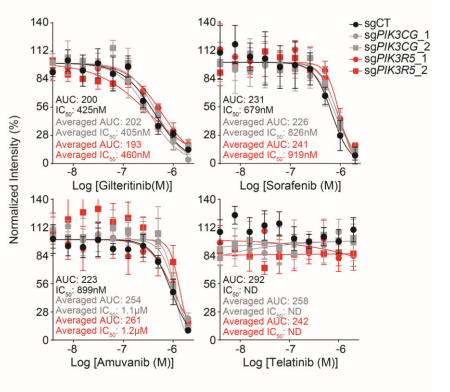
Targeting a lineage-specific PI3Kɣ-Akt signaling module in acute myeloid leukemia using a heterobifunctional degrader molecule. [Abstract]2024 Jul;5(7):1082-1101. PMID: 38816660
Sorafenib tosylate purchased from MedChemExpress. Usage Cited in: Nat Cancer. 2024 Jul;5(7):1082-1101. [Abstract]
Growth inhibition, IC50 and AUC values, of OCI-AML2 cells transduced with either a non-targeting control, two PIK3CG-directed, or two PIK3R5-directed sgRNAs and treated with increasing concentrations of the FLT3 inhibitors, Gilteritinib (10-1000 nM) and Sorafenib (10-1000 nM), or KIT inhibitors, Amuvanib and Telatinib
-
Cancer Res
The Acetyltransferase ARD1 Induces Glutathione Synthesis to Facilitate Ferroptosis Evasion in Hepatocellular Carcinoma. [Abstract]2025 Aug 21. PMID: 40838989 -
Mol Cell
EGFR promotes ALKBH5 nuclear retention to attenuate N6-methyladenosine and protect against ferroptosis in glioblastoma. [Abstract]2023 Dec 7;83(23):4334-4351.e7. PMID: 37979586 -
ACS Nano
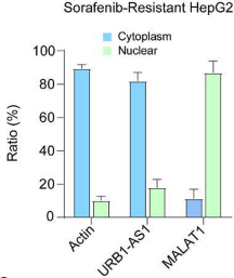
Long Noncoding RNA URB1-Antisense RNA 1 (AS1) Suppresses Sorafenib-Induced Ferroptosis in Hepatocellular Carcinoma by Driving Ferritin Phase Separation. [Abstract]2023 Nov 28;17(22):22240-22258. PMID: 37966480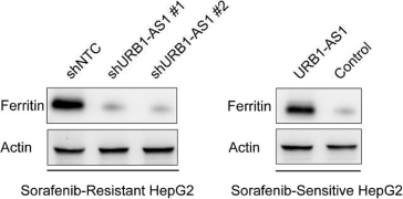
Sorafenib tosylate purchased from MedChemExpress. Usage Cited in: ACS Nano. 2023 Nov 28;17(22):22240-22258. [Abstract]
Western blot to detect the ferritin H protein level by the gain or loss of URB1-AS1 in sorafenib-sensitive or sorafenib-resistant HepG2 cells.
Sorafenib tosylate purchased from MedChemExpress. Usage Cited in: ACS Nano. 2023 Nov 28;17(22):22240-22258. [Abstract]
qPCR showing the nuclear and cytoplasmic fractions of URB1-AS1 in sorafenib-resistant HepG2 cells with β-actin and MALAT1 as cytoplasmic and nuclear controls, respectively.
-
Nat Commun
Co-delivery of sorafenib and an FSP1 inhibitor triggers dual ferroptosis in tumor cells and immunosuppressive macrophages for enhanced immunotherapy in mouse models of hepatocellular carcinoma. [Abstract]2025 Nov 18;16(1):10096. PMID: 41253833 -
Nat Commun
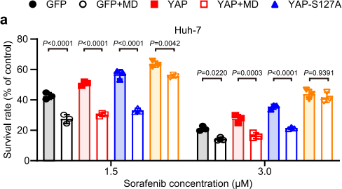
Positive feedback between arginine methylation of YAP and methionine transporter SLC43A2 drives anticancer drug resistance. [Abstract]2025 Jan 2;16(1):87. PMID: 39747898
Sorafenib tosylate purchased from MedChemExpress. Usage Cited in: Nat Commun. 2025 Jan 2;16(1):87. [Abstract]
The cell viability of YAP-, YAP-S127A-, or YAP-R124F-overexpressed Huh-7 cells cultured in methionine-deprived medium (MD) and treated with sorafenib (1.5-3 μM) for 24 h.
-
Nat Commun
2024 Sep 10;15(1):7923. PMID: 39256387 -
Nat Commun
N7-methylguanosine tRNA modification promotes esophageal squamous cell carcinoma tumorigenesis via the RPTOR/ULK1/autophagy axis. [Abstract]2022 Mar 18;13(1):1478. PMID: 35304469 -
Cell Death Differ
Branched-chain amino acid aminotransferase 2 regulates ferroptotic cell death in cancer cells. [Abstract]2021 Apr;28(4):1222-1236. PMID: 33097833 -
Bone Res
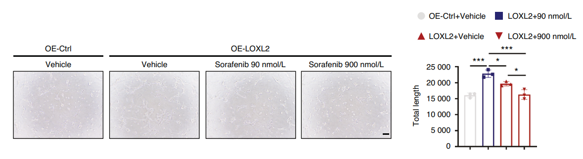
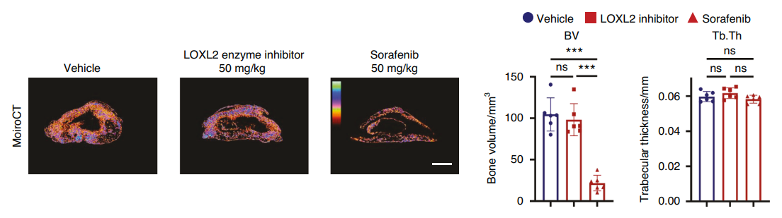
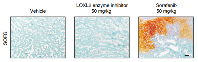
Sorafenib inhibits ossification of the posterior longitudinal ligament by blocking LOXL2-mediated vascularization. [Abstract]2024 Apr 10;12(1):24. PMID: 38594260
Sorafenib tosylate purchased from MedChemExpress. Usage Cited in: Bone Res. 2024 Apr 10;12(1):24. [Abstract]
Sorafenib tosylate (90-900 nM) markedly suppressed the upregulation of endothelial-like differentiation in ligament cells mediated by LOXL2.
Sorafenib tosylate purchased from MedChemExpress. Usage Cited in: Bone Res. 2024 Apr 10;12(1):24. [Abstract]
Sorafenib tosylate (50 mg/kg; i.g.; one daily for 14 d) significantly decreased bone volume (BV) in BMP-induced ossification mice.
Sorafenib tosylate purchased from MedChemExpress. Usage Cited in: Bone Res. 2024 Apr 10;12(1):24. [Abstract]
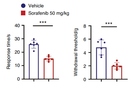
Sorafenib tosylate (50 mg/kg; i.g.; once daily for 14 d) caused sparse trabecular bone structures in BMP-induced ossification mice, accompanied by numerous cartilage structures and amorphous matrix deposition, indicating a delay in endochondral ossification.
Sorafenib tosylate purchased from MedChemExpress. Usage Cited in: Bone Res. 2024 Apr 10;12(1):24. [Abstract]
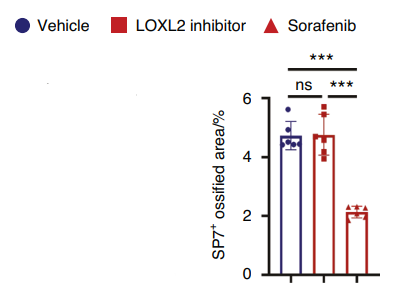
Sorafenib tosylate (50 mg/kg; i.g.; once daily for 14 d) significantly lowered the level of SP7 in BMP-induced ossification mice.
Sorafenib tosylate purchased from MedChemExpress. Usage Cited in: Bone Res. 2024 Apr 10;12(1):24. [Abstract]
Sorafenib tosylate (50 mg/kg; i.g.; once daily for 14 d)caused a shorter response time to thermal stimulation and less paw withdrawal threshold to mechanical stimulation of BMP-induced ossification mice.
-
Acta Pharm Sin B
Sorafenib promotes the E3 ubiquitin ligase FBXW7 to increase tau degradation and ameliorate tauopathies. [Abstract]2025 Nov;15(11):5817-5831. PMID: 41311387 -
Acta Pharm Sin B
A high-throughput Gaussia luciferase reporter assay for screening potential gasdermin E activators against pancreatic cancer. [Abstract]2023 Oct;13(10):4253-4272. PMID: 37799380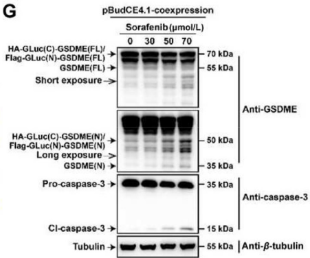
Sorafenib tosylate purchased from MedChemExpress. Usage Cited in: Acta Pharm Sin B. 2023 Oct;13(10):4253-4272. [Abstract]
PANC-1 cells were transfected with the recombinant plasmid for 24 h and treated with sorafenib (0–70 μmol/L) for another 24 h, and then total cellular extracts were subjected to Western blotting using antibodies against caspase-3, GSDME and β-tubulin.
Sorafenib tosylate purchased from MedChemExpress. Usage Cited in: Acta Pharm Sin B. 2023 Oct;13(10):4253-4272. [Abstract]
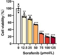
Sorafenib (12.5-125 μM; 24 h) inhibits the viability of PANC-1 cells in a dose-dependent manner.
-
Acta Pharm Sin B
The suppression of cervical cancer ferroptosis by macrophages: The attenuation of ALOX15 in cancer cells by macrophages-derived exosomes. [Abstract]2023 Jun;13(6):2645-2662. PMID: 37425043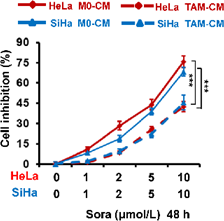
Sorafenib tosylate purchased from MedChemExpress. Usage Cited in: Acta Pharm Sin B. 2023 Jun;13(6):2645-2662. [Abstract]
HeLa and SiHa cells were treated with condition medium (CM) from human PBMC-derived M0 macrophages (Ctrl) and TAM. And then cells were treated with indicated dose of Sorafenib (Sora) for 48 h. Inhibition ratio of cell viability was detected by CCK-8 assay.
-
Sci Transl Med
PP2A inhibition is a druggable MEK inhibitor resistance mechanism in KRAS-mutant lung cancer cells. [Abstract]2018 Jul 18;10(450):eaaq1093. PMID: 30021885 -
J Extracell Vesicles
Oxidative stress induces extracellular vesicle release by upregulation of HEXB to facilitate tumour growth in experimental hepatocellular carcinoma. [Abstract]2024 Jul;13(7):e12468. PMID: 38944674 -
Autophagy
Kitasamycin overcomes ferroptosis and immunotherapy resistance by targeting the HUWE1-NCOA4-FTH1 axis. [Abstract]2026 Feb 15:1-19. PMID: 41612599 -
Autophagy
The human vault RNA enhances tumorigenesis and chemoresistance through the lysosome in hepatocellular carcinoma. [Abstract]2022 Jan;18(1):191-203. PMID: 33960270 -
Adv Sci (Weinh)
Clathrin Light Chain B Drives Hepatocellular Carcinoma Progression Through Dual Mechanisms: Small Extracellular Vesicle-Mediated Angiogenesis and the NF-κB-PCLAF Signaling Axis. [Abstract]2025 Aug 18:e08613. PMID: 40820941 -
Adv Sci (Weinh)
TRIM21-Mediated K11-Linked Ubiquitination of ID1 Suppresses Tumorigenesis and Promotes Cuproptosis in Esophageal Squamous Cell Carcinoma. [Abstract]2025 Jul 13:e02501. PMID: 40652518 -
Adv Sci (Weinh)
PP1A Modulates the Efficacy of Lenvatinib Plus ICIs Therapy by Inhibiting Ferroptosis in Hepatocellular Carcinoma. [Abstract]2025 May 8:e2501730. PMID: 40344394 -
Adv Sci (Weinh)
Stabilization of TGF-β Receptor 1 by a Receptor-Associated Adaptor Dictates Feedback Activation of the TGF-β Signaling Pathway to Maintain Liver Cancer Stemness and Drug Resistance. [Abstract]2024 Jul 9:e2402327. PMID: 38981014 -
Adv Sci (Weinh)
Micro-Engineered Organoid-on-a-Chip Based on Mesenchymal Stromal Cells to Predict Immunotherapy Responses of HCC Patients. [Abstract]2023 Sep;10(27):e2302640. PMID: 37485650 -
Adv Sci (Weinh)
Donafenib and GSK-J4 Synergistically Induce Ferroptosis in Liver Cancer by Upregulating HMOX1 Expression. [Abstract]2023 Aug;10(22):e2206798. PMID: 37330650 -
Adv Sci (Weinh)
STING Suppresses Mitochondrial VDAC2 to Govern RCC Growth Independent of Innate Immunity. [Abstract]2023 Jan;10(3):e2203718. PMID: 36445063 -
Adv Sci (Weinh)
Engineered EGCG-Containing Biomimetic Nanoassemblies as Effective Delivery Platform for Enhanced Cancer Therapy. [Abstract]2022 May;9(15):e2105894. PMID: 35486032 -
Adv Sci (Weinh)
PADI2-Catalyzed MEK1 Citrullination Activates ERK1/2 and Promotes IGF2BP1-Mediated SOX2 mRNA Stability in Endometrial Cancer. [Abstract]2021 Jan 29;8(6):2002831. PMID: 33747724 -
Exp Hematol Oncol
Effects of molecularly targeted therapies on murine thymus: highly selective mTOR inhibitors induce reversible thymic involution. [Abstract]2016 Jul 29:5:22. PMID: 27478685 -
Theranostics
Combination therapy with B7H3-redirected bispecific antibody and Sorafenib elicits enhanced synergistic antitumor efficacy. [Abstract]2020 Aug 21;10(23):10498-10512. PMID: 32929362 -
Biomaterials
2022 Oct:289:121800. PMID: 36166893 -
J Exp Clin Cancer Res
Targeting fatty acid synthase modulates sensitivity of hepatocellular carcinoma to sorafenib via ferroptosis. [Abstract]2023 Jan 6;42(1):6. PMID: 36604718 -
J Exp Clin Cancer Res
Cholesterol sensor SCAP contributes to sorafenib resistance by regulating autophagy in hepatocellular carcinoma. [Abstract]2022 Mar 30;41(1):116. PMID: 35354475 -
J Exp Clin Cancer Res
GPR119 agonist enhances gefitinib responsiveness through lactate-mediated inhibition of autophagy. [Abstract]2018 Nov 29;37(1):295. PMID: 30497501 -
Cell Discov
Dynamic O-GlcNAcylation coordinates ferritinophagy and mitophagy to activate ferroptosis. [Abstract]2022 May 3;8(1):40. PMID: 35504898 -
Redox Biol
USP20 governs tyrosine kinase inhibitors resistance through ferroptosis evasion by targeting GPX4 in cancers. [Abstract]2026 May:92:104086. PMID: 41844497 -
Redox Biol
ATF7IP inhibits Sorafenib-induced ferroptosis in hepatocellular carcinoma cells by inhibiting CYB5R2 transcription and stabilizing PARK7 protein. [Abstract]2025 Sep:85:103786. PMID: 40716153 -
Redox Biol
Reactivation of MAPK-SOX2 pathway confers ferroptosis sensitivity in KRASG12C inhibitor resistant tumors. [Abstract]2024 Nov 5:78:103419. PMID: 39527862 -
Redox Biol
MAPK15 controls cellular responses to oxidative stress by regulating NRF2 activity and expression of its downstream target genes. [Abstract]2024 Jun:72:103131. PMID: 38555711 -
Redox Biol
Increased ATF2 expression predicts poor prognosis and inhibits sorafenib-induced ferroptosis in gastric cancer. [Abstract]2023 Feb:59:102564. PMID: 36473315 -
J Hazard Mater
2025 Oct 19:499:140193. PMID: 41124733 -
Gut Microbes
Synergistic activity of Enterococcus Faeciu m-induced ferroptosis via expansion of IFN-γ+CD8+ T cell population in advanced hepatocellular carcinoma treated with sorafenib. [Abstract]2024 Jan-Dec;16(1):2410474. PMID: 39353096 -
MedComm (2020)
circRNA-SORE/UBQLN1/GPX4 Mediates the Acquisition of Sorafenib Resistance in Hepatocellular Carcinoma Through Inhibition of Ferroptosis. [Abstract]2025 Nov 23;6(12):e70488. PMID: 41287824 -
Cell Rep Med
CAN-Scan: A multi-omic phenotype-driven precision oncology platform identifies prognostic biomarkers of therapy response for colorectal cancer. [Abstract]2025 Apr 2:102053. PMID: 40187357 -
J Immunother Cancer
Hyperbaric oxygen facilitates teniposide-induced cGAS-STING activation to enhance the antitumor efficacy of PD-1 antibody in HCC. [Abstract]2022 Aug;10(8):e004006. PMID: 36002188 -
Pharmacol Res
Epigenetic regulation of the DNMT1/MT1G/KLF4/CA9 axis synergises the anticancer effects of sorafenib in hepatocellular carcinoma. [Abstract]2022 Jun;180:106244. PMID: 35550167 -
Pharmacol Res
Potential synthetic lethality for breast cancer: A selective sirtuin 2 inhibitor combined with a multiple kinase inhibitor sorafenib. [Abstract]2022 Mar:177:106050. PMID: 34973468 -
Clin Cancer Res
CDK9 inhibition by dinaciclib is a therapeutic vulnerability in epithelioid hemangioendothelioma. [Abstract]2024 Sep 13;30(18):4179-4189. PMID: 39052240 -
Cancer Lett
Induction of IL-6Rα by ATF3 enhances IL-6 mediated sorafenib and regorafenib resistance in hepatocellular carcinoma. [Abstract]2022 Jan 1:524:161-171. PMID: 34687791 -
Cancer Lett
Coupling HDAC4 with transcriptional factor MEF2D abrogates SPRY4-mediated suppression of ERK activation and elicits hepatocellular carcinoma drug resistance. [Abstract]2021 Nov 1:520:243-254. PMID: 34339801 -
Cancer Lett
PTK2 promotes cancer stem cell traits in hepatocellular carcinoma by activating Wnt/β-catenin signaling. [Abstract]2019 May 28:450:132-143. PMID: 30849480 -
Int J Biol Sci
Chemical and genetic inhibition of STAT3 sensitizes hepatocellular carcinoma cells to sorafenib induced cell death. [Abstract]2018 Apr 25;14(5):577-585. PMID: 29805309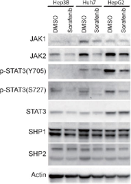
Sorafenib tosylate purchased from MedChemExpress. Usage Cited in: Int J Biol Sci. 2018 Apr 25;14(5):577-585. [Abstract]
Hep3B, HepG2 and Huh7 cells are treated with 5 μM Sorafenib. The expressing levels of JAK1, JAK2, STAT3, SHP1, SHP2, actin and phosphorylation levels of STAT3 are determined by western blot using the antibodies, respectively.
-
Cell Death Dis
SOX4-STAT6-MTHFD2 axis drives hepatocellular carcinoma progression and treatment resistance. [Abstract]2026 Jan 3. PMID: 41484064 -
Cell Death Dis
Synergistic antitumor activity of sorafenib and the NUPR1 inhibitor LZX-2-73 in multiple cancer models. [Abstract]2025 Nov 17;16(1):839. PMID: 41249113 -
Cell Death Dis
DCAF7 recruits USP2 to facilitate hepatocellular carcinoma progression by suppressing clockophagy-induced ferroptosis. [Abstract]2025 Aug 28;16(1):654. PMID: 40877242 -
Cell Death Dis
IFNγ augments TKI efficacy by alleviating protein unfolding stress to promote GSDME-mediated pyroptosis in hepatocellular carcinoma. [Abstract]2025 Jul 11;16(1):512. PMID: 40645933 -
Cell Death Dis
Pharmacological targeting of the mitochondrial phosphatase PTPMT1 sensitizes hepatocellular carcinoma to ferroptosis. [Abstract]2025 Apr 6;16(1):257. PMID: 40189563 -
Cell Death Dis
2025 Jan 26;16(1):42. PMID: 39863613 -
Cell Death Dis
Phosphorylated FOXQ1, a novel substrate of JNK1, inhibits sorafenib-induced ferroptosis by activating ETHE1 in hepatocellular carcinoma. [Abstract]2024 Jun 5;15(6):395. PMID: 38839744 -
Cell Death Dis
2024 Jan 18;15(1):66. PMID: 38238307 -
Adv Healthc Mater
A Novel Patient-Personalized Nanovector Based on Homotypic Recognition and Magnetic Hyperthermia for an Efficient Treatment of Glioblastoma Multiforme. [Abstract]2023 Jul;12(19):e2203120. PMID: 37058273 -
Cell Death Dis
SLC27A5 promotes sorafenib-induced ferroptosis in hepatocellular carcinoma by downregulating glutathione reductase. [Abstract]2023 Jan 12;14(1):22. PMID: 36635256 -
Cell Death Dis
PGK1 contributes to tumorigenesis and sorafenib resistance of renal clear cell carcinoma via activating CXCR4/ERK signaling pathway and accelerating glycolysis. [Abstract]2022 Feb 4;13(2):118. PMID: 35121728 -
Cell Death Dis
MYC-targeted WDR4 promotes proliferation, metastasis, and sorafenib resistance by inducing CCNB1 translation in hepatocellular carcinoma. [Abstract]2021 Jul 9;12(7):691. PMID: 34244479 -
Cell Death Dis
Ferritinophagy is required for the induction of ferroptosis by the bromodomain protein BRD4 inhibitor (+)-JQ1 in cancer cells. [Abstract]2019 Apr 15;10(5):331. PMID: 30988278 -
Genes Dis
PCK1 attenuates tumor stemness via activating the Hippo signaling pathway in hepatocellular carcinoma. [Abstract]2023 Sep 16;11(4):101114. PMID: 38560500 -
Cell Commun Signal
Concomitant targeting of FLT3 and SPHK1 exerts synergistic cytotoxicity in FLT3-ITD+ acute myeloid leukemia by inhibiting β-catenin activity via the PP2A-GSK3β axis. [Abstract]2024 Aug 7;22(1):391. PMID: 39113090 -
Int J Biol Macromol
KIF20A drives epithelial cell proliferation and migration in gastric adenocarcinoma, facilitating macrophage M2 polarization and subsequent immune evasion. [Abstract]2026 Mar:351:150982. PMID: 41713545 -
Int J Biol Macromol
The role of solute carrier family 16 member 3 protein in hepatocellular carcinoma and sorafenib resistance. [Abstract]2025 Oct 9;330(Pt 3):148223. PMID: 41075888 -
Acta Pharmacol Sin
Osimertinib successfully combats EGFR-negative glioblastoma cells by inhibiting the MAPK pathway. [Abstract]2021 Jan;42(1):108-114. PMID: 32398685 -
Phytomedicine
Usenamine a potentiates anti-CRC activity of sorafenib by inducing autophagy and inhibiting YAP pathway through targeting SOD2. [Abstract]2026 Jun:155:158086. PMID: 41861687 -
Phytomedicine
Arnicolide C induces ROS-mediated modulation of PI3K/Akt and MAPK pathways to suppress MYC in hepatocellular carcinoma. [Abstract]2025 Oct 20:148:157423. PMID: 41138574 -
Phytomedicine
Picropodophyllin induces ferroptosis via blockage of AKT/NRF2/SLC7A11 and AKT/NRF2/SLC40A1 axes in hepatocellular carcinoma as a natural IGF1R inhibitor. [Abstract]2025 May 10:143:156840. PMID: 40412057 -
Phytomedicine
Indole-3-carbinol inhibits PD-L1-mediated immune evasion in hepatocellular carcinoma via suppressing NF-κB p105 Ubiquitination. [Abstract]2025 Apr 1:141:156692. PMID: 40215823 -
Phytomedicine
Stigmasterol: Remodeling gut microbiota and suppressing tumor growth through Treg and CD8+ T cells in hepatocellular carcinoma. [Abstract]2024 Jul:129:155225. PMID: 38678948 -
Phytomedicine
The role of daurisoline treatment in hepatocellular carcinoma: Inhibiting vasculogenic mimicry formation and enhancing sensitivity to sorafenib. [Abstract]2021 Nov:92:153740. PMID: 34600176 -
ACS Appl Mater Interfaces
2026 Jan 25. PMID: 41582522 -
Free Radic Biol Med
The PGC1α/NRF1-MPC1 axis suppresses tumor progression and enhances the sensitivity to sorafenib/doxorubicin treatment in hepatocellular carcinoma. [Abstract]2021 Feb 1;163:141-152. PMID: 33276082 -
Free Radic Biol Med
2020 Nov 20;160:303-318. PMID: 32846217 -
ACS Appl Mater Interfaces
A New Spin on Antibody-Drug Conjugates: Trastuzumab-Fulvestrant Colloidal Drug Aggregates Target HER2-Positive Cells. [Abstract]2017 Apr 12;9(14):12195-12202. PMID: 28319364 -
Drug Deliv
Andrographolide nanophytosomes exhibit enhanced cellular delivery and pro-apoptotic activities in HepG2 liver cancer cells. [Abstract]2023 Dec;30(1):2174209. PMID: 36762548 -
Chemosphere
The influence of sunitinib and sorafenib, two tyrosine kinase inhibitors, on development and thyroid system in zebrafish larvae. [Abstract]2022 Dec;308(Pt 2):136354. PMID: 36087734 -
Clin Sci
Identifying mitigating strategies for endothelial cell dysfunction and hypertension in response to VEGF receptor inhibitors. [Abstract]2024 Sep 18;138(18):1131-1150. PMID: 39282930 -
-
Clin Sci
A novel epigenetic drug conjugating flavonoid and HDAC inhibitor confer suppression of acute myeloid leukemogenesis. [Abstract]2021 Jul 30;135(14):1751-1765. PMID: 34282832 -
Br J Pharmacol
DGT, a novel heterocyclic diterpenoid, effectively suppresses psoriasis via inhibition of STAT3 phosphorylation. [Abstract]2021 Feb;178(3):636-653. PMID: 33140855 -
Biomed Pharmacother
Decursin induces ferroptosis via the NRF2/GPX4/SLC11A2 axis and suppresses migration in hepatocellular carcinoma. [Abstract]2026 Feb:195:118941. PMID: 41496356 -
Biomed Pharmacother
Inhibition of the PI3K/AKT signaling pathway contributes to the anti-renal cell carcinoma effects of deoxyelephantopin. [Abstract]2025 May 8:187:118136. PMID: 40344699 -
J Transl Med
Endogenous protein S100A14 stabilizes glutaminase to render hepatocellular carcinoma resistant to sorafenib. [Abstract]2025 Apr 11;23(1):435. PMID: 40217256 -
J Transl Med
RPL22L1 fosters malignant features of cervical cancer via the modulation of DUSP6-ERK axis. [Abstract]2025 Feb 28;23(1):244. PMID: 40022129 -
J Transl Med
Mito-LND and (E)-Akt inhibitor-IV: novel compounds inducing endoplasmic reticulum stress and ROS accumulation against hepatocellular carcinoma. [Abstract]2024 Aug 28;22(1):792. PMID: 39198815 -
J Transl Med
Papillary thyroid cancer organoids harboring BRAFV600E mutation reveal potentially beneficial effects of BRAF inhibitor-based combination therapies. [Abstract]2023 Jan 9;21(1):9. PMID: 36624452 -
Biomed Pharmacother
Advantage of clinical colchicine concentration to promote sorafenib or regorafenib anti-cancer effects on hepatocellular carcinoma. [Abstract]2022 Sep:153:113540. PMID: 36076618 -
J Transl Med
RAF1 promotes lymphatic metastasis of hypopharyngeal carcinoma via regulating LAGE1: an experimental research. [Abstract]2022 Jun 6;20(1):255. PMID: 35668458 -
Biomed Pharmacother
TMT-based proteomics analysis of the anti-hepatocellular carcinoma effect of combined dihydroartemisinin and sorafenib. [Abstract]2020 Jun;126:109862. PMID: 32120157 -
Biomed Pharmacother
Contrary influence of clinically applied sorafenib concentrations among hepatocellular carcinoma patients. [Abstract]2017 Feb:86:27-31. PMID: 27936391 -
Cell Mol Gastroenterol Hepatol
Sorafenib Promotes Treg Cell Differentiation To Compromise Its Efficacy via VEGFR/AKT/Foxo1 Signaling in Hepatocellular Carcinoma. [Abstract]2024 Dec 30:101454. PMID: 39743020 -
Oncogene
LINC-AC092535.5 regulates MICAL2 mRNA level to inhibit p53-mediated ferroptosis in nasopharyngeal carcinoma. [Abstract]2026 Apr;45(14):1260-1274. PMID: 41832266 -
Stem Cell Res Ther
Human menstrual blood-derived stem cells reverse sorafenib resistance in hepatocellular carcinoma cells through the hyperactivation of mitophagy. [Abstract]2023 Apr 1;14(1):58. PMID: 37005657 -
Oncogene
PBLD inhibits angiogenesis via impeding VEGF/VEGFR2-mediated microenvironmental cross-talk between HCC cells and endothelial cells. [Abstract]2022 Mar;41(13):1851-1865. PMID: 35140333 -
Oncogene
Antifungal agent Terbinafine restrains tumor growth in preclinical models of hepatocellular carcinoma via AMPK-mTOR axis. [Abstract]2021 Aug;40(34):5302-5313. PMID: 34247189 -
Cell Death Discov
Engineered small extracellular vesicles loaded with miR-654-5p promote ferroptosis by targeting HSPB1 to alleviate sorafenib resistance in hepatocellular carcinoma. [Abstract]2023 Sep 30;9(1):362. PMID: 37777559 -
Cell Death Discov
Ribosomal protein L22-like1 (RPL22L1) mediates sorafenib sensitivity via ERK in hepatocellular carcinoma. [Abstract]2022 Aug 17;8(1):365. PMID: 35973992 -
Cell Rep
A shear stress-responsive pathway in monocytes drives cardiopulmonary bypass-induced inflammation via spectrin/RAF1/store-operated calcium entry. [Abstract]2026 Jan 22;45(2):116903. PMID: 41579376 -
Cell Rep
Carbonic anhydrase 2 facilitates sorafenib resistance by counteracting MCT4-mediated intracellular pH dysregulation in HCC. [Abstract]2024 Nov 27;43(12):114996. PMID: 39607826 -
Cell Rep
STUB1-mediated ubiquitination and degradation of NSUN2 promotes hepatocyte ferroptosis by decreasing m5C methylation of Gpx4 mRNA. [Abstract]2024 Oct 24;43(11):114885. PMID: 39453812 -
Cell Rep
Intratumor Mycoplasma promotes the initiation and progression of hepatocellular carcinoma. [Abstract]2023 Dec 12;42(12):113563. PMID: 38088929 -
Clin Transl Med
Oncofetal dual‑specificity phosphatase 9 drives stem-like properties through ERK1/2-PPARG-SCD axis-mediated lipid metabolism in hepatocellular carcinoma. [Abstract]2025 Dec;15(12):e70550. PMID: 41383134 -
Clin Transl Med
USP22 promotes the proliferation and Sorafenib resistance of hepatocellular carcinoma cells via its deubiquitinase activity. [Abstract]2025 May;15(5):e70324. PMID: 40341781 -
J Med Chem
Discovery of Highly Potent AKR1C3 Inhibitors Treating Sorafenib-Resistant Hepatocellular Carcinoma. [Abstract]2025 Mar 27. PMID: 40143712 -
Clin Transl Med
Loss of LncRNA DUXAP8 synergistically enhanced sorafenib induced ferroptosis in hepatocellular carcinoma via SLC7A11 de-palmitoylation. [Abstract]2023 Jun;13(6):e1300. PMID: 37337470 -
J Med Chem
2019 Nov 14;62(21):9593-9599. PMID: 31589047 -
Br J Cancer
Activation of an AKT/FOXM1/STMN1 pathway drives resistance to tyrosine kinase inhibitors in lung cancer. [Abstract]2017 Sep 26;117(7):974-983. PMID: 28850563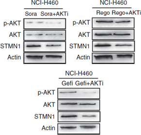
Sorafenib tosylate purchased from MedChemExpress. Usage Cited in: Br J Cancer. 2017 Sep 26;117(7):974-983. [Abstract]
The effect of the AKT inhibitor MK2206 (10 μM) on the expression levels of phosphor-AKT, AKT, and STMN1 in TKI-pretreated NCI-H460 cells. β-actin is used as a loading control.
-
Int J Nanomedicine
A Novel Triptolide Nano-Liposome with Mitochondrial Targeting for Treatment of Hepatocellular Carcinoma. [Abstract]2024 Dec 3:19:12975-12998. PMID: 39654802 -
Comput Biol Med
Liquid-liquid phase separation-related lncRNA prognostic signature and ZNF32-AS2 as a novel biomarker in hepatocellular carcinoma. [Abstract]2024 Feb:169:107975. PMID: 38199212 -
Cancer Cell Int
Establishment and characteristic analysis of a novel patient derived cell line of intrahepatic cholangiocarcinoma. [Abstract]2025 Oct 15;25(1):357. PMID: 41094661 -
Biomater Adv
Anti-tumor immunity and ferroptosis of hepatocellular carcinoma are enhanced by combined therapy of sorafenib and delivering modified GO-based PD-L1 siRNAs. [Abstract]2022 May:136:212761. PMID: 35929305 -
Cancer Cell Int
The deubiquitinating enzyme OTUD1 antagonizes BH3-mimetic inhibitor induced cell death through regulating the stability of the MCL1 protein. [Abstract]2019 Aug 27;19:222. PMID: 31467488 -
Cell Biol Toxicol
Protocatechuic acid relieves ferroptosis in hepatic lipotoxicity and steatosis via regulating NRF2 signaling pathway. [Abstract]2024 Nov 26;40(1):104. PMID: 39589556 -
Eur J Med Chem
Novel inhibitors targeting the PGK1 metabolic enzyme in glycolysis exhibit effective antitumor activity against kidney renal clear cell carcinoma in vitro and in vivo. [Abstract]2024 Mar 5:267:116209. PMID: 38354523 -
Cell Biol Toxicol
The mtDNA-STING pathway plays an important role in both navitoclax- and S63845-induced autophagy and enhances cell death. [Abstract]2023 Dec;39(6):2821-2839. PMID: 37002446 -
Int J Mol Med
STOML2 interacts with PHB to activate the MEK/ERK signaling pathway and mediates autophagy‑related proteins in the progression of hepatocellular carcinoma. [Abstract]2026 Feb;57(2):38. PMID: 41347828 -
Chin Med
Polyphyllin I exerts anti-hepatocellular carcinoma activity by targeting ZBTB16 to activate the PPARγ/RXRα signaling pathway. [Abstract]2024 Aug 24;19(1):113. PMID: 39182119 -
Biochem Pharmacol
2023 Jul:213:115593. PMID: 37196682 -
Biochem Pharmacol
ALOX5 Promotes Autophagy-dependent Ferroptosis by Activating the AMPK/mTOR Pathway in Melanoma. [Abstract]2023 Jun:212:115554. PMID: 37080437 -
Biochem Pharmacol
Organic anion transport polypeptide 1b2 selectively affects the pharmacokinetic interaction between paclitaxel and sorafenib in rats. [Abstract]2019 Nov:169:113612. PMID: 31437461 -
Mol Cancer Ther
Long Noncoding RNA MALAT1 Contributes to Sorafenib Resistance by Targeting miR-140-5p/Aurora-A Signaling in Hepatocellular Carcinoma. [Abstract]2020 May;19(5):1197-1209. PMID: 32220970 -
Lab Chip
A novel microfluidic self-perfusion chip (MSPC) for pumpless 3D cell, microtissue and organoid culture. [Abstract]2025 Apr 10. PMID: 40206017 -
World J Gastroenterol
Y-box binding protein 1 augments sorafenib resistance via the PI3K/Akt signaling pathway in hepatocellular carcinoma. [Abstract]2021 Jul 28;27(28):4667-4686. PMID: 34366628 -
J Ethnopharmacol
Modulation of the VEGF/AKT/eNOS signaling pathway to regulate liver angiogenesis to explore the anti-hepatic fibrosis mechanism of curcumol. [Abstract]2021 Nov 15:280:114480. PMID: 34358654 -
J Enzyme Inhib Med Chem
Design, synthesis, and biological evaluation of novel substituted thiourea derivatives as potential anticancer agents for NSCLC by blocking K-Ras protein-effectors interactions. [Abstract]2020 Dec;35(1):344-353. PMID: 31851852 -
Lab Chip
Development of a biomimetic liver tumor-on-a-chip model based on decellularized liver matrix for toxicity testing. [Abstract]2018 Nov 6;18(22):3379-3392. PMID: 30298144 -
Cancer Immunol Immunother
Targeting ST3GAL1 to downregulate ligands for the glycoimmune checkpoint Siglec-7 and reverse immune escape in hepatocellular carcinoma. [Abstract]2026 Apr 10;75(5):140. PMID: 41961075 -
Commun Biol
Isoacteoside alleviates hepatocellular carcinoma progression by inhibiting PDHB-mediated reprogramming of glucose metabolism. [Abstract]2025 Feb 8;8(1):205. PMID: 39922943 -
Commun Biol
USP24 promotes autophagy-dependent ferroptosis in hepatocellular carcinoma by reducing the K48-linked ubiquitination of Beclin1. [Abstract]2024 Oct 8;7(1):1279. PMID: 39379617 -
Drug Des Devel Ther
Identification of Potential Therapeutics for Infantile Hemangioma via in silico Investigation and in vitro Validation. [Abstract]2024 Sep 12:18:4065-4088. PMID: 39286286 -
Int J Mol Sci
2025 Sep 20;26(18):9185. PMID: 41009747 -
Biol Direct
L-741626 inhibits hepatocellular carcinoma progression by targeting Ref-1 to suppress MAPK/ERK signalling pathway activity. [Abstract]2025 Apr 16;20(1):54. PMID: 40241114 -
Biol Direct
Sorafenib-induced macrophage extracellular traps via ARHGDIG/IL4/PADI4 axis confer drug resistance through inhibiting ferroptosis in hepatocellular carcinoma. [Abstract]2024 Nov 11;19(1):110. PMID: 39529192 -
Int J Mol Sci
The Potential of Congo Red Supplied Aggregates of Multitargeted Tyrosine Kinase Inhibitor (Sorafenib, BAY-43-9006) in Enhancing Therapeutic Impact on Bladder Cancer. [Abstract]2023 Dec 23;25(1):269. PMID: 38203437 -
Int J Mol Sci
SHP-1/STAT3-Signaling-Axis-Regulated Coupling between BECN1 and SLC7A11 Contributes to Sorafenib-Induced Ferroptosis in Hepatocellular Carcinoma. [Abstract]2022 Sep 21;23(19):11092. PMID: 36232407 -
Chin J Nat Med
Two cardenolide glycosides from the seed fairs of Asclepias curassavica and their cytotoxic activities. [Abstract]2022 Mar;20(3):202-209. PMID: 35369964 -
Front Pharmacol
Protein and metabolic profiles of tyrosine kinase inhibitors co-resistant liver cancer cells. [Abstract]2024 May 21:15:1394241. PMID: 38835670 -
Front Pharmacol
Caryophyllene Oxide Induces Ferritinophagy by Regulating the NCOA4/FTH1/LC3 Pathway in Hepatocellular Carcinoma. [Abstract]2022 Jul 11;13:930958. PMID: 35899120 -
Eur J Pharmacol
Neoprzewaquinone A suppresses hepatocellular carcinoma through promoting the ubiquitin-related degradation of EGFR and inhibiting PI3K-AKT pathway. [Abstract]2025 Oct 21:1007:178274. PMID: 41130376 -
Bioorg Chem
Synthesis and evaluation of sulfonamide-chalcone hybrid compounds as inhibitors of VEGFR1/VEGFR2-mediated angiogenesis. [Abstract]2025 Aug 21:164:108903. PMID: 40848708 -
Int Immunopharmacol
ASIC1a regulates ferroptosis in hepatic stellate cells via the Hippo/Yap-1 pathway in liver fibrosis. [Abstract]2024 Sep 30;143(Pt 1):113226. PMID: 39353388 -
Eur J Pharmacol
2021 Sep 5:906:174217. PMID: 34087223 -
Toxicology
Sorafenib induces intestinal toxicity by disturbing gut microbiota and activating the LPS/TLR4/NF-κB signaling pathway in mice. [Abstract]2025 Jun 13:517:154220. PMID: 40518001 -
RSC Adv
Self-assembled nanoplatform-mediated co-delivery of brusatol to sensitize sorafenib for hepatocellular carcinoma treatment. [Abstract]2025 Apr 14;15(15):11675-11687. PMID: 40230634 -
Hepatol Commun
Oxyberberine sensitizes liver cancer cells to sorafenib via inhibiting NOTCH1-USP7-c-Myc pathway. [Abstract]2024 Mar 29;8(4):e0405. PMID: 38573832 -
Toxicology
Inhibitory effects of flavonoids on glucose transporter 1 (GLUT1): From library screening to biological evaluation to structure-activity relationship. [Abstract]2023 Apr:488:153475. PMID: 36870413 -
Neuropharmacology
Sorafenib promotes sensory conduction function recovery via miR-142-3p/AC9/cAMP axis post dorsal column injury. [Abstract]2019 Apr:148:347-357. PMID: 30710569 -
Molecules
Facilitating Anti-Cancer Combinatorial Drug Discovery by Targeting Epistatic Disease Genes. [Abstract]2018 Mar 23;23(4). pii: E736. PMID: 29570606 -
-
Cell Rep Methods
RECOVER identifies synergistic drug combinations in vitro through sequential model optimization. [Abstract]2023 Oct 23;3(10):100599. PMID: 37797618 -
Mol Oncol
Loss of GABARAPL1 confers ferroptosis resistance to cancer stem-like cells in hepatocellular carcinoma. [Abstract]2022 Oct;16(20):3703-3719. PMID: 36062307 -
Clin Epigenetics
2025 Jul 7;17(1):120. PMID: 40624511 -
Cancers (Basel)
Sorafenib Resistance Contributed by IL7 and MAL2 in Hepatocellular Carcinoma Can Be Overcome by Autophagy-Inducing Stapled Peptides. [Abstract]2023 Nov 3;15(21):5280. PMID: 37958451 -
Cancers
CXCR4 Inhibition Enhances Efficacy of FLT3 Inhibitors in FLT3-Mutated AML Augmented by Suppressed TGF-b Signaling. [Abstract]2020 Jun 30;12(7):1737. PMID: 32629802 -
ACS Omega
Association Study of OATP1B3 Polymorphisms on Hepatic Uptake and Drug-Drug Interaction In Vitro. [Abstract]2025 Oct 29;10(44):52562-52575. PMID: 41244417 -
FASEB J
Blocking the ADAM9/ITGAV Pathway Ameliorates Sepsis-Induced Acute Lung Injury by Promoting Macrophage Efferocytosis. [Abstract]2025 Aug 15;39(15):e70845. PMID: 40736047 -
J Clin Transl Hepatol
HBx Facilitates Drug Resistance in Hepatocellular Carcinoma via CD133-regulated Self-renewal of Liver Cancer Stem Cells. [Abstract]2025 Jan 28;13(1):15-24. PMID: 39801781 -
J Mol Med (Berl)
2022 Apr;100(4):585-598. PMID: 35122106 -
iScience
Phospho-JNK agonists show promising effects for the treatment of hepatocellular carcinoma. [Abstract]2026 May 20;29(6):116005. PMID: 42211113 -
Oncol Res
Apatinib modulates sorafenib-resistant hepatocellular carcinoma through inhibiting the EGFR/JNK/ERK signaling pathway. [Abstract]2025 May 29;33(6):1459-1472. PMID: 40486881 -
Transl Oncol
Targeting STK26 and ATG4B: miR-22-3p as a modulator of autophagy and tumor progression in HCC. [Abstract]2024 Nov 27:51:102214. PMID: 39608212 -
iScience
SMYD3 induces sorafenib resistance by activating SMAD2/3-mediated epithelial-mesenchymal transition in hepatocellular carcinoma. [Abstract]2023 May 29;26(7):106994. PMID: 37534166 -
Virol Sin
Identification of a receptor tyrosine kinase inhibitor CP-724714 inhibits SADS-CoV related swine diarrhea coronaviruses infection in vitro. [Abstract]2023 Oct;38(5):778-786. PMID: 37406816 -
Sci Rep
Chemotherapy-derived DAMPs drive reprogramming of tumor-associated macrophages toward a pro-inflammatory phenotype in hepatocellular carcinoma. [Abstract]2026 Apr 3;16(1):17440. PMID: 41933190 -
J Biotechnol
2025 Nov 12:410:1-9. PMID: 41238124 -
Sci Rep
SLC25A39 regulates Hedgehog signaling to promote tumor progression and sorafenib resistance in hepatocellular carcinoma. [Abstract]2025 Oct 15;15(1):36061. PMID: 41093994 -
Sci Rep
2025 Aug 7;15(1):28871. PMID: 40775250 -
Sci Rep
Dimethyl fumarate abrogates hepatocellular carcinoma growth by inhibiting Nrf2/Bcl-xL axis and enhances sorafenib's efficacy. [Abstract]2025 May 14;15(1):16724. PMID: 40369009 -
Sci Rep
2022 Apr 21;12(1):6120. PMID: 35449173 -
Oncol Rep
EV20‑sss‑vc/MMAF, an HER‑3 targeting antibody‑drug conjugate displays antitumor activity in liver cancer. [Abstract]2021 Feb;45(2):776-785. PMID: 33416143 -
Oncol Rep
Berberine, a natural plant alkaloid, synergistically sensitizes human liver cancer cells to sorafenib. [Abstract]2018 Sep;40(3):1525-1532. PMID: 30015938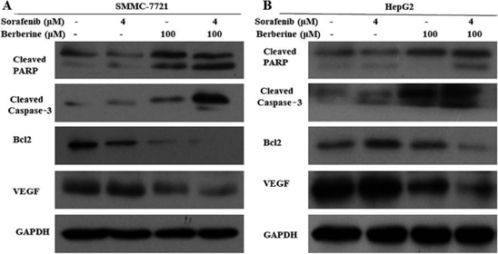
Sorafenib tosylate purchased from MedChemExpress. Usage Cited in: Oncol Rep. 2018 Sep;40(3):1525-1532. [Abstract]
SMMC-7721 and HepG2 cells are treated with 4 µM Sorafenib and 100 µM Berberine alone or in combination (4 µM Sorafenib+100 µM Berberine) for 72 h, and the expression levels of apoptosis-associated proteins are measured by western blot analysis.
-
Sci Rep
Glycosylation-dependent galectin-1/neuropilin-1 interactions promote liver fibrosis through activation of TGF-β- and PDGF-like signals in hepatic stellate cells. [Abstract]2017 Sep 8;7(1):11006. PMID: 28887481 -
Oncol Rep
Long non-coding RNA TUC338 is functionally involved in sorafenib-sensitized hepatocarcinoma cells by targeting RASAL1. [Abstract]2017 Jan;37(1):273-280. PMID: 27878301 -
Dig Liver Dis
Tumor suppressor CLCA1 inhibits angiogenesis via TGFB1/SMAD/VEGF cascade and sensitizes hepatocellular carcinoma cells to Sorafenib. [Abstract]2024 Jan;56(1):176-186. PMID: 37230858 -
J Pharmacol Exp Ther
Carboxyamidotriazole Synergizes with Sorafenib to Combat Non-Small Cell Lung Cancer through Inhibition of NANOG and Aggravation of Apoptosis. [Abstract]2017 Aug;362(2):219-229. PMID: 28515157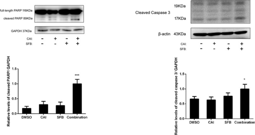
Sorafenib tosylate purchased from MedChemExpress. Usage Cited in: J Pharmacol Exp Ther. 2017 Aug;362(2):219-229. [Abstract]
The combination of sorafenib and CAI induces apoptosis in NSCLC. Effect of 10 μM CAI and/or 5 μM Sorafenib on the expression of cleaved PARP and cleaved caspase-. Protein levels of cleaved PARP and cleaved caspase-3 from treated cell lysates are normalized against GAPDH levels.
-
ACS Chem Biol
2017 Jan 20;12(1):282-290. PMID: 27983786 -
ACS Chem Biol
2016 Apr 15;11(4):992-1000. PMID: 26741163 -
Bioengineering (Basel)
Precision Oncology for High-Grade Gliomas: A Tumor Organoid Model for Adjuvant Treatment Selection. [Abstract]2025 Oct 19;12(10):1121. PMID: 41155119 -
Cell Signal
Natural exosome-like nanovesicles from Salvia chinensis Benth induce ferroptosis to suppress hepatocellular carcinoma progression via AMPK/Nrf2/xCT axis. [Abstract]2025 Jun 6:111928. PMID: 40484104 -
Cell Signal
2025 Sep:133:111872. PMID: 40389044 -
Biochim Biophys Acta Mol Cell Res
Salinomycin inhibits SREBP1 to sensitize ferroptosis and ameliorate sorafenib resistance in clear cell renal cell carcinoma. [Abstract]2025 May 11:119989. PMID: 40360020 -
PLoS Genet
TRIM21 modulates stability of pro-survival non-coding RNA vtRNA1-1 in human hepatocellular carcinoma cells. [Abstract]2025 Mar 17;21(3):e1011614. PMID: 40096176 -
Cell Signal
2024 Jun 27:111278. PMID: 38944257 -
Heliyon
Naringenin enhances the efficacy of ferroptosis inducers by attenuating aerobic glycolysis by activating the AMPK-PGC1α signalling axis in liver cancer. [Abstract]2024 May 31;10(11):e32288. PMID: 38912485 -
Heliyon
Paeoniflorin increases the anti-tumor efficacy of sorafenib in tumor-bearing mice with liver cancer via suppressing the NF-κb/PD-l1 axis. [Abstract]2024 Jan 13;10(2):e24461. PMID: 38312647 -
Biology (Basel)
2023 Oct 16;12(10):1337. PMID: 37887047 -
Med Oncol
The natural medicinal fungus Huaier promotes the anti-hepatoma efficacy of sorafenib through the mammalian target of rapamycin-mediated autophagic cell death. [Abstract]2022 Sep 29;39(12):221. PMID: 36175804 -
Exp Cell Res
12 T high static magnetic field suppresses osteosarcoma cells proliferation by regulating intracellular ROS and iron status. [Abstract]2022 Aug 15;417(2):113223. PMID: 35643180 -
Exp Cell Res
Dichloroacetate enhances the anti-tumor effect of sorafenib via modulating the ROS-JNK-Mcl-1 pathway in liver cancer cells. [Abstract]2021 Sep 1;406(1):112755. PMID: 34332981 -
Exp Cell Res
Network-based analysis with primary cells reveals drug response landscape of acute myeloid leukemia. [Abstract]2020 Aug 1;393(1):112054. PMID: 32376287 -
NPJ Syst Biol Appl
Mechanistically detailed systems biology modeling of the HGF/Met pathway in hepatocellular carcinoma. [Abstract]2019 Aug 16:5:29. PMID: 31452933 -
Eur J Med Res
Long non-coding RNA NORAD serves as a promoter of oncogenesis and inhibits ferroptosis via miR-144-3p-mTOR-ferritinophagy axis in cancer. [Abstract]2025 Aug 4;30(1):704. PMID: 40760677 -
BMC Complement Med Ther
Therapeutic efficacy of sorafenib and plant-derived phytochemicals in human colorectal cancer cells. [Abstract]2023 Jun 26;23(1):210. PMID: 37365571 -
Saudi Pharm J
Development and validation of a UPLC-MS/MS method for simultaneous detection of doxorubicin and sorafenib in plasma: Application to pharmacokinetic studies in rats. [Abstract]2023 Jul;31(7):1317-1326. PMID: 37323919 -
J Hepatocell Carcinoma
Inhibitory Effects of Esculetin on Liver Cancer Through Triggering NCOA4 Pathway-Mediation Ferritinophagy in vivo and in vitro. [Abstract]2023 Apr 11:10:611-629. PMID: 37069958 -
Phytochemistry
Five undescribed steroids from Talaromyces stipitatus and their cytotoxic activities against hepatoma cell lines. [Abstract]2021 Sep:189:112816. PMID: 34087503 -
PLoS Negl Trop Dis
Identification of anti-flaviviral drugs with mosquitocidal and anti-Zika virus activity in Aedes aegypti. [Abstract]2019 Aug 20;13(8):e0007681. PMID: 31430351 -
BMC Cancer
Sphere-forming culture enriches liver cancer stem cells and reveals Stearoyl-CoA desaturase 1 as a potential therapeutic target. [Abstract]2019 Aug 1;19(1):760. PMID: 31370822 -
Endocrinology
TRβ Agonism Induces Tumor Suppression and Enhances Drug Efficacy in Anaplastic Thyroid Cancer in Female Mice. [Abstract]2023 Aug 28;164(10):bqad135. PMID: 37702560 -
Front Oncol
Silenced LINC01134 Enhances Oxaliplatin Sensitivity by Facilitating Ferroptosis Through GPX4 in Hepatocarcinoma. [Abstract]2022 Jul 8;12:939605. PMID: 35875091 -
Front Oncol
Patient-Derived Xenograft Models for Intrahepatic Cholangiocarcinoma and Their Application in Guiding Personalized Medicine. [Abstract]2021 Jul 13:11:704042. PMID: 34327143 -
J Cancer
SREBP1-SCD1 enhanced MUFAs Biosynthesis drives Nutrient Deprived Pancreatic cancer cell Ferroptosis Resistance. [Abstract]2025 Sep 8;16(13):3960-3971. PMID: 41049011 -
NAR Cancer
2025 Aug 30;7(3):zcaf028. PMID: 40918645 -
Mol Carcinog
2025 Jul;64(7):1264-1274. PMID: 40329467 -
Mol Carcinog
Transmembrane protein 147, as a potential Sorafenib target, could expedite cell cycle process and confer adverse prognosis in hepatocellular carcinoma. [Abstract]2023 Sep;62(9):1295-1311. PMID: 37212496 -
Environ Toxicol
2022 Feb;37(2):192-200. PMID: 34661962 -
J Cancer
The Inhibitory Effects of Recombinant Hespintor Combined with Sorafenib on Transplanted Human Hepatoma in Nude Mice, and Transcriptional Regulation of Hespintor Based on RNA-Seq. [Abstract]2021 Jan 1;12(2):343-357. PMID: 33391431 -
Int J Med Sci
2021 Jan 29;18(6):1456-1464. PMID: 33628103 -
Am J Hypertens
Mineralocorticoid Receptor in Endothelial Cells Contributes to Vascular Endothelial Growth Factor Receptor Inhibitor-Induced Vascular and Kidney Damage. [Abstract]2025 Jan 16;38(2):104-110. PMID: 39514632 -
Front Biosci (Landmark Ed)
Downregulation of MTHFD2 Inhibits Proliferation and Enhances Chemosensitivity in Hepatocellular Carcinoma via PI3K/AKT Pathway. [Abstract]2024 Jan 19;29(1):35. PMID: 38287824 -
Kaohsiung J Med Sci
Limited sorafenib anticancer effects on primary cultured hepatocellular carcinoma cells with high NANOG expression. [Abstract]2022 Feb;38(2):157-164. PMID: 35142429 -
Mol Immunol
Sophora tonkinensis reprograms tumor-associated macrophages to M1-like phenotype and exerts anti-hepatocellular carcinoma effects. [Abstract]2026 Mar:191:49-59. PMID: 41666700 -
Curr Issues Mol Biol
Exploring the Molecular Mechanism of 1,25(OH)2D3 Reversal of Sorafenib Resistance in Hepatocellular Carcinoma Based on Network Pharmacology and Experimental Validation. [Abstract]2025 Apr 29;47(5):319. PMID: 40699718 -
Diseases
Examining the Effects of Dasatinib, Sorafenib, and Nilotinib on Vascular Smooth Muscle Cells: Insights into Proliferation, Migration, and Gene Expression Dynamics. [Abstract]2023 Oct 23;11(4):147. PMID: 37873791 -
Mol Cell Probes
Circular RNA ITCH increases sorafenib-sensitivity in hepatocellular carcinoma via sequestering miR-20b-5p and modulating the downstream PTEN-PI3K/Akt pathway. [Abstract]2023 Feb:67:101877. PMID: 36442661 -
Discov Oncol
Activating the Hippo pathway by nevadensin overcomes Yap-drived resistance to sorafenib in hepatocellular carcinoma. [Abstract]2023 May 27;14(1):83. PMID: 37243813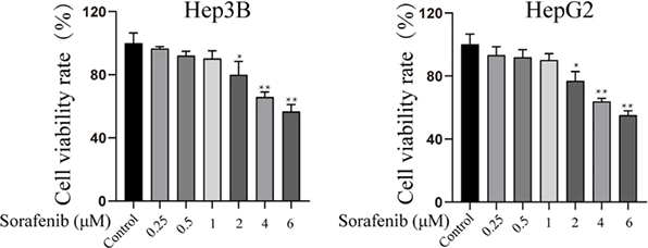
Sorafenib tosylate purchased from MedChemExpress. Usage Cited in: Discov Oncol. 2023 May 27;14(1):83. [Abstract]
Sorafenib (0.25, 0.5, 1, 2, 4, 6 µM; 24 h) significantly inhibits the cell viability of HepG2 and Hep3B.
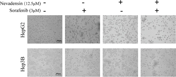
Sorafenib tosylate purchased from MedChemExpress. Usage Cited in: Discov Oncol. 2023 May 27;14(1):83. [Abstract]
Sorafenib (3 µM; 24 h) significantly inhibits the cell proliferation of HepG2 and Hep3B.
-
Am J Cancer Res
Combination of sorafenib and Valproic acid synergistically induces cell apoptosis and inhibits hepatocellular carcinoma growth via down-regulating Notch3 and pAkt. [Abstract]2017 Dec 1;7(12):2503-2514. PMID: 29312803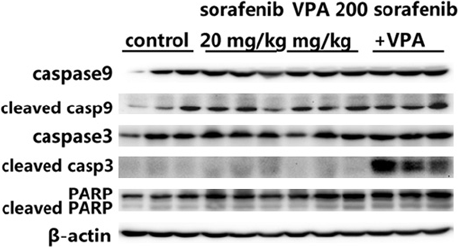
Sorafenib tosylate purchased from MedChemExpress. Usage Cited in: Am J Cancer Res. 2017 Dec 1;7(12):2503-2514. [Abstract]
VPA potentiates anti-tumor effects of Sorafenib tosylate in vivo. The expression of cleaved caspase9, cleaved caspase3, cleaved PARP from tumor tissue homogenates are analyzed by western blot.
-
J Chromatogr B Analyt Technol Biomed Life Sci
Simultaneous determination of 11 oral targeted antineoplastic drugs and 2 active metabolites by LC-MS/MS in human plasma and its application to therapeutic drug monitoring in cancer patients. [Abstract]2024 Apr 15:1237:124100. PMID: 38547701 -
Int J Biochem Cell Biol
Valproic acid overcomes sorafenib resistance by reducing the migration of Jagged2-mediated Notch1 signaling pathway in hepatocellular carcinoma cells. [Abstract]2020 Sep;126:105820. PMID: 32750425 -
J Chromatogr B Analyt Technol Biomed Life Sci
Investigation of the effect of plasma albumin levels on regorafenib-induced hepatotoxicity using a validated liquid chromatography-tandem mass spectrometry method. [Abstract]2017 Sep 1:1061-1062:220-224. PMID: 28750235 -
Toxicol Mech Methods
2025 Sep 10:1-8. PMID: 40908820 -
Toxicol In Vitro
Increased effects of 2,5-dimethylcelecoxib on sensitivity of hepatocellular carcinoma cells to sorafenib via CYP3A5 expression and activation of AMPK. [Abstract]2021 Oct:76:105226. PMID: 34293431 -
PLoS One
A novel small molecule screening assay using normal human chondrocytes toward osteoarthritis drug discovery. [Abstract]2024 Nov 1;19(11):e0308647. PMID: 39485774 -
Clin Transl Oncol
FAT4 loss promotes tumor growth and ferroptosis resistance in hepatocellular carcinoma via PI3K/AKT pathway activation. [Abstract]2026 Mar 14. PMID: 41832339 -
Fundam Clin Pharmacol
Valproic acid reverses sorafenib resistance through inhibiting activated Notch/Akt signaling pathway in hepatocellular carcinoma. [Abstract]2021 Aug;35(4):690-699. PMID: 33015852 -
Prostate
MicroRNA181c inhibits prostate cancer cell growth and invasion by targeting multiple ERK signaling pathway components. [Abstract]2018 Apr;78(5):343-352. PMID: 29341215 -
Can J Gastroenterol Hepatol
Hsa_circ_0006988 Promotes Sorafenib Resistance of Hepatocellular Carcinoma by Modulating IGF1 Using miR-15a-5p. [Abstract]2022 Dec 24:2022:1206134. PMID: 36594050 -
Biomed Res Int
A Pharmacokinetic Interaction Study of Sorafenib and Iced Teas in Rats Using UPLC-MS/MS: An Illustration of Beverage-Drug Interaction. [Abstract]2019 Nov 28;2019:2410845. PMID: 31871933 -
Biochem Biophys Res Commun
Natural exosome-like nanovesicles from Smilax China rhizome induce mitophagy-dependent ferroptosis in hepatocellular carcinoma via GPX4/ACSL4 axis. [Abstract]2026 Feb 12:800:153270. PMID: 41529459 -
BMJ Open Ophthalmol
Impact of anticancer drugs on human Tenon's fibroblast proliferation: implications for glaucoma surgery. [Abstract]2026 Jan 12;11(1):e002307. PMID: 41526033 -
Biochem Biophys Res Commun
Targeted co-delivery of PD-L1 monoclonal antibody and sorafenib to circulating tumor cells via platelet-functionalized nanocarriers. [Abstract]2023 Sep 3:671:335-342. PMID: 37327705 -
Biochem Biophys Res Commun
2022 Feb 19:593:122-128. PMID: 35063767 -
Biochem Bioph Res Co
Functional loss of TAGLN inhibits tumor growth and increases chemosensitivity of non-small cell lung cancer. [Abstract]2020 Sep 3;529(4):1086-1093. PMID: 32819569 -
-
Endocr J
Effects of sorafenib and an adenylyl cyclase activator on in vitro growth of well-differentiated thyroid cancer cells. [Abstract]2017 Nov 29;64(11):1115-1123. PMID: 28855436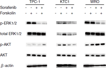
Sorafenib tosylate purchased from MedChemExpress. Usage Cited in: Endocr J. 2017 Nov 29;64(11):1115-1123. [Abstract]
Effect of Sorafenib on phosphorylation of ERK and AKT. Thyroid cancer cells are treated for 30 minutes with 10 μM Sorafenib, 10 μM Forskolin, and combination therapy of 10 μM Sorafenib with 10 μM Forskolin. The levels of ERK and AKT phosphorylation are examined by immunoblot analysis. β-actin is used as the control. Sorafenib suppresses phosphorylation of ERK, but not of AKT.
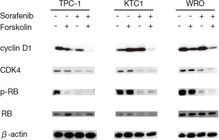
Sorafenib tosylate purchased from MedChemExpress. Usage Cited in: Endocr J. 2017 Nov 29;64(11):1115-1123. [Abstract]
Effect of Sorafenib and Forskolin on expression of CDK4 and CDK regulatory proteins. Thyroid cancer cells are treated for 24 hours with 10 μM Sorafenib, 10 μM Forskolin, and combination therapy of 10 μM Sorafenib with 10 μM Forskolin. The expression of cyclin D1, CDK4, and phosphorylation of RB are examined by immunoblot analysis. β-actin is used as the control. The combination therapy suppresses expression of cyclin D1, and Forskolin monotherapy suppresses expression of cyclin D1 in TPC-1 and W
-
Int J Environ Health Res
Investigation of synergistic effects of silver nanoparticles with sorafenib on in vitro prostate and head and neck cancer models. [Abstract]2025 Apr 24:1-9. PMID: 40270101 -
Clin Med Insights Oncol
Exploring the Clinical Implications of RPL3 Presence in BRCA-Associated Cancers: Unraveling the Interplay With Cancer Immunity. [Abstract]2024 Oct 16:18:11795549241285387. PMID: 39429685 -
Ann Med Surg (Lond)
Synergistic mechanism of Shengyang Shiyiwei Pill in enhancing bevacizumab efficacy for hepatocellular carcinoma-associated ascites. [Abstract]2025 Nov 25;88(1):290-302. PMID: 41496997 -
Scand J Immunol
IL-6 upregulates the expression of IL-6R through the JAK2/STAT3 signalling pathway to promote progression of hepatocellular carcinoma. [Abstract]2023 Jul;98(1):e13271. PMID: 38441314 -
Am J Transl Res
2023 Feb 15;15(2):896-913. PMID: 36915773 -
Am J Transl Res
Phenformin synergistically sensitizes liver cancer cells to sorafenib by downregulating CRAF/ERK and PI3K/AKT/mTOR pathways. [Abstract]2021 Jul 15;13(7):7508-7523. PMID: 34377232 -
Am J Transl Res
CXCR3 confers sorafenib resistance of HCC cells through regulating metabolic alteration and AMPK pathway. [Abstract]2020 Mar 15;12(3):825-836. PMID: 32269715 -
Braz J Med Biol Res
MicroRNA-92b augments sorafenib resistance in hepatocellular carcinoma via targeting PTEN to activate PI3K/AKT/mTOR signaling. [Abstract]2021 May 31;54(9):e10390. PMID: 34076140 -
Integr Biol (Camb)
Structural insights and influence of V599 mutations on the overall dynamics of BRAF protein against its kinase domains. [Abstract]2018 Oct 15;10(10):646-657. PMID: 30229251 -
-
Int J Clin Exp Pathol
SOX9 was involved in TKIs resistance in renal cell carcinoma via Raf/MEK/ERK signaling pathway. [Abstract]2015 Apr 1;8(4):3871-81. PMID: 26097571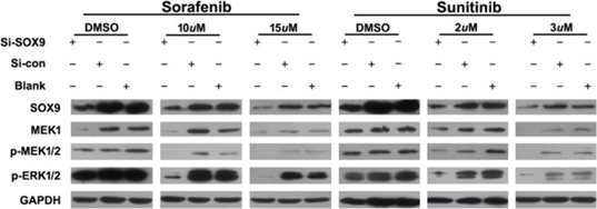
Sorafenib tosylate purchased from MedChemExpress. Usage Cited in: Int J Clin Exp Pathol. 2015 Apr 1;8(4):3871-81. [Abstract]
The relationship between SOX9 and Raf/MEK/ERK signaling pathway. Co-treatment of si-SOX9-1 and Sorafenib (10uM, 15uM)/SU 11248 (2 uM, 3 uM) significantly decreases expression of MEK1 and its phosphorylated protein (p-MEK1/2, p-ERK1/2) as assayed by Western blot (with GAPDH as internal control).
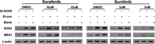
Sorafenib tosylate purchased from MedChemExpress. Usage Cited in: Int J Clin Exp Pathol. 2015 Apr 1;8(4):3871-81. [Abstract]
The relationship between SOX9 and Raf/MEK/ERK signaling pathway. Co-treatment of si-SOX9-1 and Sorafenib (10uM, 15uM)/SU 11248 (2 uM, 3 uM) significantly decreases expression of MEK1 and its phosphorylated protein (p-MEK1/2, p-ERK1/2) as assayed by RT-PCR (with β-actin as internal control).
-
-
-
-
-
-
-
-
-
-
bioRxiv
2025 Jul 12:2025.07.08.663754. PMID: 40672312 -
-
-
-
-
-
-
-
-
-
Evid Based Complement Alternat Med
Effects of Sensitized Sorafenib with a Paeoniflorin and Geniposide Mixture on Liver Cancer via the NF- κ B-HIF-2 α-SerpinB3 Pathway. [Abstract]2022 Oct 15:2022:1911311. PMID: 36285158 -
-
-
-
-
-
-
-
-
-
-
Math Biosci Eng
Bioinformatics analysis revealed hub genes and pathways involved in sorafenib resistance in hepatocellular carcinoma. [Abstract]2019 Jul 8;16(6):6319-6334. PMID: 31698564 -
-
-
Oncotarget
Role of drug-dependent transporter modulation on the chemosensitivity of cholangiocarcinoma. [Abstract]2017 Oct 6;8(52):90185-90196. PMID: 29163820 -
Oncotarget
Inhibition of the prolyl isomerase Pin1 enhances the ability of sorafenib to induce cell death and inhibit tumor growth in hepatocellular carcinoma. [Abstract]2017 May 2;8(18):29771-29784. PMID: 28404959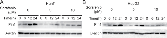
Sorafenib tosylate purchased from MedChemExpress. Usage Cited in: Oncotarget. 2017 May 2;8(18):29771-29784. [Abstract]
Sorafenib inhibits Pin1 biosynthesis and accumulation in Huh7 and HepG2 cells. Cells are treated with 5 or 10 μM Sorafenib for indicated times. Pin1 protein expression is determined by Western Blot.
-
Chem Cent J
Simultaneous determination of newly developed antiviral agents in pharmaceutical formulations by HPLC-DAD. [Abstract]2017 Jan 3:11:1. PMID: 28101128 -


Solvent & Solubility
DMSO : ≥ 100 mg/mL (156.98 mM; Hygroscopic DMSO has a significant impact on the solubility of product, please use newly opened DMSO)
* "≥" means soluble, but saturation unknown.
Please refer to the solubility information to select the appropriate solvent. Once prepared, please aliquot and store the solution to prevent product inactivation from repeated freeze-thaw cycles.
Storage method and period of stock solution: -80°C, 1 year; -20°C, 6 months (sealed storage, away from moisture). When stored at -80°C, please use it within 1 year. When stored at -20°C, please use it within 6 months.
Concentration (start) × Volume (start) = Concentration (final) × Volume (final)
Select the appropriate dissolution method based on your experimental animal and administration route.
- For the following dissolution methods, please ensure to first prepare a clear stock solution using an In Vitro approach and then sequentially add co-solvents:
- To ensure reliable experimental results, the clarified stock solution can be appropriately stored based on storage conditions. As for the working solution for In Vivo experiments, it is recommended to prepare freshly and use it on the same day.
- The percentages shown for the solvents indicate their volumetric ratio in the final prepared solution. If precipitation or phase separation occurs during preparation, heat and/or sonication can be used to aid dissolution.
Add each solvent one by one: 10% DMSO 90% (20% SBE-β-CD in Saline)
Solubility: ≥ 2.5 mg/mL (3.92 mM); Clear solution
This protocol yields a clear solution of ≥ 2.5 mg/mL (saturation unknown).
Taking 1 mL working solution as an example, add 100 μL DMSO stock solution (25.0 mg/mL) to 900 μL 20% SBE-β-CD in Saline, and mix evenly.
Preparation of 20% SBE-β-CD in Saline (4°C, storage for one week): 2 g SBE-β-CD powder is dissolved in 10 mL Saline, completely dissolve until clear.
Please enter the basic information of animal experiments:
-
-
-
-
Recommended: Prepare an additional quantity of animals to account for potential losses during experiments.
Please enter your animal formula composition:
-
%DMSO +
Recommended: Keep the proportion of DMSO in working solution below 2% if your animal is weak.
-
%+
-
+%Tween-80 + +
-
%Saline +
The co-solvents required include: DMSO, . All of co-solvents are available by MedChemExpress (MCE). , Tween 80. All of co-solvents are available by MedChemExpress (MCE).
Working solution concentration: 0.22 mg/mL
Method for preparing stock solution: mg drug dissolved in μL DMSO. Stock solution concentration: mg/mL. * In solvent : -80°C, 1 year; -20°C, 6 months (sealed storage, away from moisture)
1. Take μL DMSO stock solution;
2. Add μL .
μL , mix evenly;
3. Then add μL Tween 80, mix evenly;
4. Then add μL
Please ensure that the stock solution in the first step is dissolved to a clear state, and add co-solvents in sequence. You can use ultrasonic heating (ultrasonic cleaner, recommended frequency 20-40 kHz), vortexing, etc. to assist dissolution.


Purity & Documentation
-
Data Sheet (307 KB)
-
SDS (393 KB)
- English - EN (393 KB)
- Français - FR (393 KB)
- Deutsch - DE (393 KB)
- Norwegian - NO (393 KB)
- Español - ES (393 KB)
- Swedish - SV (393 KB)
- Italian - IT (393 KB)
- Korean - KR (393 KB)
- Portuguese - PT (393 KB)
-
Handling Instructions (2659 KB)
References
[1]. Wilhelm SM, et al. BAY 43-9006 exhibits broad spectrum oral antitumor activity and targets the RAF/MEK/ERK pathway and receptor tyrosine kinases involved in tumor progression and angiogenesis. Cancer Res. 2004 Oct 1;64(19):7099-109. [Content Brief]
[2]. Heim M, et al. The Raf kinase inhibitor BAY 43-9006 reduces cellular uptake of platinum compounds and cytotoxicity in human colorectal carcinoma cell lines. Anticancer Drugs. 2005;16(2):129-136. [Content Brief]
[3]. Gu FM, et al. Sorafenib inhibits growth and metastasis of hepatocellular carcinoma by blocking STAT3. World J Gastroenterol. 2011 Sep 14;17(34):3922-32. [Content Brief]
[4]. El-Ashmawy NE, et al. Sorafenib effect on liver neoplastic changes in rats: more than a kinase inhibitor. Clin Exp Med. 2016 Apr 16. [Content Brief]
[5]. Li M, et al. Activation of an AKT/FOXM1/STMN1 pathway drives resistance to tyrosine kinase inhibitors in lung cancer. Br J Cancer. 2017;117(7):974-983. [Content Brief]
[6]. Jin W, et al. Long non-coding RNA TUC338 is functionally involved in sorafenib-sensitized hepatocarcinoma cells by targeting RASAL1. Oncol Rep. 2017;37(1):273-280. [Content Brief]


Complete Stock Solution Preparation Table
Please refer to the solubility information to select the appropriate solvent. Once prepared, please aliquot and store the solution to prevent product inactivation from repeated freeze-thaw cycles.
Storage method and period of stock solution: -80°C, 1 year; -20°C, 6 months (sealed storage, away from moisture). When stored at -80°C, please use it within 1 year. When stored at -20°C, please use it within 6 months.
| Optional Solvent | Concentration Solvent Mass | 1 mg | 5 mg | 10 mg | 25 mg |
|---|---|---|---|---|---|
| DMSO | 1 mM | 1.5698 mL | 7.8489 mL | 15.6978 mL | 39.2446 mL |
| 5 mM | 0.3140 mL | 1.5698 mL | 3.1396 mL | 7.8489 mL | |
| 10 mM | 0.1570 mL | 0.7849 mL | 1.5698 mL | 3.9245 mL | |
| 15 mM | 0.1047 mL | 0.5233 mL | 1.0465 mL | 2.6163 mL | |
| 20 mM | 0.0785 mL | 0.3924 mL | 0.7849 mL | 1.9622 mL | |
| 25 mM | 0.0628 mL | 0.3140 mL | 0.6279 mL | 1.5698 mL | |
| 30 mM | 0.0523 mL | 0.2616 mL | 0.5233 mL | 1.3082 mL | |
| 40 mM | 0.0392 mL | 0.1962 mL | 0.3924 mL | 0.9811 mL | |
| 50 mM | 0.0314 mL | 0.1570 mL | 0.3140 mL | 0.7849 mL | |
| 60 mM | 0.0262 mL | 0.1308 mL | 0.2616 mL | 0.6541 mL | |
| 80 mM | 0.0196 mL | 0.0981 mL | 0.1962 mL | 0.4906 mL | |
| 100 mM | 0.0157 mL | 0.0785 mL | 0.1570 mL | 0.3924 mL |


 Powered by Bioz