



MRS2768
MRS2768 is a potent, selective, and metabolically stable P2Y2 receptor agonist with an EC50 of 1.89 μM for the human P2Y2 receptor. MRS2768 activates Gq/PLC/PKC signaling, leading to downstream phosphorylation of Akt, eNOS, and ERK, with effects varying by cell type. MRS2768 inhibits ENaC via Gq/PKC/Src/Akt to promote natriuresis and lower blood pressure in the kidney. MRS2768 activates eNOS to increase NO secretion in endothelial cells. MRS2768 drives proliferation via PI3K/Akt in fibroblasts and cancer cells. MRS2768 exerts anti-apoptotic effects through PKC/Src/Akt in cardiomyocytes. MRS2768 can be applied to investigate P2Y2-dependent pathological processes, including acute kidney injury, chronic kidney disease and renal fibrosis, DOCA-salt induced hypertension, myocardial infarction, pulmonary arterial hypertension, pancreatic cancer, cardiac fibrosis, dry eye disease, as well as shear stress-mediated vascular remodeling and atherosclerosis.
Nos produits utilisent uniquement pour la recherche. Nous ne vendons pas aux patients.
- CAS No.: 1047980-83-5
- Formule: C15H20N2O18P4
- Masse moléculaire:640.22
-
Stockage:
Please store the product under the recommended conditions in the Certificate of Analysis.


To place orders, for customer services and technical support, please contact: MedChemExpress USA
Tel: 609-228-6898 E-mail: [email protected] [email protected]


-
 Activité biologique
-
Chemical Information
- Protocole
- Pureté et documentation
- Références
-
 Help & FAQs


-
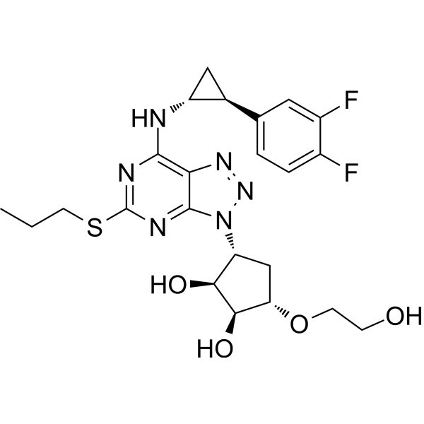
HY-10064
TicagrelorP2Y12 Antagonist -
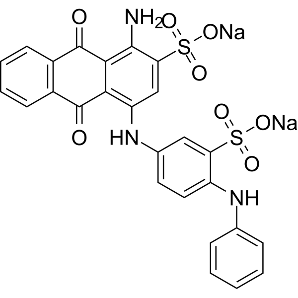
HY-108660
PSB-0739P2Y12 Receptor Antagonist -
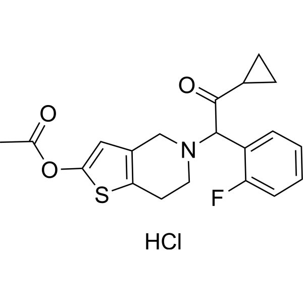
HY-15284A
Prasugrel hydrochlorideP2Y12 Receptor Antagonist -
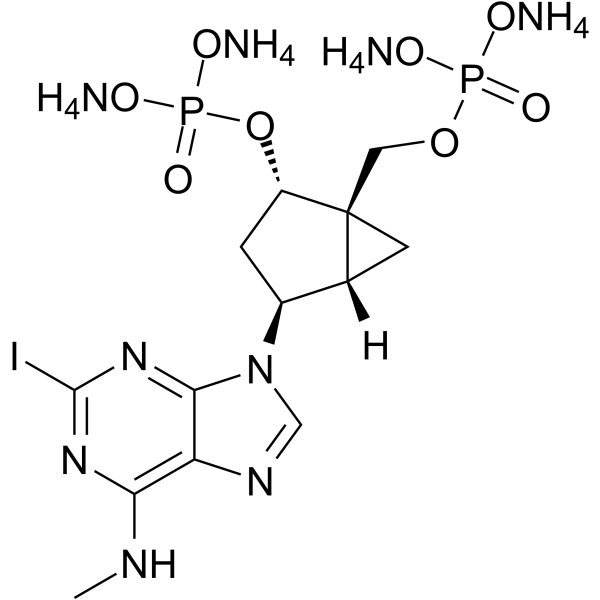
HY-108658
MRS2500 tetraammoniumP2Y1 Receptor Antagonist -
HY-15799
AZD1283P2Y12 Receptor Antagonist -
HY-W010832
Uridine-5'-diphosphate disodium saltNucleotide -
HY-13104
MRS 2578Apoptosis Inducer -

HY-10064R
Ticagrelor (Standard)P2Y12 Antagonist -
HY-108662
PITP2Y1 Receptor Antagonist -

HY-15284AR
Prasugrel hydrochloride (Standard)P2Y12 Receptor Antagonist -

HY-108660R
PSB-0739 (Standard)Reference Standard -
HY-15283
ClopidogrelAntiplatelet Agent
Voir tous les produits spécifiques à Isoform P2Y Receptor
MoreVoir tous les produits spécifiques à Isoform Phospholipase
More

Activité biologique
Description
|
P2Y2 Receptor 1.89 μM (EC50) |
PI3K |
PKC |
Akt |
CaMK II |
G-quadruplex |
Phospholipase |
Src |
eNOS |
ERK |
Sodium Channel |
In Vitro
MRS2768 (Compound 7) exerts agonistic activity against hP2Y2 (EC50 = 2.76 μM) in stably transfected astrocytoma cells, yet produces no response at hP2Y4 and hP2Y6[1].
MRS2768 shows superior resistance to both acidic hydrolysis and ectonucleotidase-mediated degradation in 1321N1 astrocytoma cell membranes compared with native UTP[1].
MRS2768 (Compound 30) (30 min) selectively stimulates PLC-mediated signaling via human P2Y2 receptors with an EC50 of 1.89 μM and produces no activation of hP2Y4 or hP2Y6 receptors in stably transfected 1321N1 human astrocytoma cells.[2].
MRS2768 (1-50 μM; 24 h) inhibits the growth of HK-2 cells at 50 μM[3].
MRS2768 (20 μM; 1 h) alone does not alter extracellular ATP levels in the culture medium of HK-2 cells, whereas TNF-α (Adalimumab) (HY-P9908) treatment and combined TNF-α and MRS2768 treatment significantly increase extracellular ATP levels[3].
MRS2768 (20 μM; 1-8 h) increases the mRNA expression level of P2Y2R in HK-2 cells[3].
MRS2768 (20 μM; 5-60 min) significantly elevates the phosphorylation of PKC, Src, and Akt in HK-2 cells[3].
MRS2768 (20 μM; 5 min) activates PKC, with Src and Akt as its downstream kinases in HK-2 cells[3].
MRS2768 (20 μM; 1 h) attenuates the upregulation of cleaved PARP, caspase-3 and caspase-8, and decreases the number of TUNEL-positive apoptotic cells as well as abnormal fragmented nuclei triggered by 16 h co-incubation of TNF-α and cycloheximide (HY-12320) in HK-2 cells[3].
MRS2768 (20 μM)-mediated P2Y2R activation protects proximal tubular cells from apoptotic stress and TNF-α stimulation through the PI3K/Akt signaling pathway[3].
MRS2768(10-50 μM; 1 h) dose-dependently reduces cardiomyocyte damage and death caused by prolonged hypoxia[4].
MRS2768 (50 μM; 1 h) exerts cardioprotective effects via P2Y2R[4].
MRS2768 (1 μM; 30 min) significantly inhibits the activity of epithelial sodium channels (ENaC) in the apical membrane of distal nephron principal cells[6].
MRS2768 (0.1 μM-1 mM; 24 h) significantly boosts PANC-1 cell proliferation in a concentration-dependent fashion, and its pro-proliferative activity at 1 μM after 24 h relies entirely on P2 purinergic receptors with no non-specific off-target effects, acting specifically through P2Y2 receptors instead of other P2 subtypes[7].
MRS2768 significantly elevates peak intracellular calcium levels in rat conjunctival goblet cells[8].
MRS2768 (1 μM)-induced peak intracellular calcium elevation is markedly reduced after P2Y2 receptor knockdown, while scrambled siRNA does not interfere with its calcium-stimulating effect in rat conjunctival goblet cells[8].
MRS2768 (10 μM; 2 h)-mediated P2Y2 receptor activation effectively stimulates tear mucin release, and its pro-secretory potency exceeds UTP and UDP in conjunctival goblet cells[8].
MRS2768 (10 μM; 1 h) induces actin stress fiber formation in human umbilical vein endothelial cells under static culture through intact P2Y2-integrin RGD binding interaction, and this cytoskeletal effect disappears after P2Y2 receptor knockdown or RGD motif mutation[9].
MRS2768 (100 nM-30 μM; 10 min) triggers concentration-dependent vasoconstriction in precontracted porcine isolated pancreatic arterial rings[10].
MRS2768 (30 μM; 7-28 days) boosts cardiac fibroblast proliferation through selective P2Y2 receptor activation[11].
MRS2768 (5 μM; 30 min) markedly reduces total ENaC activity, open probability and the number of active epithelial sodium channels in renal principal cells isolated from wild-type mice[12].
MRS2768 activates the Gαq/11-IP3 and Akt/eNOS signaling pathways in human pulmonary arterial endothelial cells, raising nitric oxide secretion in a time- and concentration-dependent way[13].
MRS2768 produces pulmonary vasodilatory effects and indicates P2Y2 as a promising therapeutic target for pulmonary arterial hypertension[13].
MedChemExpress (MCE) has not independently confirmed the accuracy of these methods. They are for reference only.
-
Cell Line:HK-2 cells
-
Concentration:1 μM, 5 μM, 10 μM, 20 μM, 50 μM
-
Incubation Time:24 h
-
Result:Decreased cell viability at 50 μM.
-
Cell Line:HK-2 cells
-
Concentration:20 μM
-
Incubation Time:1 h, 2 h, 4 h, 8 h
-
Result:Significantly upregulated P2Y2R expression at 4 h and 8 h post-treatment.
Reduced the mRNA expression levels of proinflammatory cytokines (TNF-α, MIP-2, and IL-6) in cells incubated with TNF-α.
-
Cell Line:HK-2 cells
-
Concentration:20 μM
-
Incubation Time:5 min, 10 min, 20 min, 40 min, 60 min
-
Result:Significantly boosted the phosphorylation of PKC, Src, and Akt in HK-2 cells.
Triggered the induction of p-PKC and p-Akt as early as 5 min post administration.
Sustained the elevated levels of p-PKC and p-Akt before their gradual decline at 60 min.
Induced two distinct peaks of p-Src expression at 5 min and 60 min after treatment.
Upregulated these kinases through P2Y2R activation.
Induced p-Akt was reduced by Gö6983 (HY-13689), PP2 (HY-13805), and wortmannin (HY-10197).
Induced p-Src was reduced by Gö6983 and PP2 but not affected by wortmannin.
Attenuated elevated cleaved PARP, caspase-3, and caspase-8 in TNF-α and cycloheximide-treated HK-2 cells.
Failed to exert its anti-apoptotic effect in the presence of wortmannin, which restored high levels of cleaved PARP, caspase-3, and caspase-8 in TNF-α and cycloheximide-treated cells.
-
Cell Line:HK-2 cells
-
Concentration:20 μM
-
Incubation Time:1 h
-
Result:Rescued the increase in the number of abnormally shaped nuclei after treatment with TNF-α and cyclooctanoic acid.
Rescued the increase in the number of TUNEL-positive cells after treatment with TNF-α and cyclooctanoic acid.
Lost its protective effect and exhibited apoptotic features when combined with wortmannin.
-
Cell Line:Cardiomyocytes
-
Concentration:10 μM, 50 μM
-
Incubation Time:1 h
-
Result:Significantly reduced LDH release levels.
Reduced cell death rate under hypoxic conditions.
Mediated cardioprotection against hypoxia was attenuated by the P2Y2R antagonist AR-C118925 (AR-C118925XX) (HY-110126).
-
Cell Line:PANC-1 cells
-
Concentration:1 μM
-
Incubation Time:24 h
-
Result:Induced cell proliferation is almost completely abolished after suramin pretreatment.
Lost its proliferative ability when P2Y2 receptor expression was knocked down by specific siRNA.
-
Cell Line:HUVECs
-
Concentration:10 μM
-
Incubation Time:1 h
-
Result:Markedly elevated the proportion of HUVECs with abundant actin stress fibers under static culture conditions.
Exerted cytoskeletal remodeling function via the RGD integrin-binding domain of P2Y2 receptors, and intact receptor-integrin interaction is indispensable for its downstream actin regulatory signaling.
-
Cell Line:Cardiac fibroblast
-
Concentration:30 μM
-
Incubation Time:7 days, 14 days, 21 days, 28 days
-
Result:Increased cardiac fibroblast proliferation via activating the P2Y2 receptor.
In Vivo
MRS2768 (4.44 μg/kg; i.v.; single dose) preserves left ventricular systolic function, attenuates adverse left ventricular remodeling, reduces myocardial infarct size, lowers serum Troponin T and TNF-α levels, alleviates neutrophil inflammatory infiltration, and modulates phospho-c-Jun expression after myocardial infarction[4].
MRS2768 (25 µg/kg; i.p.; once daily; until the mice were sacrificed) improves renal function, alleviates tubular injury, reduces renal fibrosis and inflammation, and normalizes abnormal JunB signaling to facilitate kidney recovery after renal ischemia-reperfusion injury[5].
MRS2768 (5 mg/kg; i.p.; once daily) induces prominent natriuresis in mice maintained on sodium-free diet via systemic P2Y2R activation[6].
MRS2768 (0.2 mg/kg; i.p.; once daily; for consecutive 6 days) induces persistent and significant elevation of 24-hour urinary sodium excretion in wild-type mice maintained on sodium-restricted diet[12].
MRS2768 (0.2 mg/kg; i.p.; once daily; given day 9 to day 12 after DOCA implantation) loses hypotensive effects in DOCA-salt hypertensive mice lacking principal cell P2Y2 receptors, verifying renal tubular P2Y2 is the core target mediating MRS2768 antihypertensive function[12].
MedChemExpress (MCE) has not independently confirmed the accuracy of these methods. They are for reference only.
-
Animal Model:IR-injured male mice (7 weeks old)[3]
-
Dosage:25 μg/kg
-
Administration:i.p., single dose
-
Result:Suppressed the elevation of plasma creatinine observed 24 hours after IR injury.
Reduced the IR-induced tubular injury and necrosis.
Markedly decreased the elevated neutrophil infiltration in tissues subjected to renal IR injury.
Attenuated the increased mRNA levels of proinflammatory cytokines (TNF-α, MIP-2, and IL-6) after IR.
Lowered the elevated number of tubular apoptotic cells detected by TUNEL staining following renal IR injury.
Significantly decreased the expression levels of cleaved caspase-3 and cleaved caspase-3 in IR-injured mice.
elevated the phosphorylation levels of PKC, Src and Akt that were Markedly induced at 1 h post renal IR.
-
Animal Model:Male C57BL/6 wild-type mice with myocardial infarction[4]
-
Dosage:4.44 μg/kg
-
Administration:i.v., single dose
-
Result:Preserved left ventricular systolic function and attenuated adverse left ventricular remodeling after myocardial infarction.
Reduced elevated serum Troponin T levels in myocardial infarction mice.
Reduced myocardial infarct size after LAD ligation.
Reduced elevated serum TNF-α levels in mice after myocardial infarction.
Attenuated inflammatory neutrophil infiltration in the myocardium after myocardial infarction.
Modulated the expression of phospho-c-Jun in the myocardium after myocardial infarction.
-
Animal Model:7-week-old male mice underwent a 1-week acclimatization period before IR injury[5]
-
Dosage:25 µg/kg
-
Administration:i.p., once daily, until the mice were sacrificed
-
Result:Reduced elevated plasma BUN levels and alleviated tubular injury after renal ischemia-reperfusion injury.
Decreased the expression of fibrogenic mediators including TGF-β, fibronectin, and α-SMA in injured kidneys.
Lowered the levels of proinflammatory cytokines including TNF-α, MCP-1, and MIP-2 after renal ischemia-reperfusion injury.
Reduced abnormally elevated JunB expression and restored normal JunB-mediated cell cycle regulation during renal recovery.
-
Animal Model:Young healthy adult (~2 mo old) male and female mice (18.60 g body wt) [6]
-
Dosage:5 mg/kg
-
Administration:i.p., once daily
-
Result:Produced remarkable urinary sodium excretion in sodium-deprived mice through systemic stimulation of P2Y2 receptors.
-
Animal Model:PC-P2ry2-KO mice[12]
-
Dosage:0.2 mg/kg
-
Administration:i.p., once daily, for consecutive 6 days
-
Result:Produced a sustained, remarkable rise in 24-hour urinary sodium excretion in wild-type mice fed a sodium-deficient diet.
-
Animal Model:DOCA-salt hypertensive PC-P2ry2-KO mice[12]
-
Dosage:0.2 mg/kg
-
Administration:i.p., once daily, given day 9 to day 12 after DOCA implantation
-
Result:Markedly reduced elevated mean arterial pressure and systolic blood pressure in DOCA-salt hypertensive wild-type mice.
- Kinase activity and phosphorylation assays
- Fibrosis/Collagen Morphometry
- Western Blot
- Collagen: Sirius Red Staining
- Detection of Apoptosis
- Nephrotoxicity Study
- Connective Tissue: Masson's Trichrome/Collagen Trichrome Staining
- Endothelial Tube Formation Assay
- Apoptosis Solutions
- Research Protocol for Cardiovascular Diseases
- Protocol for Kinase activity and phosphorylation assays
- Cell Viability Determination by MTT Colorimetric Assay


Chemical Information
-
CAS No. 1047980-83-5
-
Masse moléculaire 640.22
-
Formule C15H20N2O18P4
-
SMILES
O[C@@H]1[C@H](O)[C@@H](COP(OP(OP(OP(OC2=CC=CC=C2)(O)=O)(O)=O)(O)=O)(O)=O)O[C@H]1N3C(NC(C=C3)=O)=O
-
Livraison
Room temperature in continental US; may vary elsewhere.
-
Stockage
Please store the product under the recommended conditions in the Certificate of Analysis.


Pureté et documentation
Références
[1]. Maruoka H, et al. Pyrimidine nucleotides with 4-alkyloxyimino and terminal tetraphosphate δ-ester modifications as selective agonists of the P2Y(4) receptor. J Med Chem. 2011 Jun 23;54(12):4018-33. [Content Brief]
[2]. Ko H, et al. Synthesis and potency of novel uracil nucleotides and derivatives as P2Y2 and P2Y6 receptor agonists. Bioorg Med Chem. 2008 Jun 15;16(12):6319-32. [Content Brief]
[3]. Jeong K, et al. Activation of Purinergic P2Y2 Receptor Protects the Kidney Against Renal Ischemia and Reperfusion Injury in Mice. Int J Mol Sci. 2024 Nov 22;25(23):12563. [Content Brief]
[4]. Hochhauser E, et al. P2Y2 receptor agonist with enhanced stability protects the heart from ischemic damage in vitro and in vivo. Purinergic Signal. 2013 Dec;9(4):633-42. [Content Brief]
[5]. Jeong K, et al. Deficiency of purinergic P2Y2 receptor impairs the recovery after renal ischemia-reperfusion injury and accelerates renal fibrosis and tubular senescence in mice. Sci Rep. 2024 Dec 30;14(1):31932. [Content Brief]
[6]. Mironova E, et al. Renal Na+excretion consequent to pharmacogenetic activation of Gq-DREADD in principal cells. Am J Physiol Renal Physiol. 2019 Apr 1;316(4):F758-F767. [Content Brief]
[7]. Choi JH, et al. Uridine triphosphate increases proliferation of human cancerous pancreatic duct epithelial cells by activating P2Y2 receptor. Pancreas. 2013 May;42(4):680-6. [Content Brief]
[8]. Fjærvoll KA, et al. Pyrimidinergic P2Y1-Like Nucleotide Receptors Are Functional in Rat Conjunctival Goblet Cells. Invest Ophthalmol Vis Sci. 2025 Jan 2;66(1):46. [Content Brief]
[9]. Sathanoori R, et al. P2Y2 receptor modulates shear stress-induced cell alignment and actin stress fibers in human umbilical vein endothelial cells. Cell Mol Life Sci. 2017 Feb;74(4):731-746. [Content Brief]
[10]. Alsaqati M, et al. Investigation of the functional expression of purine and pyrimidine receptors in porcine isolated pancreatic arteries. Purinergic Signal. 2014;10(2):241-9. [Content Brief]
[11]. Certal M, et al. Calcium signaling and the novel anti-proliferative effect of the UTP-sensitive P2Y11 receptor in rat cardiac myofibroblasts. Cell Calcium. 2015 Nov;58(5):518-33. [Content Brief]
[12]. Soares AG, et al. P2Y2 receptor decreases blood pressure by inhibiting ENaC. JCI Insight. 2023 Jul 24;8(14):e167704. [Content Brief]

Calculators
Concentration (start) × Volume (start) = Concentration (final) × Volume (final)


MRS2768 Related Classifications
HY-108649 Related Classifications
 Powered by Bioz