



Momordicine I
Based on 2 publication(s) in Google Scholar
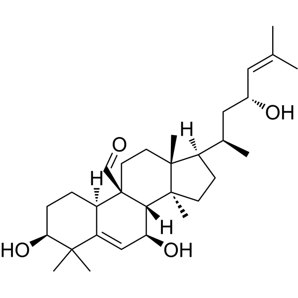
Momordicine I is a cucurbitane-type triterpenoids. Momordicine I suppresses glioma growth by promoting apoptosis and impairing mitochondrial oxidative phosphorylation. Momordicine I inhibits glycolysis, lipid metabolism, induces autophagy in HNC cells to suppress head and neck cancer growth. Momordicine I alleviates isoproterenol-induced cardiomyocyte hypertrophy through suppression of PLA2G6 and DGK-ζ. Momordicine I exerts its cardiovascular benefits by upregulating nitric oxide, inhibiting the activity of angiotensin-converting enzyme (ACE), activating the PI3K/Akt pathway, reducing oxidative stress and inflammation. Momordicine I inhibits AKT1, IL-6, and SRC, suggesting its potential application in type 2 diabetes.
For research use only. We do not sell to patients.
- Purity : 97.0%
- CAS No.: 91590-76-0
- Formula: C30H48O4
- Molecular Weight:472.70
-
Storage:
-20°C, sealed storage, away from moisture and light
* In solvent : -80°C, 6 months; -20°C, 1 month (sealed storage, away from moisture and light)

To place orders, for customer services and technical support, please contact: MedChemExpress USA
Tel: 609-228-6898 E-mail: [email protected] [email protected]


-
 Biological Activity
-
Chemical Information
- Protocols
- Purity & Documentation
- References
-
 Help & FAQs


Publications Citing Use of MedChemExpress (MCE) Momordicine I
MoreAll Endogenous Metabolite Isoforms
MoreAll AMPK Isoforms
More

Biological Activity
Description
|
PI3K |
mTOR |
IL-6 |
Akt1 |
NF-κB |
STAT3 |
In Vitro
Momordicine I (1.625-25 μg/mL, 24-25 h) has no significant effect on the activity of H9c2 cells in 1.625-12.5 μg/mL and inhibits Isoproterenol (ISO) (HY-B0468)-induced upregulations of mRNA levels and protein expressions of PLA2G6 and DGK-ζ[1].
Momordicine I exhibits selective cytotoxicity against LN229 cells, GBM 8401 cells and SVGp12 cells with IC50s of 9.2, 9.6 and 14.4 μM[2].
Momordicine I (0-10 μM, 16-48 h) impedes the migration and invasion of LN229 and GBM8401 cells via
alterations in the expression of epithelial-mesenchymal transition (EMT) markers[2].
Momordicine I (0-10 μM, 48 h) induces apoptotic death and inhibits glioma cell survival through cell cycle modulation in LN229 and GBM8401 cells[2].
Momordicine I (0-90 μM) increases intracellular ROS generation and senescence and reduces the oxidative phosphorylation (OXPHOS) capacity in LN229 and GBM8401 cells[2].
Momordicine I (0-8 μM) overcomes the tumorigenicity of Temozolomide (TMZ) (HY-17364)-resistant GBM cells[2].
Momordicine I (10-15 μg/mL) significantly reduced the expression of key glycolytic molecules (SLC2A1 (GLUT-1), HK1, PFKP, PDK3, PKM, and LDHA) and essential enzymes involved in de novo lipogenesis (including ACLY, ACC1, FASN, SREBP1, and SCD1) in Cal27 and JHU22 cells[3].
Momordicine I (10-15 μg/mL) induces autophagy, activates AMPK and inhibites mTOR and Akt signaling pathways and leading to apoptosis in Cal27 and JHU22 cells[3].
Momordicine I stably binds to the AKT1, IL-6 and SRC targets[4].
Momordicine I might reduce inflammation through the following mechanisms: inhibiting pro-inflammatory cytokines, reducing adhesion molecules expression, suppressing NF-κB activation, modulating the Nrf2 pathway and suppressing c-Met/STAT3 pathway[5].
MedChemExpress (MCE) has not independently confirmed the accuracy of these methods. They are for reference only. Further protocols information, click here.
-
Cell Line:H9c2 cells
-
Concentration:12.5 μg/mL
-
Incubation Time:Pretreated for 1 h, and then exposed to Isoproterenol (10 µM) for 24 h
-
Result:Significantly down-regulated the mRNA expressions of myocardial hypertrophy marker genes (ANP, β-MHC, α-SKA) and PLA2G6, DGK-ζ.
-
Cell Line:H9c2 cells
-
Concentration:12.5 μg/mL
-
Incubation Time:Pretreated for 1 h, and then exposed to Isoproterenol (10 µM) for 24 h
-
Result:Significantly inhibited the upregulation of PLA2G6 and DGK-ζ proteins.
-
Cell Line:LN229 cells and GBM 8401 cells
-
Concentration:0, 6, 10 μM
-
Incubation Time:16 h
-
Result:Significantly inhibited wound healing.
-
Cell Line:LN229 cells and GBM 8401 cells
-
Concentration:0, 6, 10 μM
-
Incubation Time:16 h
-
Result:Significantly inhibited transwell invasion.
-
Cell Line:LN229 cells and GBM 8401 cells
-
Concentration:0, 6, 8, 10 μM
-
Incubation Time:48 h
-
Result:Increased the rate of cell apoptosis.
-
Cell Line:LN229 cells and GBM 8401 cells
-
Concentration:0, 6, 8, 10 μM
-
Incubation Time:48 h
-
Result:Significantly increased the proportion of G1 phase cells.
-
Cell Line:LN229 cells and GBM 8401 cells
-
Concentration:0, 6, 8, 10 μM
-
Incubation Time:1, 2, 3, 4, 48 h
-
Result:Reduced the expression of interstitial markers N-cadherin and Twist.
Downregulated Ki-67 and survivin expression.
In Vivo
MedChemExpress (MCE) has not independently confirmed the accuracy of these methods. They are for reference only.
-
Animal Model:MOC2 induced xenograft model established in C57BL/6 mice (6-7 weeks)[3]
-
Dosage:30 mg/kg
-
Administration:Intraperitoneal injection (i.p.), once daily for 21 days
-
Result:Reduced the volume and weight of the tumor.
Reduced Hk1, Pdk3, Fasn, and Acly expression.


Chemical Information
-
CAS No. 91590-76-0
-
Appearance Solid
-
Molecular Weight 472.70
-
Formula C30H48O4
-
Color White to off-white
-
SMILES
O=C[C@@]([C@@](CC[C@@H]1O)([H])C(C(C)1C)=C[C@@H]2O)(CC[C@@]34C)[C@]2([H])[C@@]3(CC[C@]4([H])[C@H](C)C[C@@H](O)/C=C(C)\C)C
-
Structure Classification
-
Initial Source
-
Shipping
Room temperature in continental US; may vary elsewhere.
-
Storage
-20°C, sealed storage, away from moisture and light
* In solvent : -80°C, 6 months; -20°C, 1 month (sealed storage, away from moisture and light)


Publications (2)
-
Journal Impact Factor
-
Most Recent
-
Food Funct
Momordicine I, a triterpene from bitter melon (Momordica charantia L.), ameliorates alcohol-associated liver disease: research on the possible liver benefits. [Abstract]2026 Feb 23;17(4):2018-2036. PMID: 41637150 -
Poult Sci
PhospholipaseA2VI mediates the lipid peroxidation-ferroptosis axis to regulate rooster sperm motility. [Abstract]2025 Aug 28;104(11):105751. PMID: 40961763

Protocols
-
Western Blot
Western blotting (WB) is a commonly used experimental method in molecular biology, biochemistry, and immunogenetics for identifying and quantifying target proteins. It combines gel electrophoresis with immunoassay, enabling researchers to analyze protein expression, post-translational modifications, and molecular weight.
-
Annexin V plus membrane-impermeant dye apoptosis staining
Annexin V-based apoptosis assays rely on the detection of phosphatidylserine (PS) externalization from the inner leaflet of the plasma membrane to the outer leaflet, an early biochemical hallmark of apoptosis. Fluorescently labeled Annexin V binds PS in a calcium-dependent manner, enabling identification of early apoptotic cells by flow cytometry or fluorescence microscopy. When combined with a membrane-impermeant DNA-binding dye (e. g. , propidium iodide), this approach allows discrimination between viable (Annexin V−/dye−), early apoptotic (Annexin V+/dye−), and late apoptotic or necrotic (Annexin V+/dye+) cell populations by assessing membrane integrity and PS exposure.
-
ROS/oxidative-stress fluorescent staining
ROS/oxidative-stress fluorescent staining uses cell-permeant fluorogenic probes that become fluorescent after oxidation inside cells or tissues; commonly used examples include DCFH-DA/DCFDA for broad cellular oxidant detection, DHE for superoxide-related signal detection, MitoSOX for mitochondrial superoxide-related signal detection, and CellROX probes for oxidative-stress-associated fluorescence readouts. The assay detects probe oxidation rather than a single ROS species unless the probe and analysis method have been chemically validated for that species. DCFH-DA enters cells, is deacetylated by intracellular esterases to DCFH, and produces fluorescent DCF after oxidation, so the readout is used as an operational measure of total cellular oxidative stress rather than a species-specific ROS measurement. DHE and MitoSOX can report superoxide-related oxidation, but red fluorescence alone can include non-specific ethidium-like oxidation products; HPLC or optimized spectral approaches are
-
Research Protocol for Inflammation-related Diseases
The NLRP3 inflammasome is a cytosolic innate immune signaling platform that integrates priming signals and danger-signal activation to promote caspase-1 activation, maturation of IL-1β and IL-18, and gasdermin D-mediated pyroptotic cell death. The core experimental logic is to determine whether inflammatory disease phenotypes are driven by increased NLRP3 expression, ASC-containing inflammasome assembly, caspase-1 cleavage, GSDMD cleavage, and extracellular release of IL-1β/IL-18 rather than by nonspecific cell injury alone. The pathway is strongly linked to inflammation-related disease phenotypes because monosodium urate crystals activate NALP3/NLRP3 inflammasome signaling in gout-like crystal inflammation, cholesterol crystals activate NLRP3 inflammasomes in atherogenesis models, and DSS-induced intestinal inflammation has been reported to involve NLRP3 inflammasome activity. However, experimental colitis studies also show context-dependent protective effects of NLRP3 inflammasome co
-
Research Protocol for Metabolic Diseases
AMP-activated protein kinase, AMPK, is a conserved cellular energy sensor that responds to reduced cellular energy status and coordinates metabolism by increasing ATP-generating catabolic pathways while suppressing ATP-consuming anabolic processes. In metabolic disease research, the AMPK pathway is experimentally relevant because it regulates hepatic lipid synthesis, fatty acid oxidation, glucose production, skeletal-muscle glucose disposal, mTORC1-linked biosynthesis, autophagy, mitochondrial homeostasis, and whole-body energy balance. The central pathway logic is that energy stress, metformin, exercise-like stimulation, or direct AMPK activators increase AMPKα Thr172 phosphorylation and downstream substrate phosphorylation, including ACC and RAPTOR. Phosphorylation of ACC suppresses lipogenesis and supports fatty acid oxidation, whereas phosphorylation of RAPTOR suppresses mTORC1 signaling and links cellular energy status to growth and protein synthesis control. The pathway is linked
-
Kinase activity and phosphorylation assays
Kinase activity assays measure the ability of kinases to transfer phosphate groups from ATP to specific substrates, while phosphorylation assays detect the presence and levels of phosphorylated proteins. Common methods include radiolabeled ATP incorporation (e. g. ,), ADP release detection via bioluminescence (e. g. ,[3]), enzyme-linked immunosorbent assays (ELISA) for phospho-specific epitopes (e. g. ,[6]), and microtiter-based formats for high-throughput screening (e. g. ,[8]). The ADP-Glo assay quantifies kinase activity by measuring ADP produced during phosphorylation using a luciferase-based system. Radiometric assays involve autoradiography or scintillation counting after incorporation of 32P-labeled ATP into substrate proteins. ELISA-based approaches rely on phospho-specific antibodies to detect activated kinases in cell lysates or purified samples.
-
Apoptosis
Apoptosis, also called programmed cell death, is generally characterized by distinct morphological characteristics.
-
TUNEL staining for apoptotic DNA fragmentation
TUNEL staining detects DNA strand breaks by using terminal deoxynucleotidyl transferase to add labeled nucleotides to exposed 3′-OH DNA termini, generating either microscopic staining in fixed cells or tissue sections, or fluorescence/cytometric signal in cell suspensions. TUNEL positivity reflects DNA fragmentation but should not be interpreted alone as definitive apoptosis, because TUNEL can also label necrotic, autolytic, mechanically damaged, or DNA-repair-associated DNA breaks.
-
Autophagy
Autophagy is a process in which eukaryotic cells use lysosomes to degrade their own cytoplasmic proteins and damaged organelles under the regulation of autophagy related gene (Atg). Microtubule-associated proteins light chain 3 (LC3) is recognized as autophagy marker, which transfers from cytoplasmic LC3 (LC3-I) to membrane type (LC3-II). LC3-II/I ratio could be detected by Western Blot and fluorescence microscopy.
-
Apoptosis Solutions
Apoptosis is a regulated, generally non-lytic cell-death pathway that removes unwanted, damaged, infected, or abnormal cells through coordinated morphological changes, caspase activation, DNA fragmentation, and membrane remodeling. The intrinsic apoptosis pathway is controlled mainly by mitochondrial outer membrane permeabilization, BCL-2 family proteins, cytochrome c release, apoptosome formation, caspase-9 activation, and downstream executioner caspase-3/7 activation. The extrinsic apoptosis pathway is initiated by death receptors such as Fas, TNFR, and TRAIL receptors, which recruit adaptor proteins and activate caspase-8 before engaging executioner caspases or mitochondrial amplification through BID cleavage. Apoptosis is linked to many phenotypes, including cancer cell killing, tissue homeostasis, immune regulation, neurodegeneration, infection response, and treatment-induced cytotoxicity; unresolved questions include how apoptosis interacts with necroptosis, pyroptosis, ferroptos
-
Protocol for Kinase activity and phosphorylation assays
Kinase activity assays measure transfer of phosphate from ATP to a protein or peptide substrate, generating phosphorylated substrate, ADP, or incorporated radiolabeled phosphate as the readout; phosphorylation assays measure site-specific phosphorylation in cells or tissues as a proxy for kinase-pathway activation, inhibition, or substrate regulation. Phosphorylation can be detected by phospho-specific Western blot, immunoprecipitation kinase assay, phospho-immunofluorescence, phospho-flow cytometry, luminescent ADP detection, radiolabeled ATP incorporation, or reporter-based pathway assays, and these readouts can be applied to cancer cells, primary neurons, mouse tumors, organoids, inflammatory macrophages, ferroptosis studies, and mitophagy studies when the kinase target is biologically relevant.
-
Lysosome and acidic-vesicle live-cell staining
Lysosome and acidic-vesicle live-cell staining detects acidic intracellular compartments by using membrane-permeant acidotropic probes that accumulate in low-pH vesicles, including lysosomes, late endosomes, autolysosomes, and acidic phagosomes. LysoTracker staining is commonly used as an intensity-based readout of acidic lysosomal compartment abundance or enlargement, while acridine orange produces green fluorescence in less concentrated compartments and red fluorescence after concentration-dependent accumulation in acidic vesicular organelles. Loss or reduction of acridine-orange red signal can be used as a readout of lysosomal membrane permeabilization or reduced acidic-vesicle integrity. This protocol is designed for live cultured cells and can be adapted for fluorescence microscopy, high-content imaging, plate-reader readout, or flow cytometry when the selected literature supports the readout. Because these dyes report acidotropic accumulation rather than lysosome identity alone,
-
Macroautophagy Solutions
Macroautophagy is a conserved lysosome-dependent degradation pathway in which cytoplasmic material is sequestered into double-membrane autophagosomes and delivered to lysosomes for degradation and recycling. The pathway supports cellular homeostasis during nutrient limitation, organelle stress, protein-aggregate accumulation, infection, differentiation, and tissue remodeling by coupling cargo sequestration, autophagosome maturation, lysosomal fusion, and degradation of cargo-derived macromolecules. The core molecular sequence includes initiation by nutrient- and stress-regulated autophagy machinery, autophagosome nucleation, LC3/ATG8-family conjugation to autophagosomal membranes, cargo selection through receptors such as SQSTM1/p62, autophagosome-lysosome fusion, and lysosomal degradation. LC3 was identified as a mammalian homolog of yeast Atg8 that localizes to autophagosomal membranes after processing, and p62/SQSTM1 was shown to connect ubiquitinated cargo with autophagic degradati


Purity & Documentation
-
Data Sheet (286 KB)
-
SDS (252 KB)
- English - EN (252 KB)
- Français - FR (252 KB)
- Deutsch - DE (252 KB)
- Norwegian - NO (252 KB)
- Español - ES (252 KB)
- Swedish - SV (252 KB)
- Italian - IT (252 KB)
- Korean - KR (252 KB)
- Portuguese - PT (252 KB)
-
Handling Instructions (2659 KB)
References
[1]. Li H, et al. Momordicine I alleviates isoproterenol-induced cardiomyocyte hypertrophy through suppression of PLA2G6 and DGK-ζ. Korean J Physiol Pharmacol. 2023 Jan 1;27(1):75-84. [Content Brief]
[2]. Kao Y, et al. Momordicine I suppresses glioma growth by promoting apoptosis and impairing mitochondrial oxidative phosphorylation. EXCLI J. 2023 Jun 6;22:482-498. [Content Brief]
[3]. Bandyopadhyay D, et al. Momordicine-I suppresses head and neck cancer growth by modulating key metabolic pathways. Cell Commun Signal. 2024 Dec 18;22(1):597. [Content Brief]
[4]. Kao PF, et al. Therapeutic Potential of Momordicine I from Momordica charantia: Cardiovascular Benefits and Mechanisms. Int J Mol Sci. 2024 Sep 29;25(19):10518. [Content Brief]
[5]. Niu Y, et al. Comprehensive Studies on the Regulation of Type 2 Diabetes by Cucurbitane-Type Triterpenoids in Momordica charantia L.: Insights from Network Pharmacology and Molecular Docking and Dynamics. Pharmaceuticals (Basel). 2025 Mar 27;18(4):474. [Content Brief]

Calculators
Concentration (start) × Volume (start) = Concentration (final) × Volume (final)


Momordicine I Related Classifications
HY-122949 Related Classifications
 Powered by Bioz
Keywords
- Momordicine I
- 91590-76-0
- Reactive Oxygen Species (ROS)
- NF-κB
- Interleukin Related
- Oxidative Phosphorylation
- Src
- NO Synthase
- Autophagy
- Mitochondrial Metabolism
- DGK
- Keap1-Nrf2
- Apoptosis
- PI3K
- Endogenous Metabolite
- c-Met/HGFR
- STAT
- AMPK
- Akt
- mTOR
- Cardiomegaly
- Glycerophospholipids
- RNA sequencing
- Head and neck cancer
- Glycolysis
- Lipid metabolism
- Metabolites
- Inhibitor
- inhibitor
- inhibit