



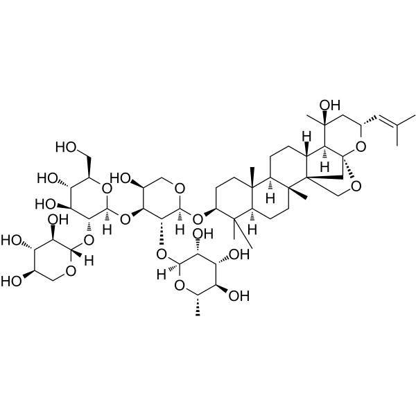
Jujuboside B

Based on 1 Customer Validation
Jujuboside B is a bioactive saponin component isolated from Ziziphi Spinosae Semen (sour jujube seed), with oral efficacy and blood-brain barrier permeability. Jujuboside B induces acute leukemia cell death and drives necroptosis apoptosis by activating the RIPK1/RIPK3/MLKL pathway. Jujuboside B upregulates the expression of NOXA, PARP and caspase-3, activates AMPK, inhibits the proliferation of breast cancer cells, and induces cell apoptosis and autophagy. Jujuboside B inhibits angiogenesis and tumor growth by blocking the VEGFR-2 signaling pathway. Jujuboside B alleviates liver injury in mice by regulating the Nrf2-STING signaling pathway. Jujuboside B alleviates liver injury by regulating anti-inflammatory responses and downregulating the expression of 11β-HSD2. Jujuboside B induces ferroptosis and overcomes radioresistance in non-small cell lung cancer via the PPARγ-ATF3-Gpx4 signaling pathway. Jujuboside B exerts inhibitory effects on platelet aggregation. Jujuboside B inhibits febrile seizures by suppressing the activity of AMPA receptors. Jujuboside B reverses chronic unpredictable mild stress-promoted tumor progression by blocking the PI3K/Akt and MAPK/ERK pathways and dephosphorylating CREB signaling. Jujuboside B is applicable to related studies on acute leukemia, breast cancer, PM2.5-induced lung injury, hepatotoxicity, liver injury, colorectal cancer, non-small cell lung cancer, thromboembolic diseases, cardiovascular diseases associated with high platelet aggregation, febrile seizures, and depressive-like phenotypes.
연구목적의 판매만을 진행합니다. 환자를 대상으로 한 판매는 하지 않습니다.
- Purity : 99.92%
- CAS No.: 55466-05-2
- 화학식: C52H84O21
- 분자량:1045.21
-
보관:
4°C, protect from light
* In solvent : -80°C, 6 months; -20°C, 1 month (protect from light)

To place orders, for customer services and technical support, please contact: MedChemExpress USA
Tel: 609-228-6898 E-mail: [email protected] [email protected]


-
 Biological Activity
-
Chemical Information
-
용액&용해도
- Protocol
- 순도&문서
- References
-
 Help & FAQs




Biological Activity
제품 설명
Cellular Effect
|
Cell Line

|
Type | Value | Description | References |
|---|---|---|---|---|
| AGS | IC50 |
107 μM
Compound: 1
|
Cytotoxicity against human AGS cells after 24 hrs by MTT assay
Cytotoxicity against human AGS cells after 24 hrs by MTT assay
|
[PMID: 24547878] |
| HCT-116 | IC50 |
114 μM
Compound: 1
|
Cytotoxicity against human HCT116 cells after 24 hrs by MTT assay
Cytotoxicity against human HCT116 cells after 24 hrs by MTT assay
|
[PMID: 24547878] |
In Vitro
Jujuboside B (40-200 μM; 24-72 h) reduces the viability of acute leukemia cell lines U937, HL-60, Jurkat and Kasumi-1 in a dose- and time-dependent manner, and decreases the survival rate of primary human acute myeloid leukemia (AML) cells[1].
Jujuboside B (0-120 μM; 2 weeks) significantly reduces the clonogenic capacity of U937 acute leukemia cells[1].
Jujuboside B (40-120 μM; 24 h) mediates necroptosis of U937 acute leukemia cells via the RIPK1/RIPK3/MLKL pathway, and upregulates the total and phosphorylated protein levels of RIPK1, RIPK3 and MLKL in U937 acute leukemia cells[1].
Jujuboside B (20-100 μM; 72 h) potently inhibits the proliferation of human breast cancer cells MDA-MB-231 (IC50 = 54.38 μM) and MCF-7 (IC50 = 74.94 μM) after a 72 h treatment[2].
Jujuboside B (25-75 μM; 10-14 days) reduces the clonogenic survival rate of MDA-MB-231 and MCF-7 human breast cancer cells[2].
Jujuboside B (25-75 μM; 16-48 h) dose-dependently inhibits the migration of human breast cancer MDA-MB-231 cells, induces dose-dependent apoptosis in human breast cancer MDA-MB-231 and MCF-7 cells, upregulates the expressions of apoptosis markers cleaved PARP and cleaved caspase-3 in human MDA-MB-231 and MCF-7 cells, upregulates NOXA expression in MDA-MB-231 and MCF-7 cells, and activates AMPK[2].
Jujuboside B (25-75 μM; 12-48 h) induces dose-dependent and time-dependent autophagy in human breast cancer cell lines MDA-MB-231 and MCF-7, enhances the conversion of LC3-I to LC3-II, and reduces the expression level of p62. It induces autophagy in an AMPK-dependent manner in human breast cancer MCF-7 cells[2].
Jujuboside B (1-100 μM; 8-48 h) dose-dependently inhibits cell viability, migration, and Matrigel-based tube formation of HUVECs, while exerting minimal effects on the viability of HCT-15 cells[6].
Jujuboside B (1-100 μM; 24 h) dose-dependently arrests HUVECs at the G0/G1 phase of the cell cycle[6].
Jujuboside B (1-100 μM; 30 min pre-incubation) dose-dependently blocks VEGF165-induced phosphorylation of VEGFR2 and its downstream mediators (Akt, FAK, Src, PLCγ1) in HUVEC[6].
Jujuboside B (100 μM; 24 h) exerts better regulatory effects on the protein levels of DR4 and DR5 at 100 ng/mL in A549 and H460 cells than the single treatment[7].
Jujuboside B (30-300 μM; 5 min) dose-dependently inhibits collagen-, thrombin-, Arachidonic acid (AA) (HY-109590)- and adenosine diphosphate (ADP)-induced platelet aggregation in rat platelet-rich plasma, with IC50 values of 92.1 μM, 201.5 μM and 95.2 μM for collagen-, thrombin- and AA-induced aggregation, respectively, and exhibits no platelet cytotoxicity at the concentration of 300 μM[8].
Jujuboside B (30-300 μM; 5 min pre-treatment followed by 6 min incubation with collagen) dose-dependently inhibits collagen-induced TXA2 production in rat platelet-rich plasma[8].
Jujuboside B (30-100 μM) inhibits the currents of recombinant GluA1/GluA2 AMPA receptors in HEK293 cells in a dose-dependent manner in vitro[9].
Jujuboside B (pre-incubated for 1 min prior to AMPA stimulation at a concentration of 10 μM) significantly inhibits AMPA-induced intracellular calcium elevation in primary cultured rat cortical neurons, and reduces the peak and cumulative values of calcium responses[9].
Jujuboside B (20-100 μM; 60 μM for viability, colony formation, pathway analysis) inhibits the viability and colony formation of A549 cells, and blocks the PI3K/Akt and MAPK/ERK signaling pathways by reducing the phosphorylation levels of key pathway components, with an IC50 of 60 μM[10].
MedChemExpress (MCE) has not independently confirmed the accuracy of these methods. They are for reference only. Further protocols information, click here.
-
Cell Line:U937, HL-60, Jurkat, Kasumi-1 acute leukemia cell lines
-
Concentration:40, 80, 100, 120, 160, 200 μM
-
Incubation Time:24 h; 48 h; 72 h
-
Result:Inhibited the growth of all four leukemia cell lines in a dose- and time-dependent manner.
Showed significant inhibitory effects after 24-h treatment, with potency increasing at longer incubation times.
-
Cell Line:U937 acute leukemia cells
-
Concentration:40, 80, 120 μM
-
Incubation Time:2 weeks
-
Result:Significantly inhibited colony formation of U937 cells at 80 μM and 120 μM, reflected by decreased colony number and size.
Did not significantly affect colony counts at 40 μM.
-
Cell Line:U937 acute leukemia cells
-
Concentration:40, 80, 120 μM
-
Incubation Time:24 h
-
Result:Significantly increased protein levels of RIPK1, p-RIPK1, RIPK3, p-RIPK3, MLKL, and p-MLKL in U937 cells in a dose-dependent manner.
Induced statistically significant upregulation of all target proteins and their phosphorylated forms at 80 μM and 120 μM.
-
Cell Line:MDA-MB-231, MCF-7 human breast cancer cell lines
-
Concentration:0, 40, 60, 80, and 100 μM
-
Incubation Time:72 h
-
Result:Significantly inhibited the proliferation of MDA-MB-231 and MCF-7 in a dose-dependent manner.
Achieved an IC50 of 54.38 μM for MDA-MB-231 and 74.94 μM for MCF-7.
-
Cell Line:MDA-MB-231 human breast cancer cell line
-
Concentration:25, 50, 75 μM
-
Incubation Time:16 h
-
Result:Inhibited the migration of MDA-MB-231 cells in a dose-dependent manner, with significant reductions in migrated cell numbers at all tested concentrations.
-
Cell Line:MDA-MB-231, MCF-7 human breast cancer cell lines
-
Concentration:25, 50, 75 μM
-
Incubation Time:48 h
-
Result:Resulted in a remarkable dose-dependent increase in the apoptotic cell population (early apoptotic Annexin V(+)/PI(−) and late apoptotic Annexin V(+)/PI(+) cells) in both cell lines.
-
Cell Line:MDA-MB-231, MCF-7 human breast cancer cell lines
-
Concentration:25, 50, 75 μM
-
Incubation Time:48 h
-
Result:Significantly increased the expression of cleaved PARP and cleaved caspase-3 in a dose-dependent manner in both cell lines.\nRemarkably elevated NOXA expression in a dose-dependent pattern in both cell lines.\nIncreased phosphorylated AMPK levels in a dose-dependent manner in both cell lines.
-
Cell Line:MDA-MB-231, MCF-7 human breast cancer cell lines
-
Concentration:25-75 μM (48 h treatment); 50 μM (12-48 h treatment)
-
Incubation Time:48 h (dose-dependent autophagy); 12-48 h (time-dependent autophagy)
-
Result:Increased the conversion of LC3-I to LC3-II in a dose-dependent manner after 48 h of treatment.
Accumulated LC3-II in a time-dependent manner from 12 to 48 h of treatment at 50 μM.
Decreased p62 expression in a dose-dependent manner after 48 h of treatment in both cell lines.
-
Cell Line:human umbilical vein endothelial cells (HUVECs), HCT-15 human colorectal adenocarcinoma cells
-
Concentration:1, 3, 10, 30, and 100 μM
-
Incubation Time:48 h
-
Result:Significantly suppressed HUVEC viability in a dose-dependent manner, with minimal effect at 1-10 μM and strong suppression at ≥30 μM.
Maintained over 65% of HCT-15 cell viability across all tested concentrations.
-
Cell Line:HUVECs
-
Concentration:1, 3, 10, 30, and 100 μM
-
Incubation Time:24 h
-
Result:Caused dose-dependent changes in the percentage of cells in G0/G1 and S phases, indicating G0/G1 phase arrest.
-
Cell Line:HUVECs
-
Concentration:1, 3, 10, 30, and 100 μM
-
Incubation Time:10 h
-
Result:Inhibited HUVEC migration in a dose-dependent manner, with ~20% inhibition at 3 μM and ~90% inhibition at 100 μM.
-
Cell Line:HUVECs
-
Concentration:1, 3, 10, 30, and 100 μM
-
Incubation Time:30 min (pre-incubation prior to VEGF165 stimulation)
-
Result:Inhibited phosphorylation of VEGFR2 in a dose-dependent manner.
Inhibited phosphorylation of downstream signaling proteins (Akt, FAK, Src, PLCγ1) in a dose-dependent manner.
-
Cell Line:A549, H460
-
Concentration:100 μM (combined with 100 ng/mL TRAIL)
-
Incubation Time:24 h
-
Result:Upregulated DR4 and DR5 protein levels more significantly than jujuboside B or TRAIL alone in A549 and H460 cells.
In Vivo
Jujuboside B (0.1-0.8 mg/kg; i.v.; 3 times within 2 days) significantly alleviates PM2.5-induced lung injury in BALB/c mice by regulating the TLR2/4-MyD88 and mTOR-autophagy pathways to reduce inflammation, apoptosis and autophagy dysfunction[3].
Jujuboside B (i.p./i.v.; once daily; consecutive 7 days/single administration, 20-40 mg/kg/0.4-1.5 mg/kg) dose-dependently ameliorates Acetaminophen (HY-66005)-induced acute liver injury in male C57BL/6 J mice, while inhibiting oxidative stress, inflammatory response, cell apoptosis and STING pathway activation, and activating the Nrf2 pathway. It also dose-dependently protects male C57BL/6 mice from cecal ligation and puncture (CLP)-induced liver injury by alleviating inflammation, enhancing antioxidant capacity, upregulating GR expression and downregulating 11β-HSD2 expression[4].
Jujuboside B (i.p., once every 2 days, 7 administrations, dose of 20 mg/kg) reduces the subcutaneous HCT-15 colorectal cancer tumor volume by 55.5% and tumor weight by 56.3% in female BALB/c nude mice through anti-angiogenic and anti-proliferative mechanisms, with no treatment-related body weight loss observed[6].
When applied topically to the chick chorioallantoic membrane, Jujuboside B (1-100 μM) inhibits angiogenesis in a dose-dependent manner, with an inhibition rate of up to 80% at the concentration of 100 μM[6].
When incorporated into subcutaneous Matrigel plugs at concentrations of 10-100 μM, Jujuboside B dose-dependently inhibits VEGF-induced angiogenesis in female BALB/c mice[6].
Jujuboside B (i.p., once every other day, 20-40 mg/kg) dose-dependently reduces the tumor volume of A549 non-small cell lung cancer in nude mice without inducing significant hepatotoxicity or nephrotoxicity[7].
Jujuboside B (10-100 mg/kg; p.o.; single administration) provides a statistically significant 63% protective effect against acute pulmonary thromboembolism induced by collagen and epinephrine in male ICR mice[8].
Jujuboside B (10-50 mg/kg; i.p.; single administration 1 hour prior to hyperthermia induction) dose-dependently inhibits the severity of febrile seizures in male P14 C57BL/6 mice[9].
Jujuboside B (40 mg/kg/day; i.p.; daily; for 2 consecutive weeks) significantly inhibits lung cancer progression in non-stressed and CUMS-stressed tumor-bearing female C57BL/6 mice by regulating apoptosis-related proteins, blocking the PI3K/Akt and MAPK/ERK signaling pathways, and reducing the levels of inflammatory cytokines[10].
Jujuboside B (40 mg/kg; i.p.; daily for 2 consecutive weeks) significantly ameliorates depression-like behaviors in female C57BL/6 mice subjected to CUMS modeling by increasing serum 5-HT and tryptophan levels, improving behavioral scores, and reducing inflammatory cytokine levels[10].
MedChemExpress (MCE) has not independently confirmed the accuracy of these methods. They are for reference only.
-
Animal Model:athymic BALB/c nude (female, 5-6-week-old)[2]
-
Dosage:20 mg/kg
-
Administration:i.p.; daily; 17,28 days
-
Result:Significantly inhibited MCF-7 tumor growth, with tumor volumes and final tumor weights markedly lower than the control group.
Showed no significant difference in animal body weight compared with the control group.
Elevated expression of cleaved PARP, cleaved caspase-3, and LC3-II in tumor tissue, indicating induction of apoptosis and autophagy.
Significantly inhibited MDA-MB-231 tumor growth, with tumor volumes and final tumor weights markedly lower than the control group.
Showed no significant difference in animal body weight compared with the control group.
Elevated expression of cleaved PARP, cleaved caspase-3, and LC3-II in tumor tissue, indicating induction of apoptosis and autophagy.
-
Animal Model:BALB/c (7-week-old; acclimated for 12 days; PM2.5-induced lung injury model)[3]
-
Dosage:0.1 mg/kg; 0.4 mg/kg; 0.8 mg/kg
-
Administration:i.v.; three times over 2 days
-
Result:Significantly reduced the PM2.5-induced increase in lung wet/dry weight ratio, total cell count in BALF, total neutrophil count in BALF, and lung injury score.
Significantly reversed PM2.5-induced changes in apoptotic protein levels: restored anti-apoptotic Bcl-2 levels, reduced pro-apoptotic Bax, cleaved caspase-3, and cleaved PARP levels, and decreased the TUNEL-positive apoptotic cell percentage.
Significantly reduced PM2.5-induced increases in pro-inflammatory cytokines IFN-γ, IL-1β, IL-6, IL-18, and TNF-α, and restored PM2.5-induced decreases in anti-inflammatory cytokines IL-2, IL-4, and IL-10.
Significantly inhibited PM2.5-induced upregulation of autophagy-related proteins LC3 II and Beclin 1, reversed PM2.5-induced increases in TLR2, TLR4, and MyD88 levels, and restored PM2.5-induced reductions in phosphorylated mTOR, Akt, and PI3K levels.
-
Animal Model:C57BL/6 J (male, 8 weeks old, Acetaminophen-induced acute hepatotoxicity)[4]
-
Dosage:20 mg/kg; 40 mg/kg
-
Administration:i.p.; daily; 7 days
-
Result:Reversed acetaminophen-induced CYP2E1 upregulation in liver tissue in a dose-dependent manner.
Reduced acetaminophen-induced mortality from 80% to 30% over 48 hours (40 mg/kg dose).
Dose-dependently reduced serum ALT, AST, and LDH levels, improved liver histopathological scores, and attenuated liver tissue necrosis compared to acetaminophen-only controls.
Dose-dependently suppressed acetaminophen-induced increases in serum and liver TNF-α, IL-6, and IFN-β levels, reduced liver macrophage infiltration (F4/80-positive cells), and lowered liver TNF-α and IL-6 protein expression.
Dose-dependently reversed acetaminophen-induced increases in liver 4-HNE and MDA levels, restored liver GSH, SOD, and CAT activities, upregulated liver SOD1 and SOD2 mRNA levels, downregulated liver NOX2 and COX2 mRNA levels, reduced hepatocyte apoptosis (increased Bcl-2/Bax ratio, decreased cleaved caspase-3), inhibited acetaminophen-induced STING pathway activation (reduced STING, p-IRF3, and p-p65 protein levels in liver tissue), and activated the Nrf2 pathway (increased total, nuclear, and cytosolic Nrf2 protein levels, upregulated liver HO-1 and NQO-1 protein and mRNA levels).
-
Animal Model:C57BL/6 (male, 8-10 weeks old, average weight 27 g, CLP-induced sepsis)[5]
-
Dosage:0.4 mg/kg; 0.75 mg/kg; 1.5 mg/kg
-
Administration:i.v.; single dose
-
Result:Significantly reduced CLP-induced hepatic histopathological damage, including hemorrhagic necrosis, portal inflammation, parenchymal necrosis, and inflammatory cell infiltration at 0.75 mg/kg and 1.5 mg/kg.
Significantly reduced elevated serum alanine aminotransferase (ALT) and aspartate aminotransferase (AST) levels at 0.75 mg/kg and 1.5 mg/kg.
Significantly reduced elevated liver tumor necrosis factor (TNF)-α, interleukin (IL)-1β, and nitric oxide (NO) levels at 0.75 mg/kg and 1.5 mg/kg; significantly reduced elevated liver TNF-α and NO levels at 0.4 mg/kg.
Reduced elevated liver malondialdehyde (MDA) levels at 0.75 mg/kg and 1.5 mg/kg.
Restored reduced liver glutathione (GSH), superoxide dismutase (SOD), and glutathione peroxidase (GSH-Px) activity at 0.75 mg/kg and 1.5 mg/kg.
Increased reduced liver glucocorticoid receptor (GR) protein expression at 0.4 mg/kg, 0.75 mg/kg, and 1.5 mg/kg.
Reduced elevated liver 11β-hydroxysteroid dehydrogenase type 2 (11β-HSD2) protein expression, with no effect on 11β-hydroxysteroid dehydrogenase type 1 (11β-HSD1) expression at 0.75 mg/kg and 1.5 mg/kg.
Did not significantly reduce serum ALT/AST levels or liver IL-1β levels at 0.4 mg/kg.
-
Animal Model:BALB/c nude (female)[6]
-
Dosage:20 mg/kg
-
Administration:i.p.; once every two days; 7 total doses
-
Result:Reduced mean tumor volume by 55.5% compared to control group.
Reduced mean tumor weight by 56.3% compared to control group.
Significantly reduced tumor microvessel density.
Significantly decreased Ki67-positive proliferative tumor cells.
Significantly increased tumor necrosis area relative to controls.
Caused no significant mouse body weight loss during treatment.
-
Animal Model:BALB/c nude (female, 6 weeks old)[7]
-
Dosage:20 mg/kg; 40 mg/kg
-
Administration:i.p.; every other day
-
Result:Reduced tumor volumes in a dose-dependent manner relative to controls, with statistically significant reductions observed over the study period.
Showed no significant difference in body weights between treated and control groups.
Exhibited no significant difference in serum levels of alanine aminotransferase (ALT), aspartate aminotransferase (AST), blood urea nitrogen (BUN), and creatinine (CRE) from controls.
-
Animal Model:ICR mice (male, 35-40 g)[8]
-
Dosage:10 mg/kg; 30 mg/kg; 100 mg/kg
-
Administration:p.o.; single dose
-
Result:Provided 17% protection against thromboembolism.
Provided 43% protection against thromboembolism.
Provided 63% protection against thromboembolism, which was statistically significant compared to vehicle.
-
Animal Model:C57BL/6 (male, postnatal day 14, 6-7 g, LPS-primed hyperthermic induction)[9]
-
Dosage:10 mg/kg; 30 mg/kg; 50 mg/kg
-
Administration:i.p.; single dose 1 hour before hyperthermic induction
-
Result:Increased seizure latency to 228.7, reduced the percentage of grade 5 seizures to 50%, and decreased seizure severity (Racine score) to 4.5.
Significantly prolonged seizure latency to 563.3s, eliminated grade 5 seizures (90% of mice exhibited maximum grade 3 seizures), decreased seizure severity (Racine score) to 2.9, prolonged latency to EEG seizure onset to 680.0s, shortened total seizure duration to 551.9s, decreased seizure spike number to 39.88, and reduced hippocampal c-Fos-positive cells to 15.50 cells/mm2.
Significantly prolonged seizure latency to 737.2 s, eliminated grade 5 seizures (80% of mice exhibited maximum grade 3 seizures), and decreased seizure severity (Racine score) to 2.8.
-
Animal Model:C57BL/6 (female, 8 weeks old, subcutaneous inoculation of LLC lung cancer cells; chronic unpredictable mild stress exposure)[10]
-
Dosage:40 mg/kg/day
-
Administration:i.p.; daily; 2 weeks
-
Result:Significantly reduced tumor volume and weight compared to vehicle controls in non-CUMS tumor-bearing mice.
Significantly reduced tumor volume and weight compared to vehicle-treated CUMS-exposed tumor-bearing mice, reversing CUMS-promoted tumor progression.
Significantly increased proapoptotic Bax protein and mRNA expression, and decreased antiapoptotic Bcl-2 protein and mRNA expression in tumor tissues compared to corresponding vehicle controls.
Significantly reduced phosphorylation levels of PI3K, Akt, MAPK, ERK, and CREB in tumor tissues compared to corresponding vehicle controls.
Significantly decreased serum levels of inflammatory cytokines TNF-α, IL-4, IL-6, and IL-10 compared to vehicle controls.
-
Animal Model:C57BL/6 (female, 8 weeks old, chronic unpredictable mild stress exposure)[10]
-
Dosage:40 mg/kg/day
-
Administration:i.p.; daily; 2 weeks
-
Result:Significantly increased serum 5-HT and tryptophan levels compared to vehicle-treated CUMS-exposed mice.
Significantly increased sucrose preference, locomotion scores, and exploratory scores compared to vehicle-treated CUMS-exposed mice, ameliorating depression-like phenotypes.
Significantly decreased serum levels of inflammatory cytokines TNF-α, IL-4, IL-6, and IL-10 compared to vehicle controls.


Chemical Information
-
CAS No. 55466-05-2
-
Appearance Solid
-
분자량 1045.21
-
화학식 C52H84O21
-
Color White to off-white
-
SMILES
C[C@]([C@]1([H])CC2)(CC[C@]3([H])[C@@]1(CC[C@H](O[C@@](OC[C@H](O)[C@@H]4O[C@@](O[C@H](CO)[C@@H](O)[C@@H]5O)([H])[C@@H]5O[C@@](OC[C@@H](O)[C@@H]6O)([H])[C@@H]6O)([H])[C@@H]4O[C@@](O[C@@H](C)[C@H](O)[C@H]7O)([H])[C@@H]7O)C3(C)C)C)[C@@]8(CO9)[C@@]2([H])[C@]([C@]%10(O)C)([H])[C@@]9(O[C@@H](/C=C(C)/C)C%10)C8
-
Structure Classification
-
Initial Source
-
선적
Room temperature in continental US; may vary elsewhere.
-
보관
4°C, protect from light
* In solvent : -80°C, 6 months; -20°C, 1 month (protect from light)


용액&용해도
In Vitro:
DMSO : 100 mg/mL (95.67 mM; Need ultrasonic; Hygroscopic DMSO has a significant impact on the solubility of product, please use newly opened DMSO)
Please refer to the solubility information to select the appropriate solvent. Once prepared, please aliquot and store the solution to prevent product inactivation from repeated freeze-thaw cycles.
Storage method and period of stock solution: -80°C, 6 months; -20°C, 1 month (protect from light). When stored at -80°C, please use it within 6 months. When stored at -20°C, please use it within 1 month.
Please refer to the solubility information to select the appropriate solvent. Once prepared, please aliquot and store the solution to prevent product inactivation from repeated freeze-thaw cycles.
Storage method and period of stock solution: -80°C, 6 months; -20°C, 1 month (protect from light). When stored at -80°C, please use it within 6 months. When stored at -20°C, please use it within 1 month.
Concentration (start) × Volume (start) = Concentration (final) × Volume (final)
In Vivo:
Select the appropriate dissolution method based on your experimental animal and administration route.
- For the following dissolution methods, please ensure to first prepare a clear stock solution using an In Vitro approach and then sequentially add co-solvents:
- To ensure reliable experimental results, the clarified stock solution can be appropriately stored based on storage conditions. As for the working solution for In Vivo experiments, it is recommended to prepare freshly and use it on the same day.
- The percentages shown for the solvents indicate their volumetric ratio in the final prepared solution. If precipitation or phase separation occurs during preparation, heat and/or sonication can be used to aid dissolution.
Add each solvent one by one: 10% DMSO 40% PEG300 5% Tween-80 45% Saline
Solubility: ≥ 2.5 mg/mL (2.39 mM); Clear solution
This protocol yields a clear solution of ≥ 2.5 mg/mL (saturation unknown).
Taking 1 mL working solution as an example, add 100 μL DMSO stock solution (25.0 mg/mL) to 400 μL PEG300, and mix evenly; then add 50 μL Tween-80 and mix evenly; then add 450 μL Saline to adjust the volume to 1 mL.
Preparation of Saline: Dissolve 0.9 g sodium chloride in ddH₂O and dilute to 100 mL to obtain a clear Saline solution.
Add each solvent one by one: 10% DMSO 90% (20% SBE-β-CD in Saline)
Solubility: ≥ 2.5 mg/mL (2.39 mM); Clear solution
This protocol yields a clear solution of ≥ 2.5 mg/mL (saturation unknown).
Taking 1 mL working solution as an example, add 100 μL DMSO stock solution (25.0 mg/mL) to 900 μL 20% SBE-β-CD in Saline, and mix evenly.
Preparation of 20% SBE-β-CD in Saline (4°C, storage for one week): 2 g SBE-β-CD powder is dissolved in 10 mL Saline, completely dissolve until clear.
In Vivo Dissolution Calculator
Please enter the basic information of animal experiments:
-
-
-
-
Recommended: Prepare an additional quantity of animals to account for potential losses during experiments.
Please enter your animal formula composition:
-
%DMSO +
Recommended: Keep the proportion of DMSO in working solution below 2% if your animal is weak.
-
%+
-
+%Tween-80 + +
-
%Saline +
The co-solvents required include: DMSO, . All of co-solvents are available by MedChemExpress (MCE). , Tween 80. All of co-solvents are available by MedChemExpress (MCE).
Working solution concentration: 0.22 mg/mL
Method for preparing stock solution: mg drug dissolved in μL DMSO. Stock solution concentration: mg/mL. * In solvent : -80°C, 6 months; -20°C, 1 month (protect from light)
1. Take μL DMSO stock solution;
2. Add μL .
μL , mix evenly;
3. Then add μL Tween 80, mix evenly;
4. Then add μL
Please ensure that the stock solution in the first step is dissolved to a clear state, and add co-solvents in sequence. You can use ultrasonic heating (ultrasonic cleaner, recommended frequency 20-40 kHz), vortexing, etc. to assist dissolution.

Protocol
-
RNA extraction experimental
By lysing cells, releasing RNA, and removing impurities such as proteins and DNA, high-purity RNA products are finally obtained. The commonly used traditional method is the guanidine isothiocyanate/phenol/chloroform method (Trizol), which is suitable for a variety of animal materials including animal tissues, microorganisms, cultured cells, etc., and most plant materials.
-
Human pluripotent stem cell endothelial-cell differentiation
Human pluripotent stem cell endothelial differentiation is based on stepwise developmental patterning: early activation of WNT/GSK3β inhibition promotes mesodermal or vascular progenitor entry, followed by endothelial specification using VEGF-related signaling, BMP4, FGF2, Notch modulation, or cAMP depending on the published protocol. Endothelial differentiation is read out by acquisition of CD31, CD34, VE-cadherin/CD144, KDR/VEGFR2, vWF, Tie2, NOS3, acetylated LDL uptake, tube/network formation, barrier function, and in vivo vessel-forming capacity where tested.
-
Breast Cancer Modeling
Breast cancer is a heterogeneous cancer, and it has been distinguished into four subtypes: luminal A, luminal B, HER2-positive and basal-like. Molecular mutations, epigenetic alterations, hormone exposure and immune microenvironment are related to the progression of breast cancer.
-
Apoptosis
Apoptosis, also called programmed cell death, is generally characterized by distinct morphological characteristics.
-
TUNEL staining for apoptotic DNA fragmentation
TUNEL staining detects DNA strand breaks by using terminal deoxynucleotidyl transferase to add labeled nucleotides to exposed 3′-OH DNA termini, generating either microscopic staining in fixed cells or tissue sections, or fluorescence/cytometric signal in cell suspensions. TUNEL positivity reflects DNA fragmentation but should not be interpreted alone as definitive apoptosis, because TUNEL can also label necrotic, autolytic, mechanically damaged, or DNA-repair-associated DNA breaks.
-
Cell Cytotoxicity Assay
Cytotoxicity assays are usually based on the assessment of cell membrane damage, which can also be indirectly detected by measuring cell viability. Detection methods include MTT assay, CKK-8 assay, LDH assay and ATP assay, etc.
-
Autophagy
Autophagy is a process in which eukaryotic cells use lysosomes to degrade their own cytoplasmic proteins and damaged organelles under the regulation of autophagy related gene (Atg). Microtubule-associated proteins light chain 3 (LC3) is recognized as autophagy marker, which transfers from cytoplasmic LC3 (LC3-I) to membrane type (LC3-II). LC3-II/I ratio could be detected by Western Blot and fluorescence microscopy.
-
Endothelial Tube Formation Assay
Endothelial tube formation assay evaluates the ability of endothelial cells to attach, migrate, align, and organize into capillary-like networks when cultured on gelled basement membrane extract or Matrigel; the readout is the morphology and quantity of tube-like networks, which reflects an in vitro endothelial morphogenesis step related to angiogenesis. Basement membrane extract/Matrigel provides laminin-rich extracellular matrix cues that support endothelial differentiation into capillary-like structures, but it can contain biologically active growth factors, so growth-factor-reduced matrix is preferred when testing defined angiogenic stimulators or inhibitors.
-
Transepithelial/transendothelial electrical resistance assay
TEER measures electrical resistance across epithelial or endothelial monolayers cultured on permeable supports, and the readout reflects ionic conductance through the cell barrier, especially the paracellular pathway regulated by junctional integrity. TEER can be measured without destroying the monolayer and is commonly used before or during transport, permeability, barrier-disruption, and barrier-maturation experiments. TEER values are influenced by biological maturation and technical conditions; reported factors include temperature, medium formulation, passage number, electrode geometry, membrane properties, and junctional length during early monolayer maturation. Therefore, TEER should be interpreted with blank-insert subtraction, area normalization, repeated readings, and, when possible, orthogonal barrier readouts such as FITC-dextran flux or tight-junction staining.
-
Apoptosis Solutions
Apoptosis is a regulated, generally non-lytic cell-death pathway that removes unwanted, damaged, infected, or abnormal cells through coordinated morphological changes, caspase activation, DNA fragmentation, and membrane remodeling. The intrinsic apoptosis pathway is controlled mainly by mitochondrial outer membrane permeabilization, BCL-2 family proteins, cytochrome c release, apoptosome formation, caspase-9 activation, and downstream executioner caspase-3/7 activation. The extrinsic apoptosis pathway is initiated by death receptors such as Fas, TNFR, and TRAIL receptors, which recruit adaptor proteins and activate caspase-8 before engaging executioner caspases or mitochondrial amplification through BID cleavage. Apoptosis is linked to many phenotypes, including cancer cell killing, tissue homeostasis, immune regulation, neurodegeneration, infection response, and treatment-induced cytotoxicity; unresolved questions include how apoptosis interacts with necroptosis, pyroptosis, ferroptos
-
Lysosome and acidic-vesicle live-cell staining
Lysosome and acidic-vesicle live-cell staining detects acidic intracellular compartments by using membrane-permeant acidotropic probes that accumulate in low-pH vesicles, including lysosomes, late endosomes, autolysosomes, and acidic phagosomes. LysoTracker staining is commonly used as an intensity-based readout of acidic lysosomal compartment abundance or enlargement, while acridine orange produces green fluorescence in less concentrated compartments and red fluorescence after concentration-dependent accumulation in acidic vesicular organelles. Loss or reduction of acridine-orange red signal can be used as a readout of lysosomal membrane permeabilization or reduced acidic-vesicle integrity. This protocol is designed for live cultured cells and can be adapted for fluorescence microscopy, high-content imaging, plate-reader readout, or flow cytometry when the selected literature supports the readout. Because these dyes report acidotropic accumulation rather than lysosome identity alone,
-
Ferroptosis Solutions
Ferroptosis is an iron-dependent, non-apoptotic form of regulated cell death characterized by lethal lipid peroxidation and sensitivity to suppression by iron chelators or lipophilic radical-trapping antioxidants. The core pathway links cystine uptake through system Xc−, glutathione availability, GPX4-dependent detoxification of phospholipid hydroperoxides, iron-dependent oxidative reactions, and polyunsaturated-phospholipid metabolism into a cell-death program that is biochemically and morphologically distinct from apoptosis, necrosis, and autophagy. The ferroptosis pathway is experimentally linked to phenotype through chemical and genetic perturbation. Erastin induces ferroptosis by inhibiting cystine uptake through system Xc− and weakening antioxidant defenses, while GPX4 inhibition or depletion causes lipid peroxide accumulation and ferroptotic cancer-cell death. ACSL4 and oxidizable arachidonoyl- or adrenoyl-containing phosphatidylethanolamines shape ferroptosis sensitivity by con
-
Hepatotoxicity Study
This protocol evaluates hepatotoxicity using complementary in vivo mouse APAP acute liver injury and in vitro hepatocyte-based cytotoxicity readouts. In vivo APAP injury is assessed by serum ALT/AST, liver histology, hepatic glutathione, protein adducts, necrosis, inflammation, and regeneration-related endpoints. In vitro hepatotoxicity is assessed by loss of viability, leakage of ALT/AST/LDH, oxidative-stress markers, mitochondrial function, nuclear morphology, intracellular calcium, and high-content imaging endpoints.
-
Inhalation Toxicity Study
Inhalation toxicity studies expose rodents to a controlled aerosol, vapor, gas, or smoke atmosphere and assess respiratory and systemic toxicity using exposure-atmosphere characterization, clinical observations, body and organ weights, bronchoalveolar lavage fluid, histopathology, blood chemistry, hematology, and, when included, molecular endpoints such as transcriptomics, proteomics, lipidomics, or tissue burden analysis. The primary biological readouts are airway irritation, pulmonary inflammation, cytotoxicity, altered surfactant or lipid homeostasis, impaired particle clearance, and tissue remodeling, reflected by BALF cell differentials, BALF protein, LDH, phosphatase activities, cytokines, lung weight, microscopic respiratory-tract lesions, and retained lung burden.
-
Macroautophagy Solutions
Macroautophagy is a conserved lysosome-dependent degradation pathway in which cytoplasmic material is sequestered into double-membrane autophagosomes and delivered to lysosomes for degradation and recycling. The pathway supports cellular homeostasis during nutrient limitation, organelle stress, protein-aggregate accumulation, infection, differentiation, and tissue remodeling by coupling cargo sequestration, autophagosome maturation, lysosomal fusion, and degradation of cargo-derived macromolecules. The core molecular sequence includes initiation by nutrient- and stress-regulated autophagy machinery, autophagosome nucleation, LC3/ATG8-family conjugation to autophagosomal membranes, cargo selection through receptors such as SQSTM1/p62, autophagosome-lysosome fusion, and lysosomal degradation. LC3 was identified as a mammalian homolog of yeast Atg8 that localizes to autophagosomal membranes after processing, and p62/SQSTM1 was shown to connect ubiquitinated cargo with autophagic degradati
-
Endothelial Cell Migration/Angiogenic Sprouting Assay
Endothelial cell migration and angiogenic sprouting assays are in vitro (and partially ex vivo-adapted) functional models that quantify the ability of endothelial cells to undergo coordinated migration, extracellular matrix invasion, and multicellular organization into capillary-like sprouts in response to pro-angiogenic stimuli such as VEGF, bFGF, or conditioned microenvironments. These assays are used to model early angiogenic events including tip-cell formation, directional migration, and lumen-like sprout extension, which collectively reflect angiogenic activation and vascular morphogenesis processes observed in vivo.
-
Mammalian live/dead viability and cytotoxicity staining
Live/dead viability and cytotoxicity staining assays are based on the simultaneous detection of intracellular esterase activity in metabolically active (viable) cells and membrane integrity loss in non-viable cells. In commonly used dual-staining approaches, membrane-permeant fluorogenic substrates are converted by intracellular esterases into fluorescent products in live cells, while impermeant DNA-binding dyes selectively enter cells with compromised plasma membranes and label nucleic acids in dead or dying cells, enabling discrimination between viable and non-viable populations by fluorescence microscopy or flow cytometry.
-
Vascular/Branching Fractal Analysis
Vascular/branching fractal analysis quantifies the geometric complexity of vessel trees or vascular networks from segmented 2D images, commonly by converting vessels into binary and/or skeletonized maps and estimating fractal dimension using box-counting or related approaches. Fractal dimension is interpreted as an image-derived readout of vascular branching complexity, space filling, or density, and has been applied to retinal photographs, fluorescein angiography, OCT angiography, capillary perfusion maps, and in vitro Matrigel angiogenesis networks. The assay readout is generated from vessel-positive pixels after image preprocessing, vessel segmentation, binarization, and optional skeletonization; reported outputs include fractal dimension, vessel density, branchpoint density, endpoint density, vessel length density, tortuosity, and generation-based branching metrics when VESGEN-style analysis is used. The biological interpretation is limited to quantitative vascular patterning and s
-
Annexin V plus membrane-impermeant dye apoptosis staining
Annexin V-based apoptosis assays rely on the detection of phosphatidylserine (PS) externalization from the inner leaflet of the plasma membrane to the outer leaflet, an early biochemical hallmark of apoptosis. Fluorescently labeled Annexin V binds PS in a calcium-dependent manner, enabling identification of early apoptotic cells by flow cytometry or fluorescence microscopy. When combined with a membrane-impermeant DNA-binding dye (e. g. , propidium iodide), this approach allows discrimination between viable (Annexin V−/dye−), early apoptotic (Annexin V+/dye−), and late apoptotic or necrotic (Annexin V+/dye+) cell populations by assessing membrane integrity and PS exposure.
-
Patient-Derived Orthotopic Xenograft (PDOX)
Patient-derived orthotopic xenograft (PDOX) modeling implants fresh patient tumor tissue or patient-derived tumor cells into the anatomically corresponding organ or tissue site of immunodeficient mice, usually by surgical orthotopic implantation, to preserve patient tumor histology, local microenvironmental context, invasion, metastatic behavior, and treatment-response features better than subcutaneous implantation. PDOX readouts include tumor engraftment, orthotopic tumor growth, local invasion, metastasis, recurrence after resection, histologic similarity to the donor tumor, biomarker retention, molecular concordance, survival, and response or resistance to therapy. PDOX models are used for preclinical drug testing and individualized therapy evaluation, but engraftment success varies by tumor type and specimen quality.
-
Research Protocol for Inflammation-related Diseases
The NLRP3 inflammasome is a cytosolic innate immune signaling platform that integrates priming signals and danger-signal activation to promote caspase-1 activation, maturation of IL-1β and IL-18, and gasdermin D-mediated pyroptotic cell death. The core experimental logic is to determine whether inflammatory disease phenotypes are driven by increased NLRP3 expression, ASC-containing inflammasome assembly, caspase-1 cleavage, GSDMD cleavage, and extracellular release of IL-1β/IL-18 rather than by nonspecific cell injury alone. The pathway is strongly linked to inflammation-related disease phenotypes because monosodium urate crystals activate NALP3/NLRP3 inflammasome signaling in gout-like crystal inflammation, cholesterol crystals activate NLRP3 inflammasomes in atherogenesis models, and DSS-induced intestinal inflammation has been reported to involve NLRP3 inflammasome activity. However, experimental colitis studies also show context-dependent protective effects of NLRP3 inflammasome co
-
Cell Viability Determination by MTT Colorimetric Assay
The following protocol uses the MTT colorimetric assay as a classic literature-established method for assessing cell viability/metabolic activity in cultured mammalian cells. MTT[3-(4,5-dimethylthiazol-2-yl)-2,5-diphenyltetrazolium bromide] is reduced by metabolically active cells to a colored formazan product; the amount of formazan is quantified spectrophotometrically and provides an indirect measure of metabolically active viable cells. Importantly, MTT reduction reflects cellular oxidoreductase/metabolic activity rather than an absolute direct count of living cells, so changes in cellular metabolism can alter the signal independently of cell number.
-
Patient-Derived Xenograft (PDX)
Patient-derived xenograft (PDX) models are generated by engrafting primary human tumor tissue directly into immunodeficient mice, allowing in vivo propagation of patient tumor biology without initial in vitro adaptation. These models are used to preserve key histopathological and molecular characteristics of the original tumor and enable assessment of tumor growth dynamics and therapeutic response in a living organism. The biological readout is tumor engraftment and subsequent growth in the murine host, which reflects the ability of human tumor cells to survive, vascularize, and expand in an immunocompromised microenvironment.
-
Subcutaneous Cell-Line-Derived Xenograft
Subcutaneous cell-line-derived xenograft (CDX) models are established by implanting cultured human cancer cell lines into immunodeficient mice, where the injected cells form localized tumors that can be monitored in vivo as a measure of tumorigenic potential, growth kinetics, and treatment response. These models are widely used in oncology research because they allow reproducible tumor formation and enable comparative assessment of tumor growth between different cell lines or genetic manipulations in a controlled in vivo microenvironment. Subcutaneous implantation of cancer cells in immunodeficient mice is a standard approach for evaluating tumor growth behavior and therapeutic response across multiple cancer types, including prostate, esophageal, pancreatic, and colon cancer models.
-
Orthotopic Cell-Line Xenograft
Orthotopic cell-line xenograft models involve implantation of human cancer cell lines into the anatomically corresponding organ of immunodeficient mice to reproduce tumor growth within a native microenvironment, enabling more clinically relevant tumor behavior compared with subcutaneous models. These models are widely used because orthotopic placement better recapitulates tumor progression, including invasion and metastatic spread, which are often underrepresented in heterotopic implantation systems. Compared with conventional xenografts, orthotopic implantation is described as more technically complex but provides improved simulation of tumor-microenvironment interactions and metastatic behavior, making it particularly valuable for translational oncology research. Surgical orthotopic implantation approaches have been emphasized as enabling faithful reproduction of clinical cancer features, including metastasis and disease progression patterns that align with the tumor’s organ of origi


순도&문서
-
Data Sheet (316 KB)
-
SDS (393 KB)
- English - EN (393 KB)
- Français - FR (393 KB)
- Deutsch - DE (393 KB)
- Norwegian - NO (393 KB)
- Español - ES (393 KB)
- Swedish - SV (393 KB)
- Italian - IT (393 KB)
- Korean - KR (393 KB)
- Portuguese - PT (393 KB)
-
Handling Instructions (2659 KB)
References
[1]. Jia MM, et al. Jujuboside B promotes the death of acute leukemia cell in a RIPK1/RIPK3/MLKL pathway-dependent manner. Eur J Pharmacol. 2020;876:173041. [Content Brief]
[2]. Guo L, et al. Jujuboside B Inhibits the Proliferation of Breast Cancer Cell Lines by Inducing Apoptosis and Autophagy. Front Pharmacol. 2021;12:668887. Published 2021 Sep 24. [Content Brief]
[3]. Ryu SH, et al. Jujuboside B post-treatment attenuates PM2.5-induced lung injury in mice. Int J Environ Health Res. 2023;33(12):1479-1489. [Content Brief]
[4]. Wang HF, et al. Jujuboside B alleviates acetaminophen-induced hepatotoxicity in mice by regulating Nrf2-STING signaling pathway. Ecotoxicol Environ Saf. 2024;269:115810. [Content Brief]
[5]. Kim C, et al. Hepatoprotective functions of jujuboside B. J Nat Med. 2023;77(1):87-95. [Content Brief]
[6]. Zhang P, et al. Jujuboside B suppresses angiogenesis and tumor growth via blocking VEGFR2 signaling pathway. Heliyon. 2023;9(6):e17072. Published 2023 Jun 7. [Content Brief]
[7]. Kim TW, et al. Jujuboside B Induces Ferroptosis and Overcomes Radioresistance Through the PPARγ-ATF3-Gpx4 Signaling Pathway in Non-Small Cell Lung Cancer. Phytother Res. 2025;39(11):5345-5364. [Content Brief]
[8]. Seo EJ, et al. Zizyphus jujuba and its active component jujuboside B inhibit platelet aggregation. Phytother Res. 2013;27(6):829-834. [Content Brief]
[9]. Jin B, et al. Jujuboside B inhibits febrile seizure by modulating AMPA receptor activity. J Ethnopharmacol. 2023;304:116048. [Content Brief]
[10]. Yang Z, et al. Jujuboside B Reverse CUMS-Promoted Tumor Progression via Blocking PI3K/Akt and MAPK/ERK and Dephosphorylating CREB Signaling. J Immunol Res. 2022;2022:5211368. Published 2022 Oct 8. [Content Brief]


Complete Stock Solution Preparation Table
Please refer to the solubility information to select the appropriate solvent. Once prepared, please aliquot and store the solution to prevent product inactivation from repeated freeze-thaw cycles.
Storage method and period of stock solution: -80°C, 6 months; -20°C, 1 month (protect from light). When stored at -80°C, please use it within 6 months. When stored at -20°C, please use it within 1 month.
| Optional Solvent | Concentration Solvent Mass | 1 mg | 5 mg | 10 mg | 25 mg |
|---|---|---|---|---|---|
| DMSO | 1 mM | 0.9567 mL | 4.7837 mL | 9.5675 mL | 23.9186 mL |
| 5 mM | 0.1913 mL | 0.9567 mL | 1.9135 mL | 4.7837 mL | |
| 10 mM | 0.0957 mL | 0.4784 mL | 0.9567 mL | 2.3919 mL | |
| 15 mM | 0.0638 mL | 0.3189 mL | 0.6378 mL | 1.5946 mL | |
| 20 mM | 0.0478 mL | 0.2392 mL | 0.4784 mL | 1.1959 mL | |
| 25 mM | 0.0383 mL | 0.1913 mL | 0.3827 mL | 0.9567 mL | |
| 30 mM | 0.0319 mL | 0.1595 mL | 0.3189 mL | 0.7973 mL | |
| 40 mM | 0.0239 mL | 0.1196 mL | 0.2392 mL | 0.5980 mL | |
| 50 mM | 0.0191 mL | 0.0957 mL | 0.1913 mL | 0.4784 mL | |
| 60 mM | 0.0159 mL | 0.0797 mL | 0.1595 mL | 0.3986 mL | |
| 80 mM | 0.0120 mL | 0.0598 mL | 0.1196 mL | 0.2990 mL |


Jujuboside B Related Classifications
HY-N0660 Related Classifications
 Powered by Bioz
Keywords
- Jujuboside B
- 55466-05-2
- Apoptosis
- PARP
- Caspase
- AMPK
- Autophagy
- VEGFR
- Keap1-Nrf2
- STING
- 11β-HSD
- Ferroptosis
- PI3K
- Akt
- p38 MAPK
- ERK
- saponin
- ferroptosis
- autophagy
- apoptosis
- U937 cells
- HL-60 cells
- Jurkat cells
- Kasumi-1cells
- MDA-MB-231 cells
- MCF-7 cells
- HUVECs
- A549 cells
- HEK293 cells
- H460 cells
- acute leukemia
- breast cancer
- PM2.5-induced lung injury
- hepatotoxicity
- liver injury
- colorectal cancer
- non-small cell lung cancer
- thromboembolic diseases
- cardiovascular diseases associated with high platelet aggregation
- febrile seizures
- depressive-like phenotypes
- Inhibitor
- inhibitor
- inhibit