



pTH-Related Protein (1-34) (human,mouse,rat)

Based on 1 Customer Validation
pTH-Related Protein (1-34) (human,mouse,rat) (pTHrP (1-34)) is a parathyroid hormone-related protein fragment. Binding of pTH-Related Protein (1-34) (human,mouse,rat) to PTH1R triggers cAMP/PKA and PKC signaling, PI3K-mediated Akt and ERK1/2 phosphorylation, NADPH oxidase-dependent ROS generation, and TGF-beta1 and p47phox expression. pTH-Related Protein (1-34) (human,mouse,rat) induces fibronectin upregulation, osteoblast differentiation, osteoclastic bone resorption, bone formation, eNOS/NO-mediated vasodilation, and sensitization of type II pneumocytes to ultraviolet-induced apoptosis (apoptosis) via caspase 3. pTH-Related Protein (1-34) (human,mouse,rat) can be used in research on diabetic nephropathy, breast cancer, acute lung injury, humoral hypercalcemia of malignancy, osteoporosis, and cartilage degeneration.
For research use only. We do not sell to patients.
- Purity : 99.64%
- CAS No.: 112540-82-6
- Formula: C180H287N57O48
- Molecular Weight:4018.00
-
Storage:
Sealed storage, away from moisture.
Powder -80°C, 2 years , -20°C, 1 year* In solvent : -80°C, 6 months; -20°C, 1 month (sealed storage, away from moisture)


To place orders, for customer services and technical support, please contact: MedChemExpress USA
Tel: 609-228-6898 E-mail: [email protected] [email protected]


-
 Biological Activity
-
Chemical Information
-
Solvent & Solubility
- Protocols
- Purity & Documentation
- References
-
 Help & FAQs


-
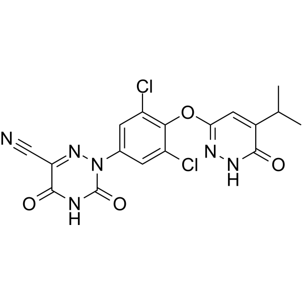
HY-12216
ResmetiromTHR-β Agonist -
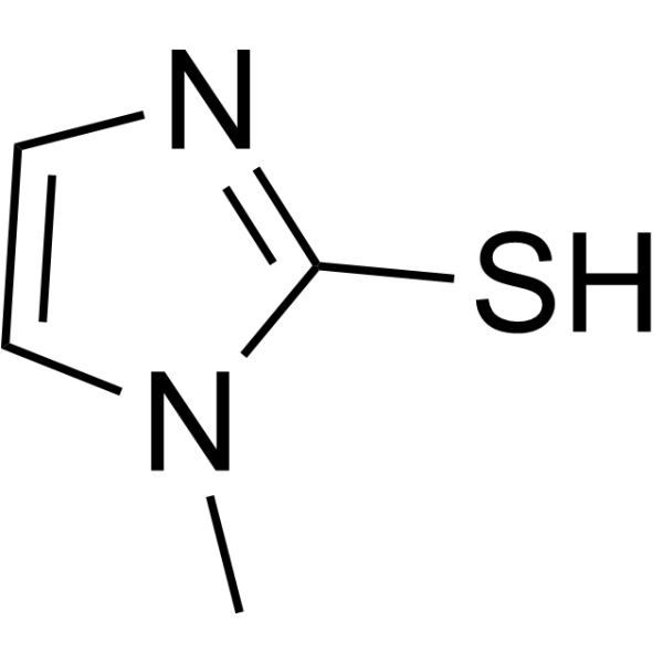
HY-B0208
MethimazoleAnti-thyroid Agent -
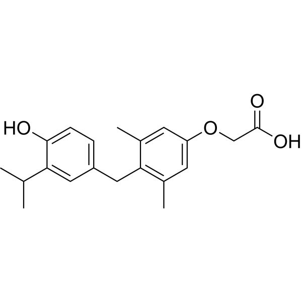
HY-14823
SobetiromeTRβ Agonist -
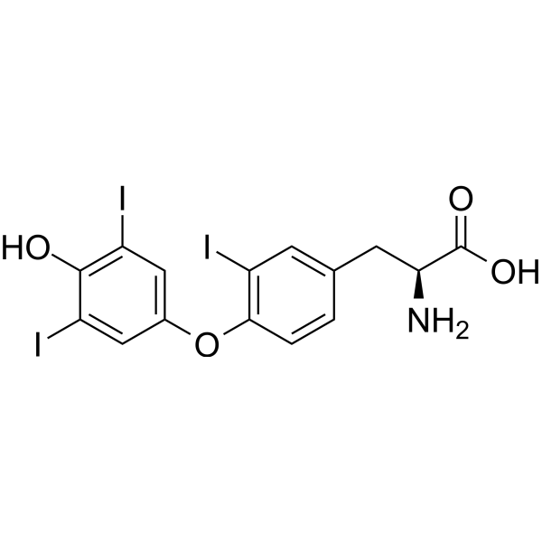
HY-W010696
Reverse T3Thyroid Hormone -
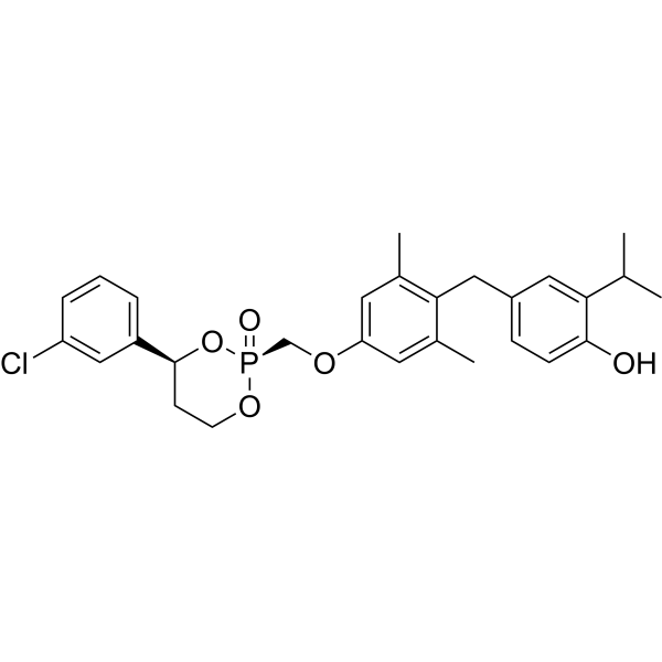
HY-111288
MB-07811 -
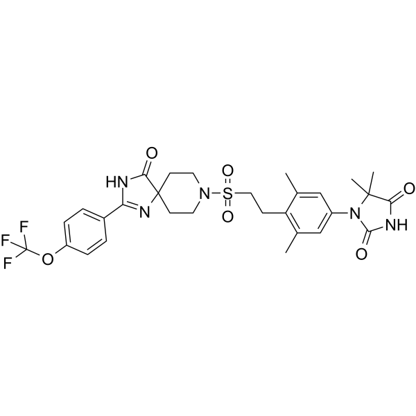
HY-100856
PCO371PTHR1 Agonist -
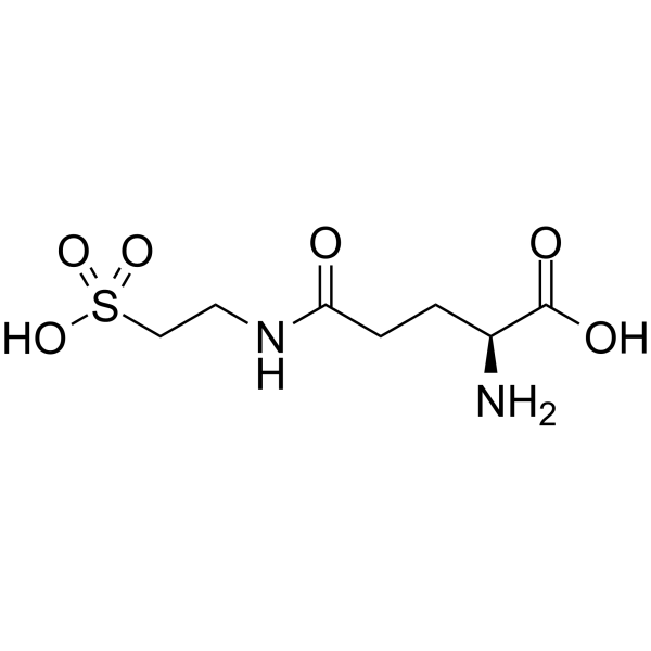
HY-106608
GlutaurineParathyroid Hormone -
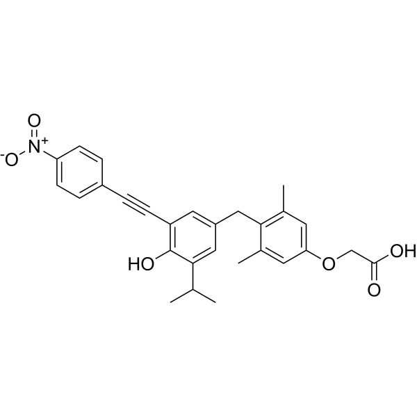
HY-141513
NH-3THR Antagonist -
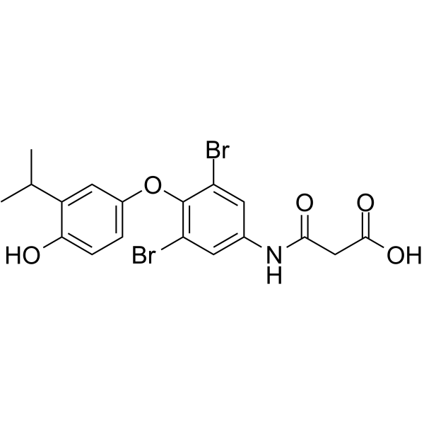
HY-10473
Eprotirome -
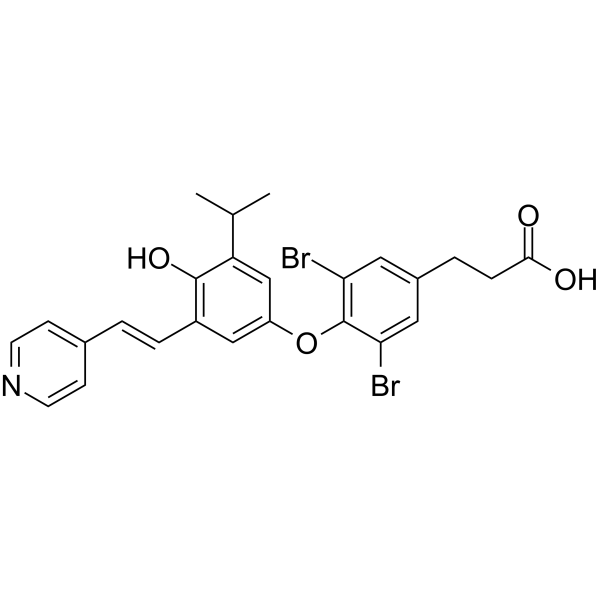
HY-111443
TR antagonist 1Thyroid Hormone Receptor Antagonist -

HY-W010696R
Reverse T3 (Standard)Thyroid Hormone -
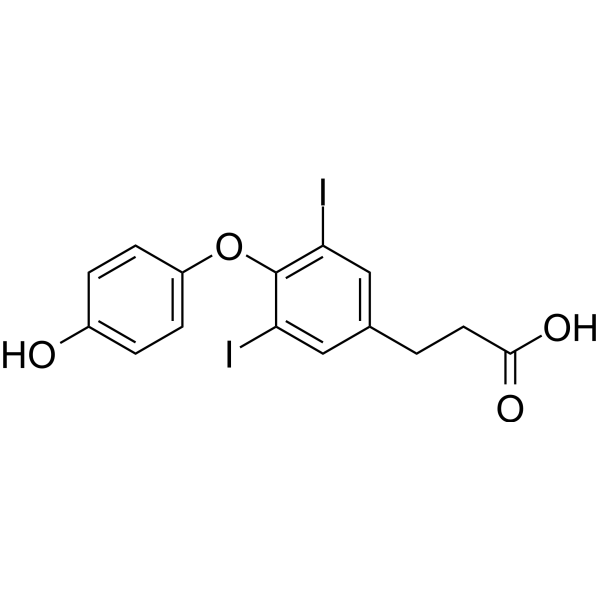
HY-126236
3,5-Diiodothyropropionic acidThyroid Hormone Analog
All Caspase Isoforms
More

Biological Activity
Description
|
PTH1R |
PKA |
cAMP |
PKC |
PI3K |
Akt |
ERK1 |
ERK2 |
TGF-beta1 |
p47phox |
eNOS |
Caspase 3 |
In Vitro
pTH-Related Protein (1-34) (human,mouse,rat) (PTHrP (1-34)) (100 nM; 3-72 h) induces fibronectin upregulation in rat MCs in a manner independent of the TGF-β1/Smad signaling pathway[1].
pTH-Related Protein (1-34) (human,mouse,rat) (100 nM; 5 min-12 h) induced Src-dependent EGFR transactivation at Y845 and subsequent Akt phosphorylation at S473 in rat MCs[1].
pTH-Related Protein (1-34) (human, mouse, rat) (100 nM; 0.5-48 h) triggers ERK1/2 activation through Src- and EGFR-dependent, PI3K-dependent mechanisms, and Akt and MEK/ERK1/2 execute parallel signaling pathways in rat MCs[1].
pTH-Related Protein (1-34) (human,mouse,rat) (1 μM; 10 min) had no effect on cAMP levels in primary rat alveolar type II epithelial cells, but increased inositol phosphate levels by 35%, suggesting that its signaling is mediated through a phospholipase C-dependent mechanism rather than a cAMP-dependent pathway[3].
pTH-Related Protein (1-34) (human,mouse,rat) (PTHrP-(1-34)) stimulates osteoclastic bone resorption in long-term 沉降 rat osteoclast cultures containing contaminating osteoblasts, with a potency comparable to that of PTH-(1-34), and its biological activity resides within the first 34 amino acids[4].
pTH-Related Protein (1-34) (human, mouse, rat) (24 h) can stimulate osteoclastic bone resorption in the rat osteoclast/UMR-106 osteoblast-like cell co-culture system, confirming that osteoblasts mediate this resorptive response[4].
pTH-Related Protein (1-34) (human,mouse,rat) (100 nM; 5 min) induces NOX-dependent ROS generation in rat MCs[1].
pTH-Related Protein (1-34) (human,mouse,rat) (100 nM) induced sustained Src activation and EGFR transactivation in rat MCs, independent of MMPs-mediated HB-EGF cleavage and release[1].
pTH-Related Protein (1-34) (human,mouse,rat) (100 nM; 24-48 h)-induced fibronectin upregulation depends on NOX-derived ROS, Src kinase, and downstream EGFR/PI3K/Akt and MEK/ERK1/2 signaling pathways in rat MCs[1].
pTH-Related Protein (1-34) (human,mouse,rat) (PTHrP (1-34)) (1.6-100 ng/mL; 24 h) dose-dependently stimulates the proliferation of quiescent MCF-7 cells, but has no mitogenic effect on Hs578T cells[2].
pTH-Related Protein (1-34) (human,mouse,rat) (PTHrP (1-34)) (100 ng/mL; 20-4 h) increases BrdU incorporation in quiescent MCF-7 cells, confirming its mitogenic effect[2].
pTH-Related Protein (1-34) (human,mouse,rat) (10-100 ng/mL; 20 min) significantly increases intracellular cAMP in MCF-7 cells, but not in Hs578T cells[2].
pTH-Related Protein (1-34) (human,mouse,rat) (pTHrP (1-34)) (1 μM; 24 h pre-treatment, maintained during irradiation and 24 h recovery) enhances ultraviolet-induced apoptosis in primary rat alveolar type II epithelial cells, manifested as increased nuclear condensation (15.6% in irradiated untreated cells versus 25.8% in treated cells) and increased cell loss, while it does not induce significant apoptosis by itself in unirradiated cells[3].
pTH-Related Protein (1-34) (human,mouse,rat) (1 μM; 24 h pretreatment, maintained during irradiation and 24 h recovery period) pretreatment enhanced UV-induced caspase 3 activity in primary rat alveolar type II epithelial cells[3].
PTHrP 1-34 induces strong vasodilation in the main nutrient artery of the rat femur, reaching 69% of maximum dilation[6].
The vasodilatory effect of PTHrP 1-34 in the main nutrient artery of the rat femur is mainly mediated through the PKA signaling pathway and requires activation of the PTH1 receptor[6].
MedChemExpress (MCE) has not independently confirmed the accuracy of these methods. They are for reference only. Further protocols information, click here.
-
Cell Line:Primary rat mesangial cells (MCs)
-
Concentration:100 nM
-
Incubation Time:3 h; 6 h; 12 h; 24 h; 48 h; 72 h
-
Result:Increased TGF-β1 mRNA and protein expression after 24 h.
Induced TβRII protein expression at 72 h.
Markedly increased fibronectin protein expression from 12 h to 48 h.
Did not affect Smad2/3 phosphorylation until 72 h.
-
Cell Line:Primary rat mesangial cells (MCs)
-
Concentration:100 nM
-
Incubation Time:5 min, 10 min, 0.5 h, 1 h, 3 h, 6 h, and 12 h
-
Result:Induced sustained EGFR transactivation via phosphorylation at Y845, but not Y1173.
Significantly increased Akt phosphorylation at S473 at 10 min.
Increased Src phosphorylation at Y416 at 5 min, lasting until 3 h.
-
Cell Line:Primary rat alveolar type II epithelial cells
-
Concentration:1 μM
-
Incubation Time:24 h pre-treatment, maintained during irradiation and 24 h recovery
-
Result:Decreased cell densities by 15% in irradiated cells.
Increased the percentage of ethidium bromide-positive cells to 2.7% in nonirradiated cells compared with 0.8% in untreated cells.
Increased the percentage of ethidium bromide-positive cells to 5.0% in irradiated cells compared with 3.4% in irradiated untreated cells.
Increased the percentage of cells with condensed or fragmented nuclei to 25.8% in ultraviolet-irradiated cells compared with 15.6% in irradiated untreated cells.\nIncreased caspase 3 activity to 8.4 RFU/min/μg protein in irradiated cells compared with 5.4 RFU/min/μg protein in irradiated untreated cells.
Augmented caspase activity to 41.3 RFU/min/μg protein in irradiated cells compared with 27.8 RFU/min/μg protein in irradiated untreated cells, representing a roughly 50% increase (149% of ultraviolet/no treatment group).\n
Increased caspase 3 activity to 14.2 RFU/min/μg protein in irradiated cells compared with 11.1 RFU/min/μg protein in irradiated untreated cells, representing a 28% increase.\n
Reduced caspase 3 activity in ultraviolet-exposed cells to 40% of the activity in nonirradiated untreated control cells.
In Vivo
MedChemExpress (MCE) has not independently confirmed the accuracy of these methods. They are for reference only.
-
Animal Model:Sprague-Dawley (male, 200-225 g, streptozotocin-induced diabetic model)[1]
-
Dosage:40, 80, 160 μg/kg
-
Administration:s.c.; once daily for 5 days per week; 3 months
-
Result:Aggravated renal hypertrophy with kidney/body weight ratios of 5.8 mg/g at 40 μg/kg, 6.0 mg/g at 80 μg/kg, and 6.4 mg/g at 160 μg/kg versus 5.4 mg/g in diabetic controls.
Increased fibronectin staining and protein levels in the 160 μg/kg group compared to diabetic rats.
Elevated p47phox protein levels in the 160 μg/kg group compared to diabetic rats.
Increased phosphorylation of EGFR Y845, Akt S473, and ERK1/2 in the 160 μg/kg group.


Chemical Information
-
CAS No. 112540-82-6
-
Appearance Solid
-
Molecular Weight 4018.00
-
Formula C180H287N57O48
-
Color White to off-white
-
Synonyms
pTHrP (1-34)
-
Sequence
Ala-Val-Ser-Glu-His-Gln-Leu-Leu-His-Asp-Lys-Gly-Lys-Ser-Ile-Gln-Asp-Leu-Arg-Arg-Arg-Phe-Phe-Leu-His-His-Leu-Ile-Ala-Glu-Ile-His-Thr-Ala
-
Sequence Shortening
AVSEHQLLHDKGKSIQDLRRRFFLHHLIAEIHTA
-
Shipping
Room temperature in continental US; may vary elsewhere.
-
Storage
Sealed storage, away from moisture
Powder -80°C 2 years -20°C 1 year * In solvent : -80°C, 6 months; -20°C, 1 month (sealed storage, away from moisture)


Solvent & Solubility
In Vitro:
H2O : ≥ 25 mg/mL (6.22 mM)
* "≥" means soluble, but saturation unknown.
Please refer to the solubility information to select the appropriate solvent. Once prepared, please aliquot and store the solution to prevent product inactivation from repeated freeze-thaw cycles.
Storage method and period of stock solution: -80°C, 6 months; -20°C, 1 month (sealed storage, away from moisture). When stored at -80°C, please use it within 6 months. When stored at -20°C, please use it within 1 month.
* Note: If you choose water as the stock solution, please dilute it to the working solution, then filter and sterilize it with a 0.22 μm filter before use.
Please refer to the solubility information to select the appropriate solvent. Once prepared, please aliquot and store the solution to prevent product inactivation from repeated freeze-thaw cycles.
Storage method and period of stock solution: -80°C, 6 months; -20°C, 1 month (sealed storage, away from moisture). When stored at -80°C, please use it within 6 months. When stored at -20°C, please use it within 1 month.
* Note: If you choose water as the stock solution, please dilute it to the working solution, then filter and sterilize it with a 0.22 μm filter before use.
Concentration (start) × Volume (start) = Concentration (final) × Volume (final)

Protocols
-
Western Blot
Western blotting (WB) is a commonly used experimental method in molecular biology, biochemistry, and immunogenetics for identifying and quantifying target proteins. It combines gel electrophoresis with immunoassay, enabling researchers to analyze protein expression, post-translational modifications, and molecular weight.
-
RNA extraction experimental
By lysing cells, releasing RNA, and removing impurities such as proteins and DNA, high-purity RNA products are finally obtained. The commonly used traditional method is the guanidine isothiocyanate/phenol/chloroform method (Trizol), which is suitable for a variety of animal materials including animal tissues, microorganisms, cultured cells, etc., and most plant materials.
-
Annexin V plus membrane-impermeant dye apoptosis staining
Annexin V-based apoptosis assays rely on the detection of phosphatidylserine (PS) externalization from the inner leaflet of the plasma membrane to the outer leaflet, an early biochemical hallmark of apoptosis. Fluorescently labeled Annexin V binds PS in a calcium-dependent manner, enabling identification of early apoptotic cells by flow cytometry or fluorescence microscopy. When combined with a membrane-impermeant DNA-binding dye (e. g. , propidium iodide), this approach allows discrimination between viable (Annexin V−/dye−), early apoptotic (Annexin V+/dye−), and late apoptotic or necrotic (Annexin V+/dye+) cell populations by assessing membrane integrity and PS exposure.
-
ROS/oxidative-stress fluorescent staining
ROS/oxidative-stress fluorescent staining uses cell-permeant fluorogenic probes that become fluorescent after oxidation inside cells or tissues; commonly used examples include DCFH-DA/DCFDA for broad cellular oxidant detection, DHE for superoxide-related signal detection, MitoSOX for mitochondrial superoxide-related signal detection, and CellROX probes for oxidative-stress-associated fluorescence readouts. The assay detects probe oxidation rather than a single ROS species unless the probe and analysis method have been chemically validated for that species. DCFH-DA enters cells, is deacetylated by intracellular esterases to DCFH, and produces fluorescent DCF after oxidation, so the readout is used as an operational measure of total cellular oxidative stress rather than a species-specific ROS measurement. DHE and MitoSOX can report superoxide-related oxidation, but red fluorescence alone can include non-specific ethidium-like oxidation products; HPLC or optimized spectral approaches are
-
Breast Cancer Modeling
Breast cancer is a heterogeneous cancer, and it has been distinguished into four subtypes: luminal A, luminal B, HER2-positive and basal-like. Molecular mutations, epigenetic alterations, hormone exposure and immune microenvironment are related to the progression of breast cancer.
-
Kinase activity and phosphorylation assays
Kinase activity assays measure the ability of kinases to transfer phosphate groups from ATP to specific substrates, while phosphorylation assays detect the presence and levels of phosphorylated proteins. Common methods include radiolabeled ATP incorporation (e. g. ,), ADP release detection via bioluminescence (e. g. ,[3]), enzyme-linked immunosorbent assays (ELISA) for phospho-specific epitopes (e. g. ,[6]), and microtiter-based formats for high-throughput screening (e. g. ,[8]). The ADP-Glo assay quantifies kinase activity by measuring ADP produced during phosphorylation using a luciferase-based system. Radiometric assays involve autoradiography or scintillation counting after incorporation of 32P-labeled ATP into substrate proteins. ELISA-based approaches rely on phospho-specific antibodies to detect activated kinases in cell lysates or purified samples.
-
Apoptosis
Apoptosis, also called programmed cell death, is generally characterized by distinct morphological characteristics.
-
TUNEL staining for apoptotic DNA fragmentation
TUNEL staining detects DNA strand breaks by using terminal deoxynucleotidyl transferase to add labeled nucleotides to exposed 3′-OH DNA termini, generating either microscopic staining in fixed cells or tissue sections, or fluorescence/cytometric signal in cell suspensions. TUNEL positivity reflects DNA fragmentation but should not be interpreted alone as definitive apoptosis, because TUNEL can also label necrotic, autolytic, mechanically damaged, or DNA-repair-associated DNA breaks.
-
Research Protocol for Endocrine Diseases
Endocrine diseases often arise from disrupted hormone production, hormone signaling, or target-tissue responsiveness; for diabetes-focused endocrine disease models, insulin signaling regulates glucose uptake, hepatic glucose output, lipid metabolism, and β-cell compensation. Type 2 diabetes develops through interacting defects in insulin resistance, β-cell dysfunction, adipose inflammation, hepatic glucose overproduction, altered incretin signaling, and ectopic lipid metabolism. A major unresolved question is whether endocrine dysfunction is driven primarily by target-tissue insulin resistance, intrinsic β-cell failure, immune/inflammatory stress, or combined multi-organ failure that differs by disease stage.
-
Apoptosis Solutions
Apoptosis is a regulated, generally non-lytic cell-death pathway that removes unwanted, damaged, infected, or abnormal cells through coordinated morphological changes, caspase activation, DNA fragmentation, and membrane remodeling. The intrinsic apoptosis pathway is controlled mainly by mitochondrial outer membrane permeabilization, BCL-2 family proteins, cytochrome c release, apoptosome formation, caspase-9 activation, and downstream executioner caspase-3/7 activation. The extrinsic apoptosis pathway is initiated by death receptors such as Fas, TNFR, and TRAIL receptors, which recruit adaptor proteins and activate caspase-8 before engaging executioner caspases or mitochondrial amplification through BID cleavage. Apoptosis is linked to many phenotypes, including cancer cell killing, tissue homeostasis, immune regulation, neurodegeneration, infection response, and treatment-induced cytotoxicity; unresolved questions include how apoptosis interacts with necroptosis, pyroptosis, ferroptos
-
Protocol for Kinase activity and phosphorylation assays
Kinase activity assays measure transfer of phosphate from ATP to a protein or peptide substrate, generating phosphorylated substrate, ADP, or incorporated radiolabeled phosphate as the readout; phosphorylation assays measure site-specific phosphorylation in cells or tissues as a proxy for kinase-pathway activation, inhibition, or substrate regulation. Phosphorylation can be detected by phospho-specific Western blot, immunoprecipitation kinase assay, phospho-immunofluorescence, phospho-flow cytometry, luminescent ADP detection, radiolabeled ATP incorporation, or reporter-based pathway assays, and these readouts can be applied to cancer cells, primary neurons, mouse tumors, organoids, inflammatory macrophages, ferroptosis studies, and mitophagy studies when the kinase target is biologically relevant.
-
Inhalation Toxicity Study
Inhalation toxicity studies expose rodents to a controlled aerosol, vapor, gas, or smoke atmosphere and assess respiratory and systemic toxicity using exposure-atmosphere characterization, clinical observations, body and organ weights, bronchoalveolar lavage fluid, histopathology, blood chemistry, hematology, and, when included, molecular endpoints such as transcriptomics, proteomics, lipidomics, or tissue burden analysis. The primary biological readouts are airway irritation, pulmonary inflammation, cytotoxicity, altered surfactant or lipid homeostasis, impaired particle clearance, and tissue remodeling, reflected by BALF cell differentials, BALF protein, LDH, phosphatase activities, cytokines, lung weight, microscopic respiratory-tract lesions, and retained lung burden.
-
Osteoclast differentiation from monocyte/macrophage precursors
Osteoclast differentiation is an in vitro induction assay in which monocyte/macrophage-lineage precursors are exposed to macrophage colony-stimulating factor (M-CSF) and receptor activator of NF-κB ligand (RANKL), generating multinucleated osteoclasts that are commonly identified by tartrate-resistant acid phosphatase (TRAP) staining and functionally confirmed by resorption pits on dentin, bone, or mineralized substrates. M-CSF supports survival and expansion of osteoclast precursors, while RANKL binding to RANK drives osteoclast commitment, fusion, maturation, and resorptive function; osteoprotegerin inhibits this pathway by binding RANKL and preventing RANK activation. The main readouts are the number of TRAP-positive multinucleated cells, formation of F-actin rings, and resorbed surface area; TRAP-positive multinucleated cells indicate osteoclast differentiation, whereas pit formation on dentin, bone, or mineralized coating indicates functional bone-resorbing activity.


Purity & Documentation
-
Data Sheet (308 KB)
-
SDS (252 KB)
- English - EN (252 KB)
- Français - FR (252 KB)
- Deutsch - DE (252 KB)
- Norwegian - NO (252 KB)
- Español - ES (252 KB)
- Swedish - SV (252 KB)
- Italian - IT (252 KB)
- Korean - KR (252 KB)
- Portuguese - PT (252 KB)
-
Handling Instructions (2659 KB)
References
[1]. Chen HM, et al. Parathyroid hormone-related protein induces fibronectin up-regulation in rat mesangial cells through reactive oxygen species/Src/EGFR signaling. Bioscience reports. 2019 Apr 30;39(4):BSR20182293. [Content Brief]
[5]. Frolik CA, et al. Comparison of recombinant human PTH(1-34) (LY333334) with a C-terminally substituted analog of human PTH-related protein(1-34) (RS-66271): In vitro activity and in vivo pharmacological effects in rats. Journal of bone and mineral research : the official journal of the American Society for Bone and Mineral Research. 1999 Feb;14(2):163-72. [Content Brief]


Complete Stock Solution Preparation Table
Please refer to the solubility information to select the appropriate solvent. Once prepared, please aliquot and store the solution to prevent product inactivation from repeated freeze-thaw cycles.
Storage method and period of stock solution: -80°C, 6 months; -20°C, 1 month (sealed storage, away from moisture). When stored at -80°C, please use it within 6 months. When stored at -20°C, please use it within 1 month.
| Optional Solvent | Concentration Solvent Mass | 1 mg | 5 mg | 10 mg | 25 mg |
|---|---|---|---|---|---|
| H2O | 1 mM | 0.2489 mL | 1.2444 mL | 2.4888 mL | 6.2220 mL |
| 5 mM | 0.0498 mL | 0.2489 mL | 0.4978 mL | 1.2444 mL |
* Note: If you choose water as the stock solution, please dilute it to the working solution, then filter and sterilize it with a 0.22 μm filter before use.


pTH-Related Protein (1-34) (human,mouse,rat) Related Classifications
HY-P3075 Related Classifications
 Powered by Bioz