



Alisol B 23-acetate
Based on 4 publication(s) in Google Scholar
Alisol B 23-acetate is an orally active prototerpane-type triterpenoid. Alisol B 23-acetate can be isolated from Alisma orientalis. Alisol B 23-acetate induces Apoptosis, promotes ROS generation, downregulates CDK4/6, MMP-2/9, upregulates cleaved PARP, activates FXR and inhibits Syk. Alisol B 23-acetate has anti-inflammatory and hepatoprotective activities. Alisol B 23-acetate protects the kidney from ischemia-reperfusion injury. Alisol B 23-acetate has anticancer activity against ovarian cancer, colon cancer, lung cancer, and gastric cancer. Alisol B 23-acetate can be used in the study of atherosclerosis and allergic asthma.
For research use only. We do not sell to patients.
- Purity : 99.65%
- CAS No.: 26575-95-1
- Formula: C32H50O5
- Molecular Weight:514.74
-
Storage:Powder -20°C, 3 years , 4°C, 2 years ; In solvent -80°C, 2 years , -20°C, 1 year

To place orders, for customer services and technical support, please contact: MedChemExpress USA
Tel: 609-228-6898 E-mail: [email protected] [email protected]


-
 Biological Activity
-
Chemical Information
-
Solvent & Solubility
- Protocols
- Purity & Documentation
- References
-
 Help & FAQs


-
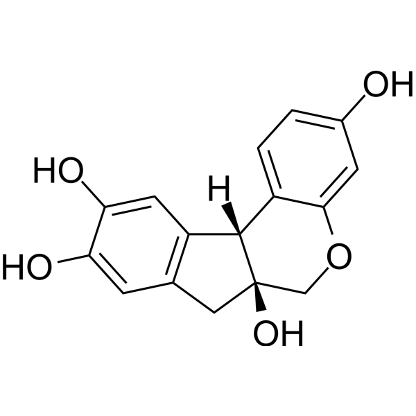
HY-N0072
BrazilinAutophagy Inducer -

HY-N0805R
Alisol B 23-acetate (Standard)Standard Of Alisol B 23-acetate -
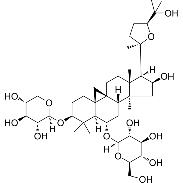
HY-N0431
Astragaloside IVMMP Inhibitor -
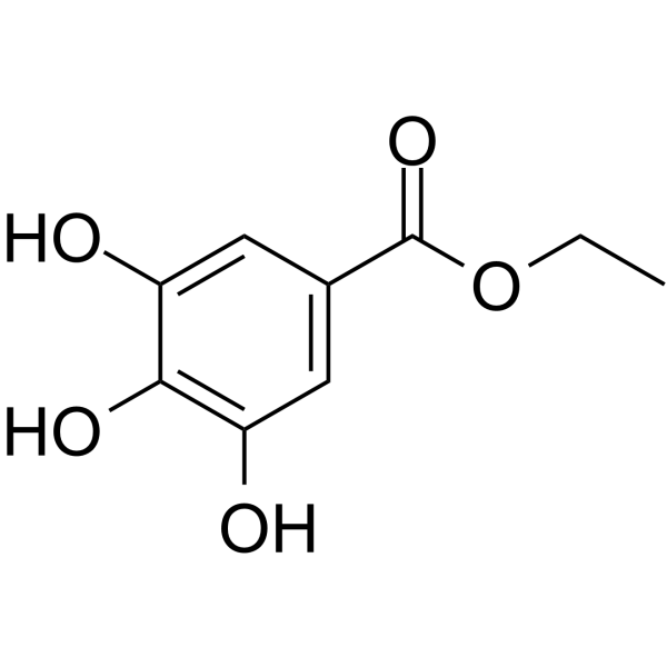
HY-N0525
Ethyl gallateBacterial Inhibitor -
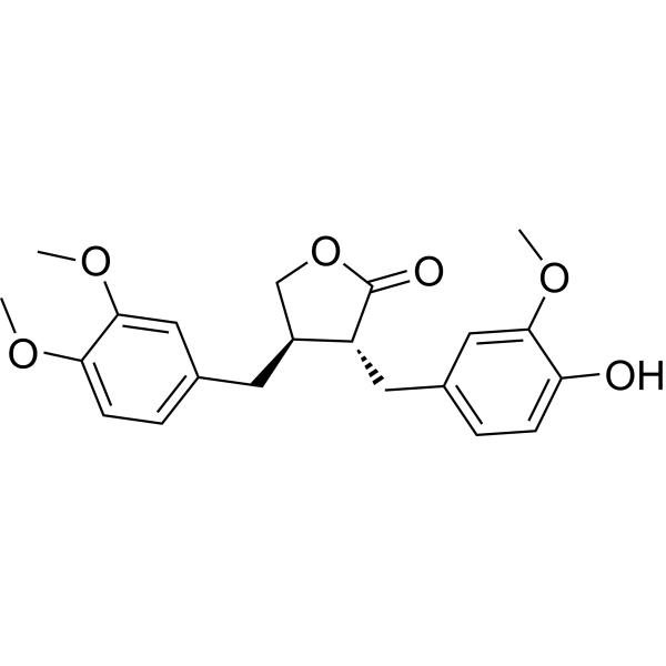
HY-N0035
ArctigeninAntitumor Agent -

HY-N0431R
Astragaloside IV (Standard)MMP Inhibitor -

HY-N0035R
Arctigenin (Standard)Antitumor Agent -

HY-N0525R
Ethyl gallate (Standard)Bacterial Inhibitor
Publications Citing Use of MedChemExpress (MCE) Alisol B 23-acetate
More

Biological Activity
Description
|
CDK4 |
CDK6 |
MMP-2 |
MMP-9 |
Cellular Effect
|
Cell Line

|
Type | Value | Description | References |
|---|---|---|---|---|
| Erythrocyte | IC50 |
12.8 μM
Compound: 57b
|
Antiplasmodial activity against Plasmodium falciparum K1 infected in human erythrocyte assessed as inhibition of parasite growth incubated for 72 hrs by [3H]-hypoxanthine incorporation based liquid scintillation counting method
Antiplasmodial activity against Plasmodium falciparum K1 infected in human erythrocyte assessed as inhibition of parasite growth incubated for 72 hrs by [3H]-hypoxanthine incorporation based liquid scintillation counting method
|
[PMID: 35985254] |
| HepG2 | EC50 |
3.57 μM
Compound: 26
|
Transactivation of FXR (unknown origin) transfected in HepG2 cells co-expressing pBSEP/pGL4.74 incubated for 24 hrs by luciferase reporter gene assay
Transactivation of FXR (unknown origin) transfected in HepG2 cells co-expressing pBSEP/pGL4.74 incubated for 24 hrs by luciferase reporter gene assay
|
[PMID: 31494470] |
In Vitro
Alisol B 23-acetate (2.5-20 µM; 24-48 h) significantly inhibits cell viability, induces G1 phase cell cycle arrest and apoptosis, suppresses migration and invasion in ovarian cancer cell lines A2780, A2780/Taxol, and HEY[1].
Alisol B 23-acetate (1-100 µM) reverses P-glycoprotein (P-gp)-mediated multidrug resistance in HepG2-DR and K562-DR cells[2].
Alisol B 23-acetate (5-20 µM; 6-24 h) induces autophagic-dependent apoptosis in human colon cancer cells (HCT116 and SW620) via ROS generation and JNK activation[3].
Alisol B 23-acetate (20-80 µM; 24 h) improves free fatty acid (FFA)-induced lipid metabolism disorders in L02 hepatocytes[5].
Alisol B 23-acetate (5-20 μM; 30 min before antigen challenge) inhibits IgE/antigen-mediated β-hexosaminidase release in RBL-2H3 mast cells in a concentration-dependent manner[14].
MedChemExpress (MCE) has not independently confirmed the accuracy of these methods. They are for reference only.
-
Cell Line:A2780, A2780/Taxol, HEY
-
Concentration:5, 6, 9, 10, 12, 15 µM
-
Incubation Time:24 h
-
Result:Downregulated protein levels of CDK4, CDK6, cyclin D1, MMP-2, MMP-9.
Upregulated Bax/Bcl-2 ratio and cleaved PARP expression.
In Vivo
Alisol B 23-acetate (50-100 mg/kg; p.o.) inhibits IgE-mediated vascular permeability and ear swelling in ICR mouse passive cutaneous anaphylaxis (PCA) model[7].
Alisol B 23-acetate (15-30 mg/kg; p.o.; daily; 9 weeks) reduces serum TG, IL-12, IFN-γ, increases HDL-C, and promotes aortic cholesterol efflux gene expression in ApoE-/- atherosclerotic mice[8].
Alisol B 23-acetate (10-40 mg/kg; p.o.; 30 days) suppresses TLR4/NOX2 pathway, reducing myocardial inflammation and ROS production in LPS-induced cardiac dysfunction mice[9].
Alisol B 23-acetate (15-60 mg/kg; p.o.; daily; 4 weeks) ameliorates hepatic steatosis, inflammation, and fibrosis, and activates FXR target genes in MCD-induced NASH mice[10].
Alisol B 23-acetate (12.5-50 mg/kg; p.o.; once daily; 7 days) promotes liver regeneration in mice after partial hepatectomy via activating farnesoid X receptor[11].
Alisol B 23-acetate (10-40 mg/kg; p.o.; once daily; 7 days) alleviates hepatotoxicity in a dose-dependent manner in CCl4 (HY-Y0298)-induced liver injury mice[12].
Alisol B 23-acetate (60 mg/kg; i.p.; 30 min before sensitization/challenge) reduces airway hyperresponsiveness and decreases IL-13 and eosinophils in BALF in OVA-induced allergic asthma BALB/c mice[14].
MedChemExpress (MCE) has not independently confirmed the accuracy of these methods. They are for reference only.
-
Animal Model:Adult male C57BL/6 mice (6-8 weeks old), LPS-induced sepsis model[9].
-
Dosage:10 mg/kg, 20 mg/kg, 40 mg/kg
-
Administration:Gavage (p.o.), 30 days
-
Result:Significantly improved the 48-hour survival rate of LPS-induced septic mice (80% in the 40 mg/kg group vs. 40% in the LPS group).
Restored left ventricular ejection fraction (LVEF), left ventricular fraction shortening (LVFS), and reduced left ventricular end-systolic diameter (LVESD) in LPS-treated mice.
Reduced inflammatory cell infiltration in myocardial tissue.
Decreased serum levels of TNF-α, IL-6, and IL-1β.
Downregulated TLR4, NOX2, p-P38, and p-ERK protein expression in myocardial tissue.
-
Animal Model:Male C57BL/6 mice (8-9 weeks old) with CCl₄-induced acute hepatotoxicity[12]
-
Dosage:10 mg/kg, 20 mg/kg, 40 mg/kg
-
Administration:Oral gavage; once daily; for 7 days
-
Result:Significantly reduced serum ALT and AST activities, decreased hepatic and serum total bile acid levels, ameliorated liver histopathological changes (e.g., congestion, vacuolization, necrosis), reduced TUNEL-positive apoptotic hepatocytes.
Increased BrdU-positive proliferating hepatocytes and mitotic index, upregulated FXR target genes (FoxM1b, Cyclin D1, Cyclin B1, Bsep, Mrp2, Shp), and downregulated Ntcp, Cyp7a1, and Cyp8b1 expression.
Enhanced STAT3 phosphorylation and upregulated anti-apoptotic genes (Bcl-xl, SOCS3).
- Detection of Apoptosis
- TUNEL staining for apoptotic DNA fragmentation
- Mitochondrial membrane-potential fluorescent assay
- Annexin V plus membrane-impermeant dye apoptosis staining
- ROS/oxidative-stress fluorescent staining
- Ovalbumin-Induced Allergic Airway Inflammation
- Mitochondrial membrane-potential and mitochondrial mass staining
- Apoptosis Solutions
- Research Protocol for Cardiovascular Diseases
- Research Protocol for Inflammation-related Diseases


Chemical Information
-
CAS No. 26575-95-1
-
Appearance Solid
-
Molecular Weight 514.74
-
Formula C32H50O5
-
Color White to off-white
-
SMILES
C[C@]([C@@]1(C2=C([C@H](C)C[C@@H]([C@]3([H])C(C)(C)O3)OC(C)=O)CC1)C)(CC[C@@]4([H])C5(C)C)[C@]([C@H](C2)O)([H])[C@]4(CCC5=O)C
-
Synonyms
23-Acetylalismol B; 23-O-Acetylalisol B; Alisol B monoacetate
-
Structure Classification
-
Initial Source
-
Shipping
Room temperature in continental US; may vary elsewhere.
-
Storage
Powder -20°C 3 years 4°C 2 years In solvent -80°C 2 years -20°C 1 year


Publications (4)
-
Journal Impact Factor
-
Most Recent
-
-
CNS Neurosci Ther
ShenQi DiHuang Decoction (SQDHD) Ameliorates Neuroinflammation and Neuropsychiatric Manifestations in Pristane Induced Lupus Mice via Blocking JAK1-STAT3 Pathway. [Abstract]2026 Mar;32(3):e70814. PMID: 41795136 -
J Integr Med
Alisol B 23-acetate promotes white adipose tissue browning to mitigate high-fat diet-induced obesity by regulating mTOR-SREBP1 signaling. [Abstract]2024 Jan;22(1):83-92. PMID: 38311542 -


Solvent & Solubility
In Vitro:
DMSO : 50 mg/mL (97.14 mM; Need ultrasonic; Hygroscopic DMSO has a significant impact on the solubility of product, please use newly opened DMSO)
Please refer to the solubility information to select the appropriate solvent. Once prepared, please aliquot and store the solution to prevent product inactivation from repeated freeze-thaw cycles.
Storage method and period of stock solution: -80°C, 2 years; -20°C, 1 year. When stored at -80°C, please use it within 2 years. When stored at -20°C, please use it within 1 year.
Please refer to the solubility information to select the appropriate solvent. Once prepared, please aliquot and store the solution to prevent product inactivation from repeated freeze-thaw cycles.
Storage method and period of stock solution: -80°C, 2 years; -20°C, 1 year. When stored at -80°C, please use it within 2 years. When stored at -20°C, please use it within 1 year.
Concentration (start) × Volume (start) = Concentration (final) × Volume (final)
In Vivo:
Select the appropriate dissolution method based on your experimental animal and administration route.
- For the following dissolution methods, please ensure to first prepare a clear stock solution using an In Vitro approach and then sequentially add co-solvents:
- To ensure reliable experimental results, the clarified stock solution can be appropriately stored based on storage conditions. As for the working solution for In Vivo experiments, it is recommended to prepare freshly and use it on the same day.
- The percentages shown for the solvents indicate their volumetric ratio in the final prepared solution. If precipitation or phase separation occurs during preparation, heat and/or sonication can be used to aid dissolution.
Add each solvent one by one: 10% DMSO 40% PEG300 5% Tween-80 45% Saline
Solubility: ≥ 2.5 mg/mL (4.86 mM); Clear solution
This protocol yields a clear solution of ≥ 2.5 mg/mL (saturation unknown).
Taking 1 mL working solution as an example, add 100 μL DMSO stock solution (25.0 mg/mL) to 400 μL PEG300, and mix evenly; then add 50 μL Tween-80 and mix evenly; then add 450 μL Saline to adjust the volume to 1 mL.
Preparation of Saline: Dissolve 0.9 g sodium chloride in ddH₂O and dilute to 100 mL to obtain a clear Saline solution.
Add each solvent one by one: 10% DMSO 90% (20% SBE-β-CD in Saline)
Solubility: ≥ 2.5 mg/mL (4.86 mM); Clear solution
This protocol yields a clear solution of ≥ 2.5 mg/mL (saturation unknown).
Taking 1 mL working solution as an example, add 100 μL DMSO stock solution (25.0 mg/mL) to 900 μL 20% SBE-β-CD in Saline, and mix evenly.
Preparation of 20% SBE-β-CD in Saline (4°C, storage for one week): 2 g SBE-β-CD powder is dissolved in 10 mL Saline, completely dissolve until clear.
In Vivo Dissolution Calculator
Please enter the basic information of animal experiments:
-
-
-
-
Recommended: Prepare an additional quantity of animals to account for potential losses during experiments.
Please enter your animal formula composition:
-
%DMSO +
Recommended: Keep the proportion of DMSO in working solution below 2% if your animal is weak.
-
%+
-
+%Tween-80 + +
-
%Saline +
The co-solvents required include: DMSO, . All of co-solvents are available by MedChemExpress (MCE). , Tween 80. All of co-solvents are available by MedChemExpress (MCE).
Working solution concentration: 0.22 mg/mL
Method for preparing stock solution: mg drug dissolved in μL DMSO. Stock solution concentration: mg/mL.
1. Take μL DMSO stock solution;
2. Add μL .
μL , mix evenly;
3. Then add μL Tween 80, mix evenly;
4. Then add μL
Please ensure that the stock solution in the first step is dissolved to a clear state, and add co-solvents in sequence. You can use ultrasonic heating (ultrasonic cleaner, recommended frequency 20-40 kHz), vortexing, etc. to assist dissolution.


Purity & Documentation
-
Data Sheet (289 KB)
-
SDS (393 KB)
- English - EN (393 KB)
- Français - FR (393 KB)
- Deutsch - DE (393 KB)
- Norwegian - NO (393 KB)
- Español - ES (393 KB)
- Swedish - SV (393 KB)
- Italian - IT (393 KB)
- Korean - KR (393 KB)
- Portuguese - PT (393 KB)
-
Handling Instructions (2659 KB)
References
[1]. Zhang LL, et al. Effects of alisol B 23-acetate on ovarian cancer cells: G1 phase cell cycle arrest, apoptosis, migration and invasion inhibition. Phytomedicine. 2016 Jul 15;23(8):800-9. [Content Brief]
[2]. Wang C, et al. Reversal of P-glycoprotein-mediated multidrug resistance by Alisol B 23-acetate. Biochem Pharmacol. 2004 Sep 1;68(5):843-55. [Content Brief]
[3]. Zhao Y, et al.Alisol B 23-acetate induces autophagic-dependent apoptosis in human colon cancer cells via ROS generation and JNK activation. Oncotarget. 2017 Jul 26;8(41):70239-70249. [Content Brief]
[4]. Wang J, et al. Alisol B-23-acetate, a tetracyclic triterpenoid isolated from Alisma orientale, induces apoptosis in human lung cancer cells via the mitochondrial pathway. Biochem Biophys Res Commun. 2018 Nov 10;505(4):1015-1021. [Content Brief]
[5]. Fu Y, et al. Alisol B 23-acetate adjusts bile acid metabolisim via hepatic FXR-BSEP signaling activation to alleviate atherosclerosis. Phytomedicine. 2022 Jul;101:154120. [Content Brief]
[6]. Kwon MJ, et al. Apoptotic effects of alisol B 23‑acetate on gastric cancer cells. Mol Med Rep. 2021 Apr;23(4):248. [Content Brief]
[7]. Shao C, et al. Alisol B 23-Acetate Inhibits IgE/Ag-Mediated Mast Cell Activation and Allergic Reaction. Int J Mol Sci. 2018 Dec 18;19(12):4092. [Content Brief]
[8]. Sun Y, et al. Alisol B 23-acetate, a new promoter for cholesterol efflux from dendritic cells, alleviates dyslipidemia and inflammation in advanced atherosclerotic mice. Int Immunopharmacol. 2021 Oct;99:107956. [Content Brief]
[9]. Wang B, et al. Alisol B 23-Acetate Ameliorates Lipopolysaccharide-Induced Cardiac Dysfunction by Suppressing Toll-Like Receptor 4 (TLR4)/NADPH Oxidase 2 (NOX2) Signaling Pathway. Med Sci Monit. 2019 Nov 10;25:8472-8481. [Content Brief]
[10]. Meng Q, et al. Alisol B 23-acetate protects against non-alcoholic steatohepatitis in mice via farnesoid X receptor activation. Acta Pharmacol Sin. 2017 Jan;38(1):69-79. [Content Brief]
[11]. Meng Q, et al. Alisol B 23-acetate promotes liver regeneration in mice after partial hepatectomy via activating farnesoid X receptor. Biochem Pharmacol. 2014 Nov 15;92(2):289-98. [Content Brief]
[12]. Meng Q, et al. Protective effects of alisol B 23-acetate from edible botanical Rhizoma alismatis against carbon tetrachloride-induced hepatotoxicity in mice. Food Funct. 2015 Apr;6(4):1241-50. [Content Brief]
[13]. Luan ZL, et al. A naturally occurring FXR agonist, alisol B 23-acetate, protects against renal ischemia-reperfusion injury. Am J Physiol Renal Physiol. 2021 Nov 1;321(5):F617-F628. [Content Brief]
[14]. Nam KH, et al. Alisol B 23-Acetate Ameliorates Ovalbumin-Induced Allergic Asthma during Sensitization and Challenge Periods. Biomol Ther (Seoul). 2023 Nov 1;31(6):611-618. [Content Brief]


Complete Stock Solution Preparation Table
Please refer to the solubility information to select the appropriate solvent. Once prepared, please aliquot and store the solution to prevent product inactivation from repeated freeze-thaw cycles.
Storage method and period of stock solution: -80°C, 2 years; -20°C, 1 year. When stored at -80°C, please use it within 2 years. When stored at -20°C, please use it within 1 year.
| Optional Solvent | Concentration Solvent Mass | 1 mg | 5 mg | 10 mg | 25 mg |
|---|---|---|---|---|---|
| DMSO | 1 mM | 1.9427 mL | 9.7136 mL | 19.4273 mL | 48.5682 mL |
| 5 mM | 0.3885 mL | 1.9427 mL | 3.8855 mL | 9.7136 mL | |
| 10 mM | 0.1943 mL | 0.9714 mL | 1.9427 mL | 4.8568 mL | |
| 15 mM | 0.1295 mL | 0.6476 mL | 1.2952 mL | 3.2379 mL | |
| 20 mM | 0.0971 mL | 0.4857 mL | 0.9714 mL | 2.4284 mL | |
| 25 mM | 0.0777 mL | 0.3885 mL | 0.7771 mL | 1.9427 mL | |
| 30 mM | 0.0648 mL | 0.3238 mL | 0.6476 mL | 1.6189 mL | |
| 40 mM | 0.0486 mL | 0.2428 mL | 0.4857 mL | 1.2142 mL | |
| 50 mM | 0.0389 mL | 0.1943 mL | 0.3885 mL | 0.9714 mL | |
| 60 mM | 0.0324 mL | 0.1619 mL | 0.3238 mL | 0.8095 mL | |
| 80 mM | 0.0243 mL | 0.1214 mL | 0.2428 mL | 0.6071 mL |


Alisol B 23-acetate Related Classifications
HY-N0805 Related Classifications
 Powered by Bioz