



3′,4′,7-Trihydroxyflavone
Based on 1 publication(s) in Google Scholar
3′,4′,7-Trihydroxyflavone is an orally active inhibitor of OXA-48 (IC50 = 1.89 μM) and COX-1 (IC50 = 36.37 μM). 3′,4′,7-Trihydroxyflavone exhibits antioxidant and anti-inflammatory properties, inhibiting the release of inflammatory cytokines such as IL-6, IL-8, and TNF-α. 3′,4′,7-Trihydroxyflavone inhibits H2O2-induced neuronal apoptosis and ROS accumulation, and exerts anti-neuroinflammatory effects by suppressing the JNK-STAT1 pathway. 3′,4′,7-Trihydroxyflavone exhibits antimicrobial and antibiotic-modifying activities against multidrug-resistant Gram-negative enteric bacteria. 3′,4′,7-Trihydroxyflavone inhibits RANKL-induced osteoclast formation via NFATc1. 3′,4′,7-Trihydroxyflavone activates the CREB-BDNF axis and restores scopolamine (HY-N0296)-induced memory deficits in mice.
For research use only. We do not sell to patients.
- Purity : 99.04%
- CAS No.: 2150-11-0
- Formula: C15H10O5
- Molecular Weight:270.24
-
Storage:
RT, protect from light.
In solvent -80°C, 1 year , -20°C, 6 months

To place orders, for customer services and technical support, please contact: MedChemExpress USA
Tel: 609-228-6898 E-mail: [email protected] [email protected]


-
 Biological Activity
-
Chemical Information
-
Solvent & Solubility
- Protocols
- Purity & Documentation
- References
-
 Help & FAQs


-
HY-14520
Tetrahydrofolic acidFolate Derivative -
HY-N0911
Rehmannioside ANeuroprotectant -
HY-113327
1,3,7-Trimethyluric acidCaffeine Metabolite -
HY-66008
N-Acetyl mesalazine5-Aminosalicylic Acid Metabolite -

HY-Z7721
(Rac)-Beraprost sodiumRacemic Isomer of Beraprost sodium -
HY-N7108
7-HydroxyflavoneAnti-inflammatory Agent -
HY-103694
NebularineAntibiotic -
HY-N2609
7,4'-DihydroxyflavoneCCL11/CBR1 Inhibitor -
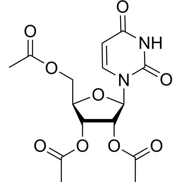
HY-14905
Uridine triacetateUridine Prodrug -
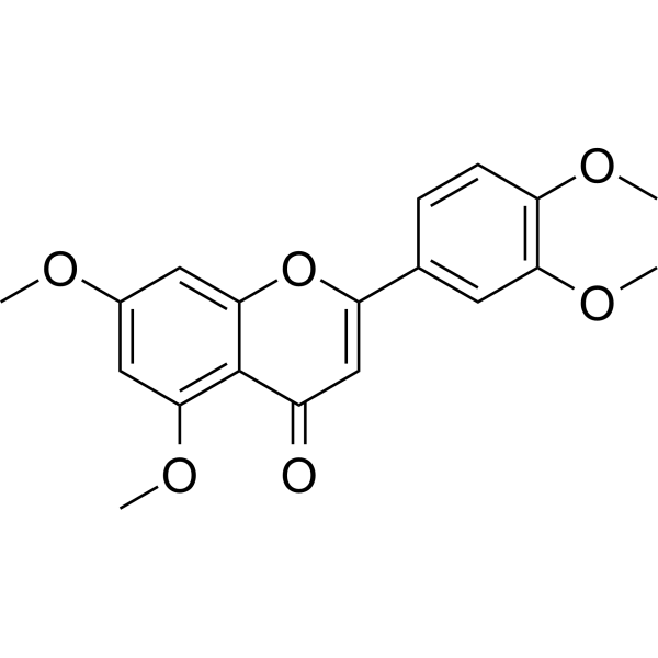
HY-N7030
5,7,3',4'-TetramethoxyflavoneParasite Inhibitor -
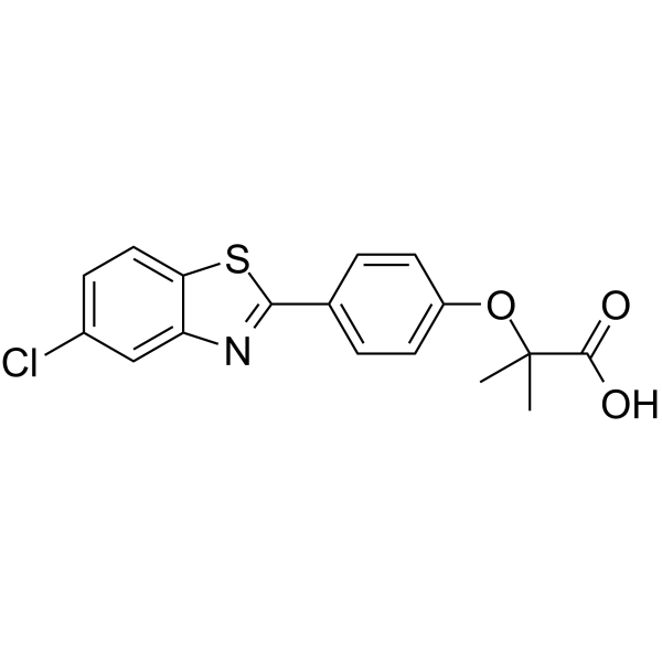
HY-117761
MHY908PPARα/γ dual agonist -
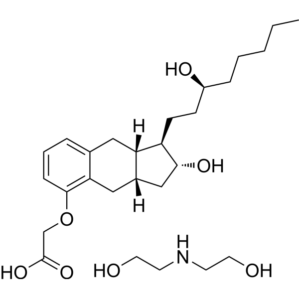
HY-B0813
Treprostinil diethanolamineEP2/DP1/IP Agonist
Publications Citing Use of MedChemExpress (MCE) 3′,4′,7-Trihydroxyflavone
MoreAll Beta-lactamase Isoforms
MoreAll Caspase Isoforms
More

Biological Activity
Description
Cellular Effect
|
Cell Line

|
Type | Value | Description | References |
|---|---|---|---|---|
| CHO | IC50 |
49.3 μM
Compound: 12
|
Cytotoxicity against CHO cells by MTT assay
Cytotoxicity against CHO cells by MTT assay
|
[PMID: 19572738] |
| HEK293 | IC50 |
>40 μM
Compound: 1d
|
Inhibition of human telomerase from HEK293 cell extracts by Flash-Plate assay
Inhibition of human telomerase from HEK293 cell extracts by Flash-Plate assay
|
[PMID: 15588081] |
| HEK293 | IC50 |
>40 μM
Compound: Fig 20, R2C1
|
Inhibition of telomerase extracted from HEK293 cells using 5'-bAATCCGTCGAGCAGAGTT as primer assessed as polymerization of telomeric repeats to the end of the primers after 2 hrs by Flash-Plate assay
Inhibition of telomerase extracted from HEK293 cells using 5'-bAATCCGTCGAGCAGAGTT as primer assessed as polymerization of telomeric repeats to the end of the primers after 2 hrs by Flash-Plate assay
|
10.1039/C0MD00241K |
| Neutrophil | IC50 |
13.5 μM
Compound: 3d
|
Inhibition of oxidative burst in PMA-stimulated human neutrophils assessed as inhibition of ROS-induced luminol oxidation incubated for 5 mins prior to PMA challenge by chemiluminescence assay
Inhibition of oxidative burst in PMA-stimulated human neutrophils assessed as inhibition of ROS-induced luminol oxidation incubated for 5 mins prior to PMA challenge by chemiluminescence assay
|
[PMID: 23871908] |
| Neutrophil | IC50 |
14.7 μM
Compound: 3d
|
Inhibition of oxidative burst in PMA-stimulated human neutrophils assessed as inhibition of superoxide anion radical-induced lucigenin oxidation incubated for 5 mins prior to PMA challenge by chemiluminescence assay
Inhibition of oxidative burst in PMA-stimulated human neutrophils assessed as inhibition of superoxide anion radical-induced lucigenin oxidation incubated for 5 mins prior to PMA challenge by chemiluminescence assay
|
[PMID: 23871908] |
| Neutrophil | IC50 |
3.7 μM
Compound: 3d
|
Inhibition of oxidative burst in PMA-stimulated human neutrophils assessed as inhibition of H2O2-induced oxidation of amplex red incubated for 5 mins prior to PMA challenge by fluorescence assay
Inhibition of oxidative burst in PMA-stimulated human neutrophils assessed as inhibition of H2O2-induced oxidation of amplex red incubated for 5 mins prior to PMA challenge by fluorescence assay
|
[PMID: 23871908] |
| Neutrophil | IC50 |
5.2 μM
Compound: 3d
|
Inhibition of oxidative burst in PMA-stimulated human neutrophils assessed as inhibition of HOCl-induced oxidation of APF after 6 mins by fluorescence assay
Inhibition of oxidative burst in PMA-stimulated human neutrophils assessed as inhibition of HOCl-induced oxidation of APF after 6 mins by fluorescence assay
|
[PMID: 23871908] |
| Peritoneal macrophage | IC50 |
26 μM
Compound: kp15
|
Inhibition of LPS-stimulated nitric oxide production in ddy mouse peritoneal macrophages measured after 20 hrs by Greiss method
Inhibition of LPS-stimulated nitric oxide production in ddy mouse peritoneal macrophages measured after 20 hrs by Greiss method
|
[PMID: 27955927] |
In Vitro
3′,4′,7-Trihydroxyflavone (0.1-20 μM, 24 h) enhances the cell viability and survival against Scopolamine (HY-N0296)-induced damage in SH-SY5Y cells[1].
3′,4′,7-Trihydroxyflavone (0.1-100 µM) reduces the secretion of IL-6 and IL-8 in TNFα-stimulated HaCaT cells, with IC50s of 17.8, 126.2 µg/mL, with maximum inhibition rates of 74.4, 40.2 %, reduces HaCaT cell viability to 74%[2].
3′,4′,7-Trihydroxyflavone (50 µM, 64 μg/mL) inhibits OXA-48 activity by 80% and exhibits antimicrobial activity against BW25113 ∆acrA∆bamB (OXA-48) in combination with β-lactam antibiotics[3].
3′,4′,7-Trihydroxyflavone (1-20 μM, 30 min) prevents H2O2-induced cell death and lactate dehydrogenase (LDH) release in SH-SY5Y and hippocampal neuronal cells[4].
3′,4′,7-Trihydroxyflavone (1-20 µM, 30 min) inhibits H2O2-induced apoptotic features of SH-SY5Y cells, reverses induced nuclear necrosis, and condenses cell shrinkage[4].
3′,4′,7-Trihydroxyflavone (0.5-20 µM, 30 min) inhibits H2O2-induced ROS accumulation, SOD, CAT, and GSH reduction, and activation of F-κB p65 translocation from the cytosol to the nucleus in SH-SY5Y cells[4].
3′,4′,7-Trihydroxyflavone (1-20 µM, 30 min) inhibits H2O2-induced phosphorylation of JNK, p38, ERK 1/2 MAPKs and PI3K/Akt levels, upregulation of Bax, caspase-3, caspase-9 and PARP levels, downregulation of Bcl-2 and Bcl-xL levels, release of cytochrome c and loss of MMP in SH-SY5Y cells[4].
3′,4′,7-Trihydroxyflavone shows antibacterial activity against 12 Gram-negative bacteria E. coli, E. aerogenes, K. pneumonia, with MIC values ranging from 4 to 256 μg/mL[5].
3′,4′,7-Trihydroxyflavone improves the activity of Tetracycline (HY-A0107) and Erythromycin (HY-B0220) on 80% of the tested bacteria, with FIC values ranging from 0.5 to < 0.062[5].
3′,4′,7-Trihydroxyflavone (0.1-100 μM) has no effect at 0.1–30 µM, but decreases cell viability at 100 µM for MG6 cells[6].
3′,4′,7-Trihydroxyflavone (0.1-10 μM, 6-48 h) reduces NO, TNF-α, and iNOS levels, inhibits STAT1 phosphorylation and total STAT1 expression in LPS (HY-D1056)-induced MG6 cells[6].
3′,4′,7-Trihydroxyflavone (0.1-10 μM, 0.5 h) inhibits JNK phosphorylation but not p38 or ERK phosphorylation in LPS-induced MG6 cells[6].
3′,4′,7-Trihydroxyflavone (0.1-10 μM, 24-48 h) inhibits NO release, iNOS expression, STAT1 phosphorylation in IFN-γ-induced MG6 cells[6].
3′,4′,7-Trihydroxyflavone (0-5 μg/mL, 1-7 days) inhibits RANKL-induced osteoclast formation and bone resorption in BMMs[7].
3′,4′,7-Trihydroxyflavone (5 μg/mL, 7 days) suppresses RANKL-induced NFATc1 expression via Blimp-1 and p38 MAPK pathway in BMMs[7].
MedChemExpress (MCE) has not independently confirmed the accuracy of these methods. They are for reference only. Further protocols information, click here.
-
Cell Line:SH-SY5Y cells
-
Concentration:0.1, 1, 5, 10, and 20 μM
-
Incubation Time:24 h
-
Result:Enhanced the cell viability and survival against Scopolamine (HY-N0296)-induced damage.
-
Cell Line:SH-SY5Y cells
-
Concentration:0.1, 1, 5, 10, and 20 μM
-
Incubation Time:30 min
-
Result:Inhibited apoptosis, reversed H2O2-induced nuclear necrosis and condensation, and cell shrinkage.
-
Cell Line:SH-SY5Y cells
-
Concentration:0.5, 1, 5, 10, and 20 μM
-
Incubation Time:30 min
-
Result:Reduced ROS, SOD, CAT and GSH fluorescence intensity.
Increased the fluorescence intensity of MMP and rhodamine 123, eliminated cytochrome c diffusion caused by H2O2.
-
Cell Line:SH-SY5Y cells
-
Concentration:1, 5, 10, and 20 μM
-
Incubation Time:30 min
-
Result:Reduced the phosphorylation of Akt, JNK, p38 MAPK, and Akt to 40.64%, 111%, 105%, and 95% of the control value, respectively.
Inhibited the upregulation or downregulation of Bax and Bcl-xL to 118% and 62% of control values, respectively at 1 µM, inhibited the upregulation or downregulation of Bax, Bcl-2, and Bcl-xL to 111%, 87%, and 82% of control values, respectively, at 5 µM, inhibited the upregulation or downregulation of Bax, Bcl-2, and Bcl-xL to 102%, 98%, and 93% of control values, respectively at 20 µM.
Reduced the release of cytochrome c from mitochondria to the cytosol.
Significantly inhibited the increase in cleaved PARP, cleaved caspase-3 and cleaved caspase-9.
-
Cell Line:LPS-induced MG6 cells
-
Concentration:0.1, 1, and 10 μM
-
Incubation Time:0.5, 6, 24 h
-
Result:Reduced iNOS levels.
Inhibited STAT1 phosphorylation and total STAT1 expression.
Inhibited JNK phosphorylation but not p38 or ERK phosphorylation.
-
Cell Line:IFN-γ-induced MG6 cells
-
Concentration:0.1, 1, and 10 μM
-
Incubation Time:24 h
-
Result:Inhibited NO release and increased iNOS expression.
Inhibited STAT1 phosphorylation but did not inhibit the increase in total STAT1 levels.
-
Cell Line:BMMs
-
Concentration:5 μg/mL with RANKL (100 ng/mL) and M-CSF (30 ng/mL)
-
Incubation Time:7 days
-
Result:Suppressed the mRNA expression levels of CTR and cathepsin K.
Suppressed the expression of DC-STAMP and ATP6v0d2 induced by RANKL. Decreased the mRNA level of Blimp1.
-
Cell Line:BMMs
-
Concentration:5 μg/mL with RANKL (200 ng/mL)
-
Incubation Time:30 min
-
Result:Abolished RANKL-induced NFATc1 expression, but not c-Fos protein expression.
Suppressed phosphorylation of p38.
In Vivo
MedChemExpress (MCE) has not independently confirmed the accuracy of these methods. They are for reference only.
-
Animal Model:Scopolamine-induced cognitive deficits mice (male C57BL/6J, 8 weeks old) model[1]
-
Dosage:10/50 mg/kg
-
Administration:p.o., once a day, 3 days/three times per week, 28 days
-
Result:Improved cognitive abilities at 50 mg/kg.
Had no acute cognitive improvement after three days.
Restored ACh and ChAT levels and reduced AChE activity, increased BDNF mRNA and protein levels, and restored pCREB levels at 50 mg/kg.
Increased the number of neurons and restored astrocyte levels (GFAP marker).
-
Animal Model:Scopolamine-induced cognitive deficits mice (male C57BL/6J, 8 weeks old) model[1]
-
Dosage:10 ng
-
Administration:ICV, twice a week, 28 days
-
Result:Improved spontaneous change, novel object recognition index and passive avoidance, acetylcholine activity, and restored acetylcholinesterase and choline acetyltransferase activities.
Induced the enhancement of the CREB-BDNF signaling pathway and increased LTP.


Chemical Information
-
CAS No. 2150-11-0
-
Appearance Solid
-
Molecular Weight 270.24
-
Formula C15H10O5
-
Color Off-white to light yellow
-
SMILES
O=C1C2=CC=C(O)C=C2OC(C3=CC(O)=C(O)C=C3)=C1
-
Structure Classification
-
Initial Source
-
Shipping
Room temperature in continental US; may vary elsewhere.
-
Storage
RT, protect from light
In solvent -80°C 1 year -20°C 6 months


Publications (1)
-
Journal Impact Factor
-
Most Recent
-
Spectrochim Acta A Mol Biomol Spectrosc
Communication of bifunctional G-quadruplex chaperone with aptamer for highly selective adenosine signaling by an ESIPT fluorogen. [Abstract]2026 Jun 5:354:127637. PMID: 41762796


Solvent & Solubility
In Vitro:
DMSO : 2.5 mg/mL (9.25 mM; ultrasonic and warming and heat to 60°C; Hygroscopic DMSO has a significant impact on the solubility of product, please use newly opened DMSO)
Please refer to the solubility information to select the appropriate solvent. Once prepared, please aliquot and store the solution to prevent product inactivation from repeated freeze-thaw cycles.
Storage method and period of stock solution: -80°C, 1 year; -20°C, 6 months. When stored at -80°C, please use it within 1 year. When stored at -20°C, please use it within 6 months.
Please refer to the solubility information to select the appropriate solvent. Once prepared, please aliquot and store the solution to prevent product inactivation from repeated freeze-thaw cycles.
Storage method and period of stock solution: -80°C, 1 year; -20°C, 6 months. When stored at -80°C, please use it within 1 year. When stored at -20°C, please use it within 6 months.
Concentration (start) × Volume (start) = Concentration (final) × Volume (final)

Protocols
-
Research Protocol for Infectious Diseases
Infectious-disease experiments test how pathogens interact with host barriers, innate immune receptors, inflammatory signaling, pathogen replication, and tissue injury; pattern-recognition receptors such as TLRs, RIG-I-like receptors, NOD-like receptors, and inflammasomes detect microbial molecules and activate NF-κB, interferon, and cytokine responses. The central hypothesis is that infection severity reflects the balance between pathogen burden and host response: protective inflammation restricts pathogen growth, whereas excessive or mislocalized inflammation contributes to tissue damage and disease phenotype. Unresolved questions include which host pathways are protective versus pathogenic, why some infection models fail to translate to human disease, and which combined readouts best predict clinically relevant infection outcomes.
-
Apoptosis
Apoptosis, also called programmed cell death, is generally characterized by distinct morphological characteristics.
-
TUNEL staining for apoptotic DNA fragmentation
TUNEL staining detects DNA strand breaks by using terminal deoxynucleotidyl transferase to add labeled nucleotides to exposed 3′-OH DNA termini, generating either microscopic staining in fixed cells or tissue sections, or fluorescence/cytometric signal in cell suspensions. TUNEL positivity reflects DNA fragmentation but should not be interpreted alone as definitive apoptosis, because TUNEL can also label necrotic, autolytic, mechanically damaged, or DNA-repair-associated DNA breaks.
-
Cell Cytotoxicity Assay
Cytotoxicity assays are usually based on the assessment of cell membrane damage, which can also be indirectly detected by measuring cell viability. Detection methods include MTT assay, CKK-8 assay, LDH assay and ATP assay, etc.
-
LPS-Induced Endotoxemia/Systemic Inflammation
Lipopolysaccharide (LPS)-induced endotoxemia is a widely used in vivo model of acute systemic inflammation in which LPS, a Gram-negative bacterial endotoxin, activates innate immune signaling primarily through TLR4, leading to rapid and transient induction of pro-inflammatory cytokines such as TNF-α, IL-6, and IL-1β in circulation and tissues. This cytokine surge is commonly used as a measurable readout of systemic inflammatory activation and immune dysregulation, and is typically assessed within hours after intraperitoneal LPS administration in mouse models of endotoxemia. The model captures key features of systemic inflammatory response syndrome, including cytokine release, immune cell activation, and downstream tissue responses, and has been used to evaluate anti-inflammatory interventions such as cytokine modulation, lipid mediators, and immune cell-targeting therapies.
-
Annexin V plus membrane-impermeant dye apoptosis staining
Annexin V-based apoptosis assays rely on the detection of phosphatidylserine (PS) externalization from the inner leaflet of the plasma membrane to the outer leaflet, an early biochemical hallmark of apoptosis. Fluorescently labeled Annexin V binds PS in a calcium-dependent manner, enabling identification of early apoptotic cells by flow cytometry or fluorescence microscopy. When combined with a membrane-impermeant DNA-binding dye (e. g. , propidium iodide), this approach allows discrimination between viable (Annexin V−/dye−), early apoptotic (Annexin V+/dye−), and late apoptotic or necrotic (Annexin V+/dye+) cell populations by assessing membrane integrity and PS exposure.
-
ROS/oxidative-stress fluorescent staining
ROS/oxidative-stress fluorescent staining uses cell-permeant fluorogenic probes that become fluorescent after oxidation inside cells or tissues; commonly used examples include DCFH-DA/DCFDA for broad cellular oxidant detection, DHE for superoxide-related signal detection, MitoSOX for mitochondrial superoxide-related signal detection, and CellROX probes for oxidative-stress-associated fluorescence readouts. The assay detects probe oxidation rather than a single ROS species unless the probe and analysis method have been chemically validated for that species. DCFH-DA enters cells, is deacetylated by intracellular esterases to DCFH, and produces fluorescent DCF after oxidation, so the readout is used as an operational measure of total cellular oxidative stress rather than a species-specific ROS measurement. DHE and MitoSOX can report superoxide-related oxidation, but red fluorescence alone can include non-specific ethidium-like oxidation products; HPLC or optimized spectral approaches are
-
Apoptosis Solutions
Apoptosis is a regulated, generally non-lytic cell-death pathway that removes unwanted, damaged, infected, or abnormal cells through coordinated morphological changes, caspase activation, DNA fragmentation, and membrane remodeling. The intrinsic apoptosis pathway is controlled mainly by mitochondrial outer membrane permeabilization, BCL-2 family proteins, cytochrome c release, apoptosome formation, caspase-9 activation, and downstream executioner caspase-3/7 activation. The extrinsic apoptosis pathway is initiated by death receptors such as Fas, TNFR, and TRAIL receptors, which recruit adaptor proteins and activate caspase-8 before engaging executioner caspases or mitochondrial amplification through BID cleavage. Apoptosis is linked to many phenotypes, including cancer cell killing, tissue homeostasis, immune regulation, neurodegeneration, infection response, and treatment-induced cytotoxicity; unresolved questions include how apoptosis interacts with necroptosis, pyroptosis, ferroptos
-
Research Protocol for Inflammation-related Diseases
The NLRP3 inflammasome is a cytosolic innate immune signaling platform that integrates priming signals and danger-signal activation to promote caspase-1 activation, maturation of IL-1β and IL-18, and gasdermin D-mediated pyroptotic cell death. The core experimental logic is to determine whether inflammatory disease phenotypes are driven by increased NLRP3 expression, ASC-containing inflammasome assembly, caspase-1 cleavage, GSDMD cleavage, and extracellular release of IL-1β/IL-18 rather than by nonspecific cell injury alone. The pathway is strongly linked to inflammation-related disease phenotypes because monosodium urate crystals activate NALP3/NLRP3 inflammasome signaling in gout-like crystal inflammation, cholesterol crystals activate NLRP3 inflammasomes in atherogenesis models, and DSS-induced intestinal inflammation has been reported to involve NLRP3 inflammasome activity. However, experimental colitis studies also show context-dependent protective effects of NLRP3 inflammasome co
-
Gram Staining of Tissue Sections
Gram staining of tissue sections is a histochemical technique used to differentiate Gram-positive and Gram-negative bacteria within histological specimens based on differences in bacterial cell wall structure and dye retention, adapted from classical bacteriological Gram staining into tissue-compatible “histological Gram stain” variants. In tissue applications, modifications of the Brown-Hopps and Brown-Brenn methods are commonly used to improve differentiation of microorganisms embedded within host connective tissue and to reduce overstaining or loss of Gram-negative signal, which are known limitations of earlier approaches. The principle relies on crystal violet-iodine complex retention in Gram-positive organisms and subsequent decolorization and counterstaining steps that allow contrast visualization of Gram-negative organisms against tissue background.
-
Osteoclast differentiation from monocyte/macrophage precursors
Osteoclast differentiation is an in vitro induction assay in which monocyte/macrophage-lineage precursors are exposed to macrophage colony-stimulating factor (M-CSF) and receptor activator of NF-κB ligand (RANKL), generating multinucleated osteoclasts that are commonly identified by tartrate-resistant acid phosphatase (TRAP) staining and functionally confirmed by resorption pits on dentin, bone, or mineralized substrates. M-CSF supports survival and expansion of osteoclast precursors, while RANKL binding to RANK drives osteoclast commitment, fusion, maturation, and resorptive function; osteoprotegerin inhibits this pathway by binding RANKL and preventing RANK activation. The main readouts are the number of TRAP-positive multinucleated cells, formation of F-actin rings, and resorbed surface area; TRAP-positive multinucleated cells indicate osteoclast differentiation, whereas pit formation on dentin, bone, or mineralized coating indicates functional bone-resorbing activity.


Purity & Documentation
-
Data Sheet (291 KB)
-
SDS (394 KB)
- English - EN (394 KB)
- Français - FR (394 KB)
- Deutsch - DE (394 KB)
- Norwegian - NO (394 KB)
- Español - ES (394 KB)
- Swedish - SV (394 KB)
- Italian - IT (394 KB)
- Korean - KR (394 KB)
- Portuguese - PT (394 KB)
-
Handling Instructions (2659 KB)
References
[1]. Kim SS, et al. 3',4',7-trihydroxyflavone activates the CREB-BDNF axis and restores scopolamine-induced memory deficit in mice. Eur J Pharmacol. 2025 Jul 15;999:177645 [Content Brief]
[2]. Codo Toafode NM, et al. Anti-Inflammatory Potential of Phenolic Compounds Isolated From Entada africana Guill. & Perr. Used in the Republic of Benin. Front Pharmacol. 2022 Jun 30;13:931240. [Content Brief]
[3]. Zhang Y, et al. Discovery of Quercetin and Its Analogs as Potent OXA-48 Beta-Lactamase Inhibitors. Front Pharmacol. 2022 Jun 22;13:926104. [Content Brief]
[4]. Kwon SH, et al. 3',4',7-Trihydroxyflavone prevents apoptotic cell death in neuronal cells from hydrogen peroxide-induced oxidative stress. Food Chem Toxicol. 2015 Jun;80:41-51. [Content Brief]
[5]. Dzotam JK, et al. In vitro antibacterial and antibiotic modifying activity of crude extract, fractions and 3',4',7-trihydroxyflavone from Myristica fragrans Houtt against MDR Gram-negative enteric bacteria. BMC Complement Altern Med. 2018 Jan 15;18(1):15. [Content Brief]
[6]. Akaishi T, et al. 3',4',7-Trihydroxyflavone Downregulates NO Production in LPS- or IFN-γ-Activated MG6 Microglial Cells by Attenuating the JNK-STAT1 Pathway. Biol Pharm Bull. 2022;45(3):301-308. [Content Brief]
[7]. Kang JH, et al. 3'4'7-Trihydroxyflavone inhibits RANKL-induced osteoclast formation via NFATc1. Pharmazie. 2015 Oct;70(10):661-7. [Content Brief]
[8]. Selvam C, et al. Cyclooxygenase inhibitory flavonoids from the stem bark of Semecarpus anacardium Linn. Phytother Res. 2004 Jul;18(7):582-4. [Content Brief]
[9]. Jung MJ, et al. Antioxidant activity from the stem bark of Albizzia julibrissin. Arch Pharm Res. 2003 Jun;26(6):458-62. [Content Brief]


Complete Stock Solution Preparation Table
Please refer to the solubility information to select the appropriate solvent. Once prepared, please aliquot and store the solution to prevent product inactivation from repeated freeze-thaw cycles.
Storage method and period of stock solution: -80°C, 1 year; -20°C, 6 months. When stored at -80°C, please use it within 1 year. When stored at -20°C, please use it within 6 months.
| Optional Solvent | Concentration Solvent Mass | 1 mg | 5 mg | 10 mg | 25 mg |
|---|---|---|---|---|---|
| DMSO | 1 mM | 3.7004 mL | 18.5021 mL | 37.0041 mL | 92.5104 mL |
| 5 mM | 0.7401 mL | 3.7004 mL | 7.4008 mL | 18.5021 mL |


3′,4′,7-Trihydroxyflavone Related Classifications
HY-N2736 Related Classifications
 Powered by Bioz
Keywords
- 3′,4′,7-Trihydroxyflavone
- 2150-11-0
- Beta-lactamase
- COX
- Interleukin Related
- Bacterial
- JNK
- ERK
- p38 MAPK
- STAT
- Apoptosis
- NO Synthase
- Nuclear Factor of activated T Cells (NFAT)
- Lactate Dehydrogenase
- Reactive Oxygen Species (ROS)
- SOD
- Akt
- Caspase
- Bcl-2 Family
- flavonoid aglycone
- cognitive deficits mice model
- SH-SY5Y cells
- HaCaT cell
- MG6 cells
- BMMs
- Inhibitor
- inhibitor
- inhibit