



2'-Hydroxyflavanone

Based on 1 Customer Validation
2'-Hydroxyflavanone is an orally active antiproliferative agent. 2'-Hydroxyflavanone inhibits STAT3 phosphorylation, nuclear translocation, and transactivation, and inhibits NF-κB, p38 MAPK, and SAPK/JNK activation, LPS-induced ROS, nitric oxide, lipid peroxidation, loss of mitochondrial membrane potential, cytokine release, and macrophage apoptosis. 2'-Hydroxyflavanone induces Egr-1/NAG-1 -related apoptosis involving Bax, p21, caspase-3, and caspase-7, inhibits AKR1C3 and suppresses androgen receptor, β-catenin, and AKT signaling, and reduces GSK-3β inhibitory phosphorylation. 2'-Hydroxyflavanone can be used for research on pain, cutaneous leishmaniasis, pancreatic ductal adenocarcinoma, colon cancer, and prostate cancer.
For research use only. We do not sell to patients.
- Purity : 99.84%
- CAS No.: 17348-76-4
- Formula: C15H12O3
- Molecular Weight:240.25
-
Storage:
Store at room temperature 3 years.
In solvent -80°C, 2 years , -20°C, 1 year

To place orders, for customer services and technical support, please contact: MedChemExpress USA
Tel: 609-228-6898 E-mail: [email protected] [email protected]


-
 Biological Activity
-
Chemical Information
-
Solvent & Solubility
- Protocols
- Purity & Documentation
- References
-
 Help & FAQs


-
HY-N8495
4'-HydroxyflavanoneSREBP Maturation Inhibitor -
HY-W028263
6-HydroxyflavanoneNatural Product -
HY-111803
4'-MethoxyflavonolAnti-stress Agent -
HY-N1461
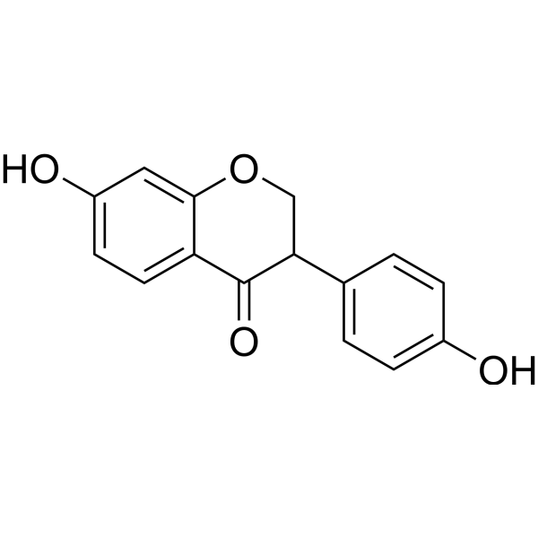
DihydrodaidzeinPhytoestrogen -
HY-N0112
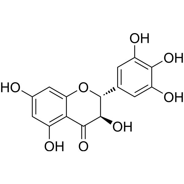
DihydromyricetinAutophagy Inducer -

HY-N8495R
4'-Hydroxyflavanone (Standard)SREBP Maturation Inhibitor -
HY-N8852
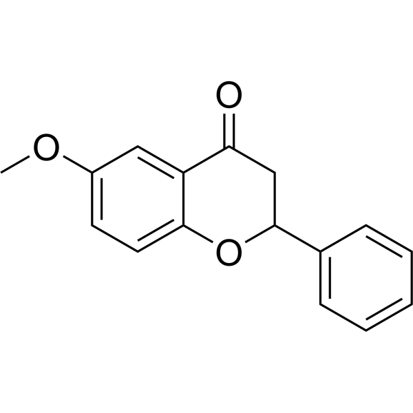
6-MethoxyflavanoneGABAA Receptor Modulator -

HY-W285243
3′-HydroxyflavanoneFlavonoids -
HY-W087008
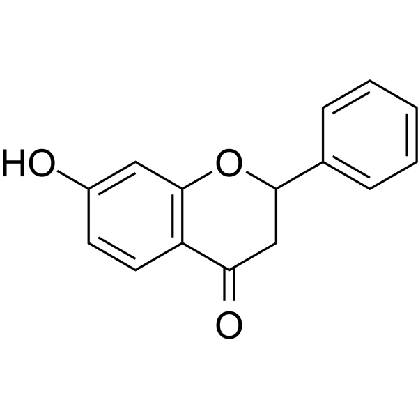
7-HydroxyflavanoneCYP19 Inhibitor -
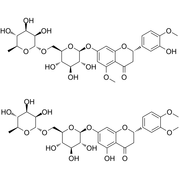
HY-N0165
Methyl-HesperidinVasodilating Agent -

HY-N0112R
Dihydromyricetin (Standard)Autophagy Inducer -
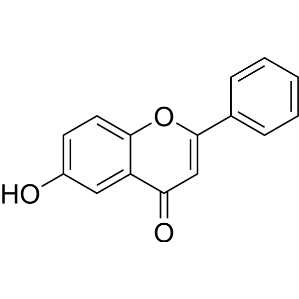
HY-N7110
6-HydroxyflavoneAnti-inflammatory Agent


Biological Activity
Description
|
STAT3 |
SAPK |
NF-κB |
p38 MAPK |
JNK |
Bax |
Caspase-3 |
p21 |
Caspase-7 |
EGR-1 |
NAG-1 |
Akt |
AKR1C3 |
androgen recepto |
β-catenin |
GSK3β |
Cellular Effect
|
Cell Line

|
Type | Value | Description | References |
|---|---|---|---|---|
| DU-145 | IC50 |
4.11 μM
Compound: 1; 2'-HF
|
Antiproliferative activity against human DU145 cells assessed as reduction in cell growth treated every 2 days for 4 days by hemocytometry
Antiproliferative activity against human DU145 cells assessed as reduction in cell growth treated every 2 days for 4 days by hemocytometry
|
[PMID: 30189396] |
| J774.A1 | IC50 |
71.3 μM
Compound: 1
|
Inhibition of IL-1beta production in mouse J774A.1 cells incubated for 12 hrs by sandwich immunoassay
Inhibition of IL-1beta production in mouse J774A.1 cells incubated for 12 hrs by sandwich immunoassay
|
[PMID: 25824662] |
| J774.A1 | IC50 |
71.3 μM
Compound: 1
|
Inhibition of LPS-induced nitric oxide production in mouse J774A.1 cells pre-incubated for 1 hr followed by LPS stimulation for 24 hrs by Griess reagent based assay
Inhibition of LPS-induced nitric oxide production in mouse J774A.1 cells pre-incubated for 1 hr followed by LPS stimulation for 24 hrs by Griess reagent based assay
|
[PMID: 25824662] |
| J774.A1 | IC50 |
71.3 μM
Compound: 1
|
Inhibition of TNFalpha production in mouse J774A.1 cells incubated for 6 hrs by sandwich immunoassay
Inhibition of TNFalpha production in mouse J774A.1 cells incubated for 6 hrs by sandwich immunoassay
|
[PMID: 25824662] |
| LNCaP | IC50 |
5.45 μM
Compound: 1; 2'-HF
|
Antiproliferative activity against human LNCAP cells assessed as reduction in cell growth treated every 2 days for 4 days by hemocytometry
Antiproliferative activity against human LNCAP cells assessed as reduction in cell growth treated every 2 days for 4 days by hemocytometry
|
[PMID: 30189396] |
| PC-3 | IC50 |
2.56 μM
Compound: 1; 2'-HF
|
Antiproliferative activity against human PC3 cells assessed as reduction in cell growth treated every 2 days for 4 days by hemocytometry
Antiproliferative activity against human PC3 cells assessed as reduction in cell growth treated every 2 days for 4 days by hemocytometry
|
[PMID: 30189396] |
| RAW264.7 | IC50 |
71.3 μM
Compound: 1
|
Inhibition of IL-1beta production in mouse RAW264.7 cells incubated for 12 hrs by sandwich immunoassay
Inhibition of IL-1beta production in mouse RAW264.7 cells incubated for 12 hrs by sandwich immunoassay
|
[PMID: 25824662] |
| RAW264.7 | IC50 |
71.3 μM
Compound: 1
|
Inhibition of LPS-induced nitric oxide production in mouse RAW264.7 cells pre-incubated for 1 hr followed by LPS stimulation for 24 hrs by Griess reagent based assay
Inhibition of LPS-induced nitric oxide production in mouse RAW264.7 cells pre-incubated for 1 hr followed by LPS stimulation for 24 hrs by Griess reagent based assay
|
[PMID: 25824662] |
| RAW264.7 | IC50 |
71.3 μM
Compound: 1
|
Inhibition of TNFalpha production in mouse RAW264.7 cells incubated for 6 hrs by sandwich immunoassay
Inhibition of TNFalpha production in mouse RAW264.7 cells incubated for 6 hrs by sandwich immunoassay
|
[PMID: 25824662] |
In Vitro
2'-Hydroxyflavanone (30-50 µM; 48 h) reduces the clonogenic capacity of BxPC-3 and MIA PaCa-2 cells[3].
2'-Hydroxyflavanone (12-24 h) reduces wound healing in BxPC-3 and MIA PaCa-2 cells[3].
The wound-healing inhibitory effect of 2'-Hydroxyflavanone (12-24 h) is reversed by IL6-induced STAT3 activation in BxPC-3 and MIA PaCa-2 cells[3].
2'-Hydroxyflavanone (10-20 µM; 24-36 h) reduces Transwell migration and invasion of BxPC-3 and MIA PaCa-2 cells[3].
2'-Hydroxyflavanone (10-20 µM; 48 h) reverses the expression of EMT markers and reduces MMP9 in BxPC-3 and MIA PaCa-2 cells[3].
2'-Hydroxyflavanone combined with Erlotinib reduces colony formation in BxPC-3 and MIA PaCa-2 cells[3].
2'-Hydroxyflavanone (2′-HF) (5-20 µM; 7 days) is the strongest inhibitor of HCT116 colony formation among the tested hydroxyflavanones[5].
2'-Hydroxyflavanone (10-40 µM; 48 h) shows the strongest effect in reducing HCT116 viability among the flavanones tested[5].
2'-Hydroxyflavanone increases the binding of Egr-1 to the NAG-1 promoter in HCT116 cells[5].
2'-Hydroxyflavanone (40 µM=) rapidly induces Egr-1 in HCT116 cells, with expression peaking at approximately 2 h[5].
2'-Hydroxyflavanone (2HF) (5-40 μM; 48 h) reduces the MTT viability of DU145 and PC-3 cells[6].
2'-Hydroxyflavanone (5-10 μM; 12-24 h) reduces wound closure in DU145 and PC-3 cells[6].
2'-Hydroxyflavanone (5-10 μM; 24 h) inhibits Transwell migration and invasion of DU145 and PC-3 cells[6].
2'-Hydroxyflavanone (5-10 μM; 48 h) reverses EMT marker expression in DU145 and PC-3 cells by increasing E-cadherin and decreasing N-cadherin, vimentin, and MMP9[6].
2'-Hydroxyflavanone (5-10 μM; 48 h) inhibits the expression of β-catenin and p-GSK-3β (Ser9) in DU145 and PC-3 cells[6].
2'-Hydroxyflavanone (2HF) (10-60 μM; 48 h) reduces the viability of PC-3, DU145, and C4-2 prostate cancer cells in a dose-dependent manner[7].
2'-Hydroxyflavanone (10-40 μM; 48 h) inhibits the clonogenic ability of PC-3 and DU145 cells[7].
2'-Hydroxyflavanone (10-40 μM; 36 h) inhibits the phosphorylation of AKT Ser473 and STAT3 Tyr705 in PC-3 and DU145 cells[7].
2'-Hydroxyflavanone (10-40 μM; 36 h) inhibits STAT3-responsive luciferase activity in PC-3 and DU145 cells[7].
2'-Hydroxyflavanone (10-40 µM; 24-48 h) activates caspase-3, caspase-7, and PARP cleavage in HCT116 cells in a dose- and time-dependent manner[5].
2'-Hydroxyflavanone (40 µM; 24 h) increases in situ cleaved caspase-7 staining in HCT116 cells[5].
2'-Hydroxyflavanone (40 µM; 12 h) increases annexin V-positive cells more strongly in p53-deficient HCT116 cells than in wild-type HCT116 cells[5].
2'-Hydroxyflavanone (40 µM; 12 h) increases annexin V positivity in HCT116 cells; NAG-1 knockdown reduces this effect[5].
2'-Hydroxyflavanone increases annexin V positivity in HCT116 cells; Egr-1 knockdown reduces this effect[5].
2'-Hydroxyflavanone (10-40 μM; 24 h) induces apoptosis in PC-3 and DU145 cells and increases the Annexin V/PI-positive population in C4-2 cells[7].
2'-Hydroxyflavanone (10-40 μM; 36 h) modulates BCL-2 family proteins in PC-3 and DU145 cells and promotes PARP/caspase-3 cleavage[7].
2'-Hydroxyflavanone combined with PI3K or STAT3 inhibition further suppresses Bcl-2 and AKT/STAT3 signaling in PC-3 cells[7].
2'-Hydroxyflavanone-induced apoptosis in PC-3 cells is enhanced by PI3K or STAT3 inhibition[7].
2'-Hydroxyflavanone (30-50 µM; 48 h) dose-dependently inhibits STAT3 phosphorylation in BxPC-3 and MIA PaCa-2 cells[3].
2'-Hydroxyflavanone (20 µM; 24-48 h) induces apoptosis in HCT116 cells, increasing the sub-G0/G1 population to 34.64% at 48 h[5].
2'-Hydroxyflavanone (20-40 µM; 24 h) induces p53 and p21 in wild-type HCT116 cells, whereas apoptosis markers are more strongly induced in p53-deficient HCT116 cells[5].
2'-Hydroxyflavanone induces NAG-1, Bax, p21, caspase-7, and PARP responses in HCT116 cells; Egr-1 knockdown attenuates these responses[5].
2'-Hydroxyflavanone (40-50 µM) inhibits IL6-induced STAT3 phosphorylation and nuclear translocation in BxPC-3 and MIA PaCa-2 cells[3].
The viability-inhibitory effect of 2'-Hydroxyflavanone is reversed by IL6-induced STAT3 activation in BxPC-3 and MIA PaCa-2 cells[3].
2'-Hydroxyflavanone combined with Erlotinib (HY-50896) reduces the viability of BxPC-3 and MIA PaCa-2[3].
2'-Hydroxyflavanone's inhibitory effect on the migration and invasion of BxPC-3 and MIA PaCa-2 cells is reversed by IL6-induced STAT3 activation[3].
2'-Hydroxyflavanone (40 µM) inhibits STAT3 phosphorylation and, in combination with Erlotinib, reduces EGFR phosphorylation in BxPC-3 and MIA PaCa-2 cells[3].
2'-Hydroxyflavanone (30 μM) prevents LPS-induced NF-κB nuclear translocation in RAW 264.7 macrophages at 30, 60, and 120 min, but has no such effect at 15 min[4].
2'-Hydroxyflavanone (30 μM) prevents LPS-induced p38MAPK and SAPK/JNK activation in RAW 264.7 macrophages but does not affect ERK1/2 phosphorylation[4].
2'-Hydroxyflavanone (20-50 μM) prevents LPS-induced lipid peroxidation in RAW 264.7 macrophages[4].
2'-Hydroxyflavanone (20-50 μM) dose-dependently prevents LPS-induced NO release in RAW 264.7 macrophages[4].
2'-Hydroxyflavanone (72 h) shows no cytotoxicity to BALB/c peritoneal macrophages at infection-related concentrations, with a CC50 of 88.15 μM, and its selectivity indices against wild-type and antimony-resistant L. amazonensis are 28.5 and 26.2, respectively[2].
2'-Hydroxyflavanone (5-10 μM)-induced changes in EMT markers and PC-3 morphological changes are reversed by CA-β-catenin overexpression[6].
2'-Hydroxyflavanone (5-10 μM)-mediated inhibition of wound healing, migration, and invasion is reversed by CA-β-catenin overexpression in DU145 and PC-3 cells[6].
MedChemExpress (MCE) has not independently confirmed the accuracy of these methods. They are for reference only.
-
Cell Line:BxPC-3 and MIA PaCa-2 cells
-
Concentration:10, 20, 30, 40, 50, 60, 70, 80, 90 μM
-
Incubation Time:24, 48 h
-
Result:Inhibited the viability of BxPC-3 and MIA PaCa-2.
-
Cell Line:BxPC-3 and MIA PaCa-2 cells
-
Concentration:30, 40, 50 µM
-
Incubation Time:48 h
-
Result:Reduced the colony-forming ability of BxPC-3 and MIA PaCa-2 cells.
-
Cell Line:BxPC-3 and MIA PaCa-2 cells
-
Concentration:30, 40, 50 µM
-
Incubation Time:24 h
-
Result:Late apoptotic rates in BxPC-3 cells were 23.04% at 30 µM, 32.87% at 40 µM, and 43.09% at 50 µM.
In MIA PaCa-2 cells, late apoptotic rates were 14.59% at 30 µM, 23.62% at 40 µM, and 33.28% at 50 µM.
-
Cell Line:BxPC-3 and MIA PaCa-2 cells
-
Concentration:30, 40, 50 µM
-
Incubation Time:24 h
-
Result:Caused G1/G2 phase arrest in both BxPC-3 and MIA PaCa-2 cells in a dose-dependent manner.
-
Cell Line:BxPC-3 and MIA PaCa-2 cells
-
Concentration:30, 40, 50 µM
-
Incubation Time:48 h
-
Result:Increased cleaved PARP, cleaved Caspase3, Bax, and p27, and decreased total Caspase3, Bcl-2, and CyclinD1 in both cell lines.\n
Decreased p-STAT3 in a dose-dependent manner.
-
Cell Line:BxPC-3 and MIA PaCa-2 cells
-
Concentration:10, 20 µM
-
Incubation Time:48 h
-
Result:Inhibited the migration of BxPC-3 and MIA PaCa-2 cells.
-
Cell Line:RAW 264.7 murine macrophages
-
Concentration:10, 20, 30, 50 μM
-
Incubation Time:24 h; 48 h
-
Result:At 10, 20, 30, and 50 μM, no observable changes in RAW 264.7 viability occurred at 48 h; 24-h viability differences were not significant.
LPS decreased viability at 24 and 48 h.
At 24 h, no observable protection against LPS-induced viability loss occurred.
At 48 h, 30 and 50 μM prevented the LPS-induced decrease in viability.
-
Cell Line:HCT116 human colon cancer cells
-
Concentration:5, 10, 15, 20 µM
-
Incubation Time:7 days
-
Result:Inhibited HCT116 colony formation at 5, 10, 15, and 20 µM after 7 days.
Was the most potent among the tested flavanones.
-
Cell Line:HCT116 human colon cancer cells
-
Concentration:10, 15, 20, 40 µM
-
Incubation Time:48 h
-
Result:Produced the most prominent inhibition of HCT116 viability among the tested flavanones at 10, 15, 20, and 40 µM after 48 h.
-
Cell Line:HCT116 human colon cancer cells
-
Concentration:20 µM
-
Incubation Time:24 or 48 h
-
Result:Sub-G0/G1 populations were 1.34% at 0 h, 9.48% at 24 h, and 34.64% at 48 h.
G2/M populations were 27.64% at 0 h, 62.91% at 24 h, and 42.63% at 48 h.
G0/G1 populations were 43.14% at 0 h, 24.24% at 24 h, and 15.74% at 48 h.
S-phase populations were 28.39% at 0 h, 3.63% at 24 h, and 7.39% at 48 h.
PI-stained dead cells were substantially increased at 40 µM after 24 h.
-
Cell Line:HCT116 human colon cancer cells
-
Concentration:10, 20, 40 µM
-
Incubation Time:24 and 48 h
-
Result:Gradually increased cleavage of caspase-3 and caspase-7 in a dose- and time-dependent manner and increased PARP cleavage.
-
Cell Line:HCT116 human colon cancer cells
-
Concentration:40 µM
-
Incubation Time:24 h
-
Result:Cleaved caspase-7 immunofluorescence was increased in HCT116 cells.
-
Cell Line:Wild-type and p53-null HCT116 cells
-
Concentration:20, 40 µM
-
Incubation Time:24 h
-
Result:Dose-dependently increased p53 and p21 in wild-type HCT116 cells but not in p53-null HCT116 cells.
Cleaved caspase-7 and PARP levels were higher in p53-null HCT116 cells than in wild-type cells after exposure.
-
Cell Line:Wild-type and p53-null HCT116 cells
-
Concentration:40 µM
-
Incubation Time:12 h
-
Result:The annexin V-stained population was more strongly increased in p53-null HCT116 cells than in wild-type HCT116 cells after exposure.
-
Cell Line:HCT116/siCont and HCT116/siNAG1 cells
-
Concentration:40 µM
-
Incubation Time:12 h
-
Result:HCT116/siNAG1 cells displayed reduced appearance of phosphatidylserine on the outer membrane compared with HCT116/siCont cells.
-
Cell Line:DU145 and PC-3 cell lines
-
Concentration:5, 10, 20, 40 μM
-
Incubation Time:48 h
-
Result:Lower cytotoxicity was observed at 5 and 10 μM, whereas 20 and 40 μM reduced MTT OD values in DU145 and PC-3 cells.
-
Cell Line:DU145 and PC-3 cell lines
-
Concentration:5, 10 μM
-
Incubation Time:12 and 24 h
-
Result:Cell migration and invasion significantly decreased in a dose-dependent manner in DU145 and PC-3 cells.
-
Cell Line:DU145 and PC-3 cell lines
-
Concentration:5, 10 μM
-
Incubation Time:48 h
-
Result:E-cadherin expression increased and N-cadherin, vimentin, and MMP9 expression decreased in DU145 and PC-3 cells.\n
β-catenin and p-GSK-3β (Ser9) expression were downregulated in DU145 and PC-3 cells.
-
Cell Line:PC-3, DU145, and C4-2 prostate cancer cells
-
Concentration:20, 40, 60 μM (PC-3); 10, 20, 40 μM (DU145; C4-2)
-
Incubation Time:48 h
-
Result:Cell viability of PC-3, DU145, and C4-2 was inhibited in a dose-dependent manner.
-
Cell Line:PC-3 and DU145 prostate cancer cells
-
Concentration:10, 20, 40 μM
-
Incubation Time:48 h
-
Result:The number of colonies formed by PC-3 and DU145 cells was significantly decreased in a dose-dependent manner.
-
Cell Line:PC-3, DU145, and C4-2 prostate cancer cells
-
Concentration:10, 20, 40 μM
-
Incubation Time:24 h exposure
-
Result:Induced apoptosis in PC-3 and DU145 cells.
Increased the Annexin V/PI-positive population in C4-2 cells.
-
Cell Line:PC-3 and DU145 prostate cancer cells
-
Concentration:10, 20, 40 μM
-
Incubation Time:36 h
-
Result:In PC-3 and DU145 cells, Mcl-1 and Bcl-2 were downregulated and Bax was upregulated in a dose-dependent manner.
Cleavage of PARP was increased and total caspase-3 was decreased, indicating caspase-mediated apoptosis.
In Vivo
2'-Hydroxyflavanone (50 mg/kg/day; p.o.; once daily, seven days per week; from day 7 to day 42 post-infection) reduces lesion size and parasite burden by 99% in BALB/c mice with antimony-resistant Leishmania amazonensis cutaneous infection, indicating that it is active and lacks cross-resistance[2].
2′-Hydroxyflavanone (30-45 mg/kg; i.p.; single administration) shows antinociceptive effects in the hot plate test, which are attenuated by Naloxone (HY-17417A), supporting the involvement of opioidergic mechanisms in mice[1].
2′-Hydroxyflavanone (30-45 mg/kg; i.p.; single administration) shows an antinociceptive effect in the hot plate test, which is attenuated by Pentylenetetrazol, supporting the involvement of GABAergic mechanisms in mice[1].
2′-Hydroxyflavanone (15-45 mg/kg; i.p.; single administration) reduces acetic acid (HY-Y0319)-induced writhing responses in mice[1].
2′-Hydroxyflavanone (15-45 mg/kg; i.p.; single administration; assessed at 30, 60, and 90 min) increases tail-flick latency in mice[1].
2′-Hydroxyflavanone (15-45 mg/kg; i.p.; single administration) reduces Carrageenan (HY-125474)-induced paw edema in mice[1].
2′-Hydroxyflavanone (15-45 mg/kg; i.p.; paw volume assessed at 1-5 h) reduces Histamine (HY-B1204)-induced paw edema in mice[1].
2′-Hydroxyflavanone (15-45 mg/kg; i.p.; paw volume assessed at 1-5 h) reduces Serotonin (HY-B1473A)-induced paw edema in mice[1].
2′-Hydroxyflavanone (15-45 mg/kg; i.p.; single administration) reduces Xylene-induced ear edema in mice[1].
2′-Hydroxyflavanone (15-45 mg/kg; i.p.; single administration) improves static mechanical allodynia in Cisplatin (HY-17394)-treated mice[1].
2′-Hydroxyflavanone (15-45 mg/kg; i.p.; single administration) improves cold allodynia in Cisplatin-treated mice[1].
2′-Hydroxyflavanone (15-45 mg/kg; i.p.; single administration) ameliorates thermal hyperalgesia in Cisplatin-treated mice[1].
2′-Hydroxyflavanone (15-45 mg/kg; i.p.; single administration) ameliorates dynamic allodynia in Cisplatin-treated mice[1].
MedChemExpress (MCE) has not independently confirmed the accuracy of these methods. They are for reference only.
-
Animal Model:Balb/c (18-22 g; sex not reported)[1]
-
Dosage:15, 30, and 45 mg/kg
-
Administration:i.p.; single dose; latency assessed at 30, 60, and 90 min
-
Result:Hot-plate latency was 8.3 s at 30 min, 9.25 s at 60 min, and 10.36 s at 90 min at 15 mg/kg.
Hot-plate latency was 9.01 s at 30 min, 10.04 s at 60 min, and 11.5 s at 90 min at 30 mg/kg.
Hot-plate latency was 9.5 s at 30 min, 12.8 s at 60 min, and 14.08 s at 90 min at 45 mg/kg.
-
Animal Model:Balb/c (18-22 g; sex not reported)[1]
-
Dosage:30 and 45 mg/kg
-
Administration:i.p.; single dose
-
Result:Naloxone 1 mg/kg antagonized the hot-plate anti-nociceptive activity at both 30 and 45 mg/kg.
-
Animal Model:Balb/c (18-22 g; sex not reported)[1]
-
Dosage:30 and 45 mg/kg
-
Administration:i.p.; single dose
-
Result:Pentylenetetrazol 15 mg/kg reduced the hot-plate anti-nociceptive activity at both 30 and 45 mg/kg.
-
Animal Model:Albino mice (18-24 g)
[1] -
Dosage:15, 30, and 45 mg/kg
-
Administration:i.p.; single dose
-
Result:The number of writhes was 48.66 at 15 mg/kg, 33.33 at 30 mg/kg, and 22.16 at 45 mg/kg.
-
Animal Model:Albino mice (18-22 g)[1]
-
Dosage:15, 30, and 45 mg/kg
-
Administration:i.p.; single dose; assessed at 30, 60, and 90 min
-
Result:Tail withdrawal latency was 5 s at 30 min, 6.58 s at 60 min, and 6.16 s at 90 min at 15 mg/kg.
Tail withdrawal latency was 6.003 s at 30 min and 7.8 s at 60 min at 30 mg/kg.
Tail withdrawal latency was 6.9 s at 30 min and 8.6 s at 60 min at 45 mg/kg.
-
Animal Model:Albino mice (25 to 30 gm)[1]
-
Dosage:15, 30, and 45 mg/kg
-
Administration:i.p.; single dose
-
Result:Carrageenan-induced paw edema was reduced at 1, 3, and 5 h at 15 mg/kg.
The initial and final phases of carrageenan-induced inflammation were reduced at 1-3 h at 30 mg/kg.
Carrageenan-induced paw edema was suppressed across the tested doses.
-
Animal Model:Albino mice both male and female (25-30 gm)[1]
-
Dosage:15, 30, and 45 mg/kg
-
Administration:i.p.; paw volume assessed over 1-5 h
-
Result:Histamine-induced paw edema was reduced.
Histamine-induced paw volume showed a time-dependent decline at 45 mg/kg.
-
Animal Model:Albino mice both male and female (25-30 gm)[1]
-
Dosage:15, 30, and 45 mg/kg
-
Administration:i.p.; paw volume assessed over 1-5 h
-
Result:Serotonin-induced paw edema was reduced.
Serotonin-induced paw volume showed a time-dependent decline at 45 mg/kg.
-
Animal Model:Albino mice both male and female (25-30 gm)[1]
-
Dosage:15, 30, and 45 mg/kg
-
Administration:i.p.; single dose
-
Result:Ear edema volume was reduced at 30 mg/kg.
Xylene-induced ear edema showed a marked decline at 45 mg/kg.
-
Animal Model:Albino mice (20 to 30 gm)[1]
-
Dosage:15, 30, and 45 mg/kg
-
Administration:i.p.; single dose mg/kg in combination groups
-
Result:Paw withdrawal threshold increased at 15, 30, and 45 mg/kg at 1 and 3 h relative to Cisplatin control.
Gabapentin 75 mg/kg in combination increased paw withdrawal threshold.
-
Animal Model:Albino mice (20 to 30 gm)[1]
-
Dosage:15, 30, and 45 mg/kg
-
Administration:i.p.; single dose
-
Result:Paw withdrawal latency after acetone application increased at 15, 30, and 45 mg/kg at 1 and 3 h.
Co-administration with gabapentin 75 mg/kg produced prominent anti-allodynic effects.
-
Animal Model:Albino mice (20 to 30 gm)[1]
-
Dosage:15, 30, and 45 mg/kg
-
Administration:i.p.; single dose
-
Result:Paw withdrawal latency increased at 30 mg/kg combined with Cisplatin at 1 and 3 h.
Comparable increases occurred at 45 mg/kg combined with Cisplatin during 1-3 h.
Latency did not increase at 15 mg/kg.
-
Animal Model:Albino mice (20 to 30 gm)[1]
-
Dosage:15, 30, and 45 mg/kg
-
Administration:i.p.; single dose
-
Result:Paw withdrawal latency increased at 45 mg/kg combined with Cisplatin at 1 and 3 h.
Paw withdrawal latency also increased at 30 mg/kg combined with Cisplatin at 1 and 3 h.
Latency did not increase at 15 mg/kg.
-
Animal Model:BALB/c (female, 8-10 weeks old)[2]
-
Dosage:50 mg/kg/day
-
Administration:p.o. via orogastric tube; once daily seven times per week; from day 7 post-infection until day 42
-
Result:At 50 mg/kg/day, reduced lesion size from day 21 post-infection and inhibited parasite load by 98.8% relative to vehicle.
-
Animal Model:BALB/c (female, 8-10 weeks old)[2]
-
Dosage:50 mg/kg/day
-
Administration:p.o. via orogastric tube; once daily seven times per week; from day 7 post-infection until day 42
-
Result:At 50 mg/kg/day, reduced lesion size starting from day 25 post-infection and inhibited parasite load by 99% relative to vehicle.
-
Animal Model:KPC (LSL-KrasG12D/+; Trp53fl/+; Pdx1-Cre; 8 weeks old at randomization)[3]
-
Dosage:100 mg/kg/day
-
Administration:oral gavage (i.g.); daily; for 2 months
-
Result:Reduced tumor volume compared with control.
Reduced tumor/pancreas weight (%) compared with control.
Local invasion or distant metastasis occurred in 4 of 6 control mice and 1 of 6 mice.
IHC of KPC tumor tissue showed decreased p-STAT3 and CyclinD1 positivity, decreased Bcl-2 area, increased E-cadherin area, and decreased N-cadherin area compared with control.
-
Animal Model:BALB/c-nu (male, 3-4 weeks old, 15-20 g)[7]
-
Dosage:100 mg/kg body weight in 100 µl corn oil
-
Administration:p.o. (oral gavage); once daily; for 30 days
-
Result:Reduced tumor burden of subcutaneous PC-3 xenografts at 100 mg/kg/day; average tumor volume was smaller than in the control group.
IHC of xenograft tissues showed lower p-AKT (Ser473), p-STAT3 (Tyr705), and Bcl-2 expression and increased cleaved caspase-3 compared with control.
- Western Blot
- Mitochondrial membrane-potential fluorescent assay
- Annexin V plus membrane-impermeant dye apoptosis staining
- ROS/oxidative-stress fluorescent staining
- Research Protocol for Inflammation-related Diseases
- Cell Viability Determination by MTT Colorimetric Assay
- Kinase activity and phosphorylation assays
- Phagocytosis Functional Assay
- Cytoplasmic-Nuclear Fractionated Protein Extraction
- Detection of Apoptosis
- TUNEL staining for apoptotic DNA fragmentation
- Research Protocol for Endocrine Diseases
- Fluorescent plasma-membrane potential dye assay
- Mitochondrial membrane-potential and mitochondrial mass staining
- Apoptosis Solutions
- Protocol for Kinase activity and phosphorylation assays
- Ferroptosis Solutions


Chemical Information
-
CAS No. 17348-76-4
-
Appearance Solid
-
Molecular Weight 240.25
-
Formula C15H12O3
-
Color White to off-white
-
SMILES
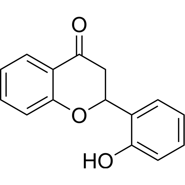
O=C1CC(C2=CC=CC=C2O)OC3=CC=CC=C13
-
Structure Classification
-
Initial Source
-
Shipping
Room temperature in continental US; may vary elsewhere.
-
Storage
Store at room temperature 3 years
In solvent -80°C 2 years -20°C 1 year


Solvent & Solubility
In Vitro:
DMSO : 100 mg/mL (416.23 mM; Need ultrasonic; Hygroscopic DMSO has a significant impact on the solubility of product, please use newly opened DMSO)
Please refer to the solubility information to select the appropriate solvent. Once prepared, please aliquot and store the solution to prevent product inactivation from repeated freeze-thaw cycles.
Storage method and period of stock solution: -80°C, 2 years; -20°C, 1 year. When stored at -80°C, please use it within 2 years. When stored at -20°C, please use it within 1 year.
Please refer to the solubility information to select the appropriate solvent. Once prepared, please aliquot and store the solution to prevent product inactivation from repeated freeze-thaw cycles.
Storage method and period of stock solution: -80°C, 2 years; -20°C, 1 year. When stored at -80°C, please use it within 2 years. When stored at -20°C, please use it within 1 year.
Concentration (start) × Volume (start) = Concentration (final) × Volume (final)
In Vivo Dissolution Calculator
Please enter the basic information of animal experiments:
-
-
-
-
Recommended: Prepare an additional quantity of animals to account for potential losses during experiments.
Working solution concentration: 0.22 mg/mL


Purity & Documentation
-
Data Sheet (326 KB)
-
SDS (393 KB)
- English - EN (393 KB)
- Français - FR (393 KB)
- Deutsch - DE (393 KB)
- Norwegian - NO (393 KB)
- Español - ES (393 KB)
- Swedish - SV (393 KB)
- Italian - IT (393 KB)
- Korean - KR (393 KB)
- Portuguese - PT (393 KB)
-
Handling Instructions (2659 KB)
References


Complete Stock Solution Preparation Table
Please refer to the solubility information to select the appropriate solvent. Once prepared, please aliquot and store the solution to prevent product inactivation from repeated freeze-thaw cycles.
Storage method and period of stock solution: -80°C, 2 years; -20°C, 1 year. When stored at -80°C, please use it within 2 years. When stored at -20°C, please use it within 1 year.
| Optional Solvent | Concentration Solvent Mass | 1 mg | 5 mg | 10 mg | 25 mg |
|---|---|---|---|---|---|
| DMSO | 1 mM | 4.1623 mL | 20.8117 mL | 41.6233 mL | 104.0583 mL |
| 5 mM | 0.8325 mL | 4.1623 mL | 8.3247 mL | 20.8117 mL | |
| 10 mM | 0.4162 mL | 2.0812 mL | 4.1623 mL | 10.4058 mL | |
| 15 mM | 0.2775 mL | 1.3874 mL | 2.7749 mL | 6.9372 mL | |
| 20 mM | 0.2081 mL | 1.0406 mL | 2.0812 mL | 5.2029 mL | |
| 25 mM | 0.1665 mL | 0.8325 mL | 1.6649 mL | 4.1623 mL | |
| 30 mM | 0.1387 mL | 0.6937 mL | 1.3874 mL | 3.4686 mL | |
| 40 mM | 0.1041 mL | 0.5203 mL | 1.0406 mL | 2.6015 mL | |
| 50 mM | 0.0832 mL | 0.4162 mL | 0.8325 mL | 2.0812 mL | |
| 60 mM | 0.0694 mL | 0.3469 mL | 0.6937 mL | 1.7343 mL | |
| 80 mM | 0.0520 mL | 0.2601 mL | 0.5203 mL | 1.3007 mL | |
| 100 mM | 0.0416 mL | 0.2081 mL | 0.4162 mL | 1.0406 mL |


2'-Hydroxyflavanone Related Classifications
HY-134556 Related Classifications
 Powered by Bioz
Keywords
- 2'-Hydroxyflavanone
- 17348-76-4
- STAT
- NF-κB
- p38 MAPK
- JNK
- Reactive Oxygen Species (ROS)
- Apoptosis
- Bcl-2 Family
- Caspase
- Androgen Receptor
- β-catenin
- Akt
- GSK-3
- Parasite
- Nuclear factor-κB
- Nuclear factor-kappaB
- c-Jun N-terminal kinase
- Beta catenin
- PKB
- Protein kinase B
- Glycogen synthase kinase-3
- Glycogen synthase kinase 3
- AKR1C3
- AKT
- Egr-1
- NAG-1
- SAPK/JNK
- STAT3
- androgen receptor
- p38MAPK
- Inhibitor
- inhibitor
- inhibit