



Notoginsenoside R2
Based on 1 publication(s) in Google Scholar
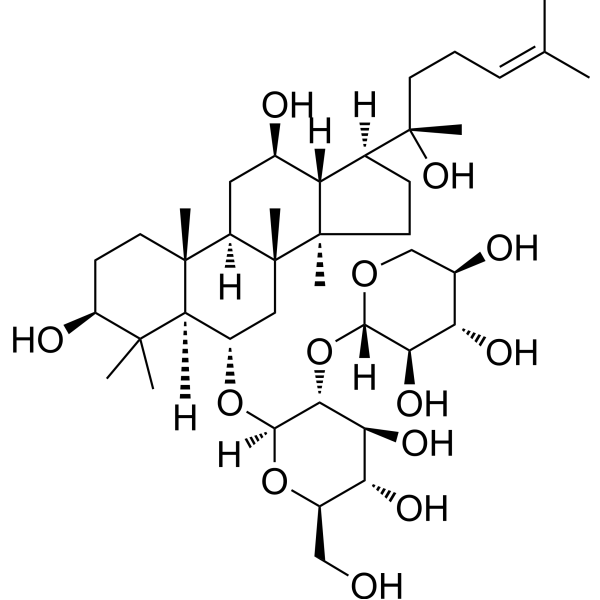
Notoginsenoside R2 (20(S)-Notoginsenoside R2; Ginsenoside Ng-R2) is an orally active notoginsenoside. Notoginsenoside R2 activates P90RSK and Nrf2 via the MEK1/2-ERK1/2 pathway to inhibit 6-OHDA-induced apoptotic damage in nerve cells. Notoginsenoside R2 upregulates SOX8/β-catenin by reducing miR-27a, thereby suppressing Aβ25-35-induced neuronal apoptosis and inflammatory responses. Notoginsenoside R2 alleviates lipid accumulation and mitochondrial dysfunction in diabetic nephropathy by inhibiting c-Src. Notoginsenoside R2 alleviates hepatic fibrosis by inducing hepatic stellate cell senescence and inhibiting the inflammatory microenvironment via JAK/STAT3 suppression. Notoginsenoside R2 can be used in research related to Parkinson's disease, Alzheimer's disease, diabetic nephropathy and hepatic fibrosis.
연구목적의 판매만을 진행합니다. 환자를 대상으로 한 판매는 하지 않습니다.
- Purity : 99.80%
- CAS No.: 80418-25-3
- 화학식: C41H70O13
- 분자량:770.99
-
보관:
-20°C, protect from light
* In solvent : -80°C, 6 months; -20°C, 1 month (protect from light)

To place orders, for customer services and technical support, please contact: MedChemExpress USA
Tel: 609-228-6898 E-mail: [email protected] [email protected]


-
 Biological Activity
-
Chemical Information
-
용액&용해도
- Protocol
- 순도&문서
- References
-
 Help & FAQs


-
HY-N0044
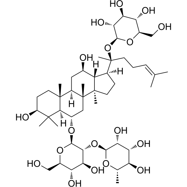
Ginsenoside ReEndogenous Metabolite -
HY-N0602
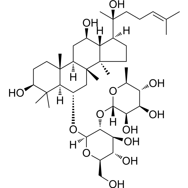
Ginsenoside Rg2NF-κB Inhibitor -
HY-N2530
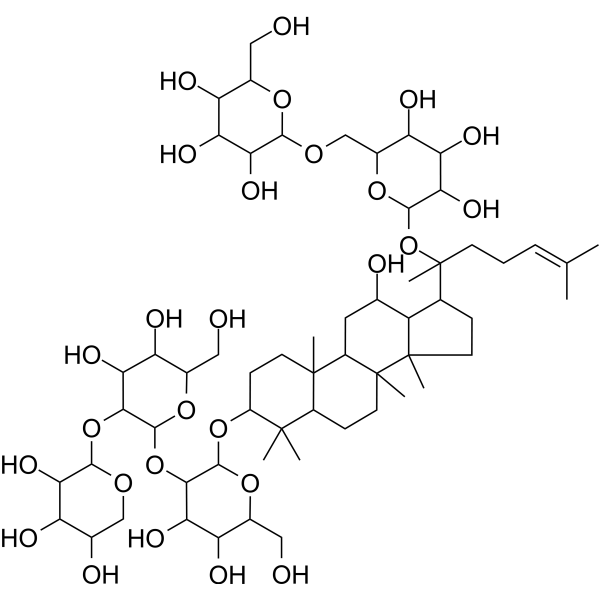
Notoginsenoside FaProtopanaxadiol (ppd)-Type Saponin -
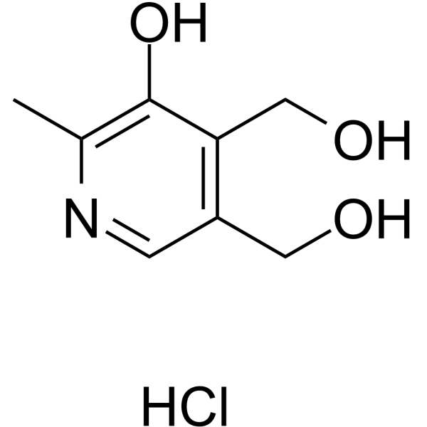
HY-N0682
Pyridoxine hydrochloridePyridine Derivative -

HY-N0682R
Pyridoxine hydrochloride (Standard)Pyridine Derivative -

HY-N0909R
Notoginsenoside R2 (Standard)Triterpenoid Saponin
Publications Citing Use of MedChemExpress (MCE) Notoginsenoside R2
MoreAll MEK Isoforms
MoreAll Caspase Isoforms
More

Biological Activity
제품 설명
In Vitro
Notoginsenoside R2 (20 μM; 24 h) protects SH-SY5Y cells against 6-OHDA (HY-B1081)-induced cell death, with reduced counts of TUNEL-positive cells and decreased overall apoptosis rate[1].
Notoginsenoside R2 (20 μM; 24 h) reduces 6-OHDA-induced LDH release in SH-SY5Y cells, PC12 cells, and primary cortical neurons[1].
Notoginsenoside R2 (20 μM; 24 h) inhibits 6-OHDA-induced oxidative stress in SH-SY5Y cells, as evidenced by decreased intracellular ROS production and MDA levels, and reverses mitochondrial membrane depolarization in cells[1].
Notoginsenoside R2 (20 μM) increases the activities of HO-1, GSH-PX and GR in SH-SY5Y cells, and pre-incubation for 24 h reverses the 6-OHDA-induced decreases in the activities of these enzymes and the reduced nuclear accumulation level of Nrf2[1].
Notoginsenoside R2 (20 μM; 4-24 h) activates the MEK1/2-ERK1/2 signaling pathway in SH-SY5Y cells, thereby inducing the activation of downstream P90RSK and Nrf2. The latter mediates the inhibition of 6-OHDA-induced apoptotic protein expression. Notably, the activation of the aforementioned pathway depends on MEK1/2-ERK1/2 signal transduction, but is independent of JNK, p38 or PI3K/Akt signal transduction[1].
Notoginsenoside R2 (10-30 μM) dose-dependently attenuates Aβ25-35 (HY-P0128)-induced apoptosis of primary rat cortical neurons, reduces the expression levels of activated caspase-3 and miR-27a, downregulates COX-2 expression, upregulates the mRNA and protein expression of SOX8, and increases the expression of total β-catenin protein and nuclear β-catenin protein[2].
Notoginsenoside R2 (10-40 μM; 24 h) attenuates lipid accumulation, oxidative stress, mitochondrial dysfunction and apoptosis in HK-2 cells stimulated with high glucose and palmitic acid (HGPA) by inhibiting c-Src activation[3].
Notoginsenoside R2 (10-100 μM; 24 h) inhibits the proliferation of HSC-T6 cells[5].
Notoginsenoside R2 (20-30 μM; 24 h) downregulates the mRNA and protein expressions of α-SMA and COL-I in HSC-T6 cells[5].
Notoginsenoside R2 (20 μM; 24 h) downregulates fibrosis markers (α-SMA, COL-I, Desmin, TIMP-1) and upregulates senescence markers (p16, p21, p53) in HSC-T6 cells[5].
Notoginsenoside R2 (20 μM; 24 h) inhibits the JAK/STAT3 signaling pathway in HSC-T6 cells[5].
Notoginsenoside R2 (20 μM; 24 h) inhibits the mRNA expression of proinflammatory cytokines in HSC-T6 cells[5].
Notoginsenoside R2 (20 μM; 24 h) upregulates the mRNA expression of SASP components in HSC-T6 cells[5].
Notoginsenoside R2 (20 μM; 24 h) reduces intracellular ROS levels in HSC-T6 cells[5].
Notoginsenoside R2 (20 μM; 24 h) induces senescence in HSC-T6 cells[5].
MedChemExpress (MCE) has not independently confirmed the accuracy of these methods. They are for reference only. Further protocols information, click here.
-
Cell Line:SH-SY5Y human neuroblastoma cells
-
Concentration:20 μM
-
Incubation Time:24 h (pre-incubation)
-
Result:Significantly reduced the number of TUNEL-positive cells and the overall apoptosis rate induced by 6-OHDA.
Had no effect on apoptosis rate or TUNEL-positive cell count when treated alone.
-
Cell Line:SH-SY5Y human neuroblastoma cells
-
Concentration:20 μM
-
Incubation Time:4 h, 8 h, 16 h, 20 h, 24 h
-
Result:Increased phosphorylation of P90RSK, BAD, MEK1/2, and ERK1/2, and enhanced nuclear Nrf2 accumulation in a time-dependent manner when treated alone, with MEK1/2 phosphorylation peaking at 16 h and ERK1/2 phosphorylation peaking at 20 h.
Blocked 6-OHDA-induced increases in cytochrome c release, cleaved caspase-9, and cleaved caspase-3, and reversed 6-OHDA-induced reductions in p-BAD levels when pre-incubated for 24 h.
Induced P90RSK phosphorylation and nuclear Nrf2 accumulation were prevented by pre-incubation with MEK1/2 inhibitor PD98059, while JNK, p38, or PI3K/Akt inhibitors had no effect.
Induced phosphorylation of ERK1/2, P90RSK, and BAD, as well as nuclear Nrf2 accumulation and increases in HO-1, GSH-PX, and GR activities were blocked by MEK1/2 or ERK1/2 siRNA transfection.
Induced increases in HO-1, GSH-PX, and GR activities were blocked by Nrf2 siRNA transfection.
-
Cell Line:HSC-T6 hepatic stellate cells
-
Concentration:10, 20, 40, 80, 100 μM
-
Incubation Time:24 h
-
Result:Suppressed HSC-T6 cell proliferation in a dose-dependent manner at concentrations of 40-100 μM.
Caused statistically significant reductions in cell viability at 80 μM and 100 μM relative to the normal control.
-
Cell Line:HSC-T6 cells
-
Concentration:20, 30 μM
-
Incubation Time:24 h
-
Result:Caused a marked reduction in mRNA expression of α-SMA relative to the normal control.
Caused a marked reduction in mRNA expression of COL-I relative to the normal control.
-
Cell Line:HSC-T6 cells
-
Concentration:20 μM
-
Incubation Time:24 h
-
Result:Significantly decreased the area of α-SMA staining in HSC-T6 cells relative to the normal control.
Significantly decreased the area of COL-I staining in HSC-T6 cells relative to the normal control.
-
Cell Line:HSC-T6 cells
-
Concentration:20 μM
-
Incubation Time:24 h
-
Result:Significantly reduced protein expression of α-SMA relative to the normal control.
Significantly reduced protein expression of COL-I) relative to the normal control.
Significantly reduced protein expression of Desmin relative to the normal control.
Significantly reduced protein expression of TIMP-1 relative to the normal control.
Significantly increased protein expression of p16 relative to the normal control.
Significantly increased protein expression of p21 relative to the normal control.
Significantly increased protein expression of p53 relative to the normal control.\nSignificantly reduced the phosphorylation of JAK relative to the normal control.
Significantly reduced the phosphorylation of STAT3 relative to the normal control.
Left total JAK and STAT3 protein levels unchanged.
-
Cell Line:HSC-T6 cells
-
Concentration:20 μM
-
Incubation Time:24 h
-
Result:Significantly reduced mRNA expression of pro-inflammatory mediators including IL-6, IL-1β, CCL2, IL-8, and TNF-α relative to the normal control.\nSignificantly upregulated mRNA expression of SASP components including p16, p21, p53, and MMP10 relative to the normal control.
In Vivo
Notoginsenoside R2 (250 mg/kg; i.p.; once daily; for 20 consecutive weeks) improves cognitive function in Alzheimer's disease rats induced by Aβ25-35, reduces hippocampal neuronal apoptosis, and regulates the expression of apoptosis- and inflammation-related proteins[2].
Notoginsenoside R2 (20-40 mg/kg, oral administration daily for 8 weeks) improves renal dysfunction, histopathological damage, lipid accumulation, oxidative stress, mitochondrial dysfunction and apoptosis in db/db mice, a model of diabetic nephropathy. This effect is partially achieved by inhibiting the activation of c-Src[3].
Notoginsenoside R2 (6.25-25 μM; administered 72 hours after TAA pretreatment) alleviates TAA (HY-Y0698)-induced liver fibrosis in zebrafish in a dose-dependent manner by reducing oxidative stress, suppressing inflammatory responses, and inducing hepatic stellate cell senescence via a STAT3-dependent pathway[5].
MedChemExpress (MCE) has not independently confirmed the accuracy of these methods. They are for reference only.
-
Animal Model:SAMP8 (male, 3-month-old, 20-30 g, spontaneous Alzheimer's disease model); SAMR1 (male, normal aging control)[2]
-
Dosage:250 mg/kg
-
Administration:i.p.; daily; 20 consecutive weeks
-
Result:Reduced learning reaction time to 25.23 s.
Reduced learning error time to 4.52.
Increased memory reaction time to 180 s.
Reduced memory error time to 5.41.
Alleviated hippocampal neuronal disorganization and loss of pyramidal cells.
Reduced TUNEL-positive apoptotic cells compared to the AD group.
Decreased cleaved caspase-3 and COX-2 protein expression compared to the AD group.
Increased SOX8 and β-catenin protein expression compared to the AD group.
-
Animal Model:Sprague-Dawley (male, 18-20 months-old, 200-220 g, Aβ25-35-induced Alzheimer's disease model via bilateral hippocampal injection); Sprague-Dawley (male, 3-6 months-old, control)[2]
-
Dosage:250 mg/kg
-
Administration:i.p.; daily; 20 consecutive weeks
-
Result:Reduced learning reaction time to 25.23 s.
Reduced learning error time to 3.98.
Increased memory reaction time to 182.32 s.
Reduced memory error time to 6.23.
Restored hippocampal neuronal structure compared to the Aβ25-35-treated group.
Reduced TUNEL-positive apoptotic cells compared to the Aβ25-35-treated group.
Decreased cleaved caspase-3 and COX-2 protein expression compared to the Aβ25-35-treated group.
Increased SOX8 and β-catenin protein expression compared to the Aβ25-35-treated group.
-
Animal Model:C57BKS/Lepr db/db (male, 7-8 weeks old, spontaneous diabetic nephropathy with elevated urine albumin-to-creatinine ratios)[3]
-
Dosage:20 mg/kg/day; 40 mg/kg/day
-
Administration:p.o.; daily; 8 weeks
-
Result:Reduced urine albumin-to-creatinine ratios (ACR) levels compared to untreated db/db mice, with 40 mg/kg dose causing greater reduction than positive control Losartan.
Significantly lowered serum creatinine (Scr) and blood urea nitrogen (BUN) levels relative to untreated db/db mice at 40 mg/kg dose; significantly reduced Scr levels at 20 mg/kg dose.
Significantly reduced serum total cholesterol (TC) levels compared to untreated db/db mice at 20 mg/kg dose; reduced TC levels to a greater, statistically significant extent at 40 mg/kg dose.
Left serum triglyceride (TG) levels unchanged with either dose.
Unaffected liver function markers (ALT, AST), indicating no liver toxicity.
Ameliorated renal histopathological abnormalities including mesangial expansion, glycogen deposition, collagen fiber deposition, and podocyte foot process effacement, and significantly reduced renal expression of fibrosis-related proteins (fibronectin, vimentin, α-SMA) at both doses.
Significantly reduced intrarenal lipid accumulation (measured by Oil Red O staining and renal triglyceride content), and suppressed renal expression of phosphorylated c-Src (Tyr416), phosphorylated JNK, phosphorylated STAT1, and CD36 at both doses.
Reduced renal malondialdehyde (MDA) levels, increased superoxide dismutase (SOD), glutathione (GSH), glutathione reductase (GR), catalase (CAT), and ATP levels relative to untreated db/db mice at both doses.
Downregulated renal expression of mitochondrial fission proteins Drp1 and Fis1, and upregulated the mitochondrial fusion protein Mfn2 at both doses.
Reduced renal TUNEL-positive apoptotic cells, lowered expression of pro-apoptotic proteins (Bax, cleaved-caspase-3), and increased expression of the anti-apoptotic protein Bcl-2 at both doses.
-
Animal Model:wild-type AB strain; transgenic Tg(lfabp10α:eGFP) strain (2-day post-fertilization larvae)[5]
-
Dosage:6.25 μM; 12.5 μM; 25 μM (general); 25 μM (STAT3-knockdown zebrafish)
-
Administration:administered following 72-hour TAA preconditioning
-
Result:Caused no notable morphological abnormalities, developmental delays, or hatching rate differences compared to untreated controls at 6.25 μM, 12.5 μM, and 25 μM.
Significantly mitigated TAA-induced hepatic damage (reduced intercellular gaps, hepatocellular enlargement, and vacuolation) as observed via H&E staining, and dose-dependently reduced mRNA levels of fibrotic markers α-SMA and COL-I at 6.25 μM, 12.5 μM, and 25 μM.
Significantly reduced hepatic reactive oxygen species (ROS) levels, reduced mRNA expression of pro-inflammatory cytokines (IL-6, IL-1β, CCL2, IL-8, TNF-α), reduced STAT3 protein levels and JAK/STAT3 pathway gene expression, and significantly upregulated protein and mRNA levels of senescence biomarkers p16 and p21, plus SASP components including p53 at 25 μM.
Lost antifibrotic effects in STAT3-knockdown zebrafish: no longer mitigated hepatic vacuolation/tissue disorganization, failed to reduce α-SMA and COL-I mRNA levels, showed diminished ROS suppression, lost anti-inflammatory activity, and no longer upregulated senescence biomarkers p16 and p21 or SASP components at 25 μM.


Chemical Information
-
CAS No. 80418-25-3
-
Appearance Solid
-
분자량 770.99
-
화학식 C41H70O13
-
Color White to off-white
-
SMILES
C[C@]([C@@](C[C@H]1O)([H])[C@]2(CC[C@@H]3O)C)(C[C@H](O[C@@](O[C@H](CO)[C@@H](O)[C@@H]4O)([H])[C@@H]4O[C@@](OC[C@@H](O)[C@@H]5O)([H])[C@@H]5O)[C@@]2([H])C3(C)C)[C@]6([C@@]1([H])[C@]([C@@](C)(O)CC/C=C(C)/C)([H])CC6)C
-
Synonyms
20(S)-Notoginsenoside R2; Ginsenoside Ng-R2
-
Structure Classification
-
선적
Room temperature in continental US; may vary elsewhere.
-
보관
-20°C, protect from light
* In solvent : -80°C, 6 months; -20°C, 1 month (protect from light)


Publications (1)
-
Journal Impact Factor
-
Most Recent
-
J Ginseng Res
Notoginsenoside R2 attenuates hepatic fibrosis via STAT3-dependent hepatic stellate cells senescence induction and inflammatory microenvironment suppression. [Abstract]2025 Sep;49(5):574-584. PMID: 40843014


용액&용해도
In Vitro:
DMSO : 100 mg/mL (129.70 mM; Need ultrasonic; Hygroscopic DMSO has a significant impact on the solubility of product, please use newly opened DMSO)
Please refer to the solubility information to select the appropriate solvent. Once prepared, please aliquot and store the solution to prevent product inactivation from repeated freeze-thaw cycles.
Storage method and period of stock solution: -80°C, 6 months; -20°C, 1 month (protect from light). When stored at -80°C, please use it within 6 months. When stored at -20°C, please use it within 1 month.
Please refer to the solubility information to select the appropriate solvent. Once prepared, please aliquot and store the solution to prevent product inactivation from repeated freeze-thaw cycles.
Storage method and period of stock solution: -80°C, 6 months; -20°C, 1 month (protect from light). When stored at -80°C, please use it within 6 months. When stored at -20°C, please use it within 1 month.
Concentration (start) × Volume (start) = Concentration (final) × Volume (final)
In Vivo:
Select the appropriate dissolution method based on your experimental animal and administration route.
- For the following dissolution methods, please ensure to first prepare a clear stock solution using an In Vitro approach and then sequentially add co-solvents:
- To ensure reliable experimental results, the clarified stock solution can be appropriately stored based on storage conditions. As for the working solution for In Vivo experiments, it is recommended to prepare freshly and use it on the same day.
- The percentages shown for the solvents indicate their volumetric ratio in the final prepared solution. If precipitation or phase separation occurs during preparation, heat and/or sonication can be used to aid dissolution.
Add each solvent one by one: 10% DMSO 40% PEG300 5% Tween-80 45% Saline
Solubility: ≥ 2.5 mg/mL (3.24 mM); Clear solution
This protocol yields a clear solution of ≥ 2.5 mg/mL (saturation unknown).
Taking 1 mL working solution as an example, add 100 μL DMSO stock solution (25.0 mg/mL) to 400 μL PEG300, and mix evenly; then add 50 μL Tween-80 and mix evenly; then add 450 μL Saline to adjust the volume to 1 mL.
Preparation of Saline: Dissolve 0.9 g sodium chloride in ddH₂O and dilute to 100 mL to obtain a clear Saline solution.
Add each solvent one by one: 10% DMSO 90% (20% SBE-β-CD in Saline)
Solubility: ≥ 2.5 mg/mL (3.24 mM); Clear solution
This protocol yields a clear solution of ≥ 2.5 mg/mL (saturation unknown).
Taking 1 mL working solution as an example, add 100 μL DMSO stock solution (25.0 mg/mL) to 900 μL 20% SBE-β-CD in Saline, and mix evenly.
Preparation of 20% SBE-β-CD in Saline (4°C, storage for one week): 2 g SBE-β-CD powder is dissolved in 10 mL Saline, completely dissolve until clear.
In Vivo Dissolution Calculator
Please enter the basic information of animal experiments:
-
-
-
-
Recommended: Prepare an additional quantity of animals to account for potential losses during experiments.
Please enter your animal formula composition:
-
%DMSO +
Recommended: Keep the proportion of DMSO in working solution below 2% if your animal is weak.
-
%+
-
+%Tween-80 + +
-
%Saline +
The co-solvents required include: DMSO, . All of co-solvents are available by MedChemExpress (MCE). , Tween 80. All of co-solvents are available by MedChemExpress (MCE).
Working solution concentration: 0.22 mg/mL
Method for preparing stock solution: mg drug dissolved in μL DMSO. Stock solution concentration: mg/mL. * In solvent : -80°C, 6 months; -20°C, 1 month (protect from light)
1. Take μL DMSO stock solution;
2. Add μL .
μL , mix evenly;
3. Then add μL Tween 80, mix evenly;
4. Then add μL
Please ensure that the stock solution in the first step is dissolved to a clear state, and add co-solvents in sequence. You can use ultrasonic heating (ultrasonic cleaner, recommended frequency 20-40 kHz), vortexing, etc. to assist dissolution.

Protocol
-
Collagen: Sirius Red Staining
Sirius Red or picrosirius red staining is a histochemical method for visualizing collagen-rich extracellular matrix in tissue sections, and collagen fibers are detected as red-stained structures under bright-field microscopy with enhanced birefringence under polarized light. Picrosirius red is useful for assessing total collagen organization, distribution, and fibrosis burden, but polarized color should not be interpreted as a definitive collagen type I versus type III readout because color is affected by fiber orientation, thickness, and packing.
-
Annexin V plus membrane-impermeant dye apoptosis staining
Annexin V-based apoptosis assays rely on the detection of phosphatidylserine (PS) externalization from the inner leaflet of the plasma membrane to the outer leaflet, an early biochemical hallmark of apoptosis. Fluorescently labeled Annexin V binds PS in a calcium-dependent manner, enabling identification of early apoptotic cells by flow cytometry or fluorescence microscopy. When combined with a membrane-impermeant DNA-binding dye (e. g. , propidium iodide), this approach allows discrimination between viable (Annexin V−/dye−), early apoptotic (Annexin V+/dye−), and late apoptotic or necrotic (Annexin V+/dye+) cell populations by assessing membrane integrity and PS exposure.
-
Connective Tissue: Masson's Trichrome/Collagen Trichrome Staining
Masson’s Trichrome (collagen/trichrome staining) is a histological technique that differentially stains tissue compartments using sequential acidic dyes to distinguish collagen from muscle and cytoplasmic components based on dye affinity and tissue permeability differences, enabling visualization of fibrosis and connective tissue architecture in histological sections. The classical formulation typically uses Weigert's iron hematoxylin for nuclear staining, Biebrich scarlet-acid fuchsin for cytoplasm and muscle, and aniline blue (or light green variants) for collagen, producing a characteristic blue/green collagen signal contrasted against red cytoplasm and dark nuclei. The staining principle relies on selective displacement of smaller dye molecules by larger anionic dyes in collagen-rich regions under controlled acidified conditions, which enhances collagen-specific dye retention. This property makes the method widely used for fibrosis assessment in organs such as heart, liver, lung, a
-
Senescence-associated β-galactosidase staining
Senescence-associated β-galactosidase staining detects β-galactosidase activity that is histochemically visible at pH 6. 0 in senescent cells, where X-gal cleavage produces an insoluble blue precipitate observable by bright-field microscopy. This activity reflects increased lysosomal β-galactosidase/lysosomal mass rather than a senescence-essential enzyme, because GLB1 depletion or genetic lysosomal β-galactosidase deficiency can abolish SA-β-gal staining while cells still undergo senescence. SA-β-gal was originally reported in senescent but not presenescent fibroblasts and keratinocytes, absent from quiescent fibroblasts and terminally differentiated keratinocytes, and increased with donor age in human skin samples. Because SA-β-gal can also appear in some non-senescent or tissue-specific contexts, interpretation should be paired with experimental controls and, when possible, independent senescence markers.
-
Alzheimer’s Disease Modeling
Alzheimer’s Disease (AD) is a neurodegenerative disorder characterized by a progressive decline in cognitive functions and loss of specific types of neurons and synapses. Alzheimer's symptoms can be simulated in mice by injecting drugs (such as Aβ) or genetically modified.
-
Fibrosis/Collagen Morphometry
Fibrosis and collagen morphometry is based on the quantitative visualization of fibrillar collagen deposition in tissue sections using histochemical stains such as Sirius Red (Picrosirius Red) or Masson's trichrome, followed by image-based or polarization-enhanced analysis to estimate collagen proportional area as a surrogate of extracellular matrix accumulation during fibrotic remodeling. Sirius Red combined with polarized light microscopy enhances detection of collagen fibers due to birefringence properties, enabling more specific visualization of collagen type I and III fibrils compared to conventional bright-field histology, while whole-section or region-restricted digital morphometry reduces field-selection bias in fibrosis assessment. Alternative quantitative approaches include second harmonic generation (SHG) and two-photon excited fluorescence microscopy, which enable label-free detection of fibrillar collagen and have been validated against histological staining and biochemica
-
3T3-L1 preadipocyte-to-adipocyte differentiation
3T3-L1 preadipocytes are induced to differentiate after growth arrest using adipogenic media containing insulin, dexamethasone, and IBMX; differentiation is assessed by lipid-droplet accumulation, triglyceride increase, Oil Red O staining, and adipocyte-marker induction such as PPARγ and C/EBPα.
-
Apoptosis
Apoptosis, also called programmed cell death, is generally characterized by distinct morphological characteristics.
-
TUNEL staining for apoptotic DNA fragmentation
TUNEL staining detects DNA strand breaks by using terminal deoxynucleotidyl transferase to add labeled nucleotides to exposed 3′-OH DNA termini, generating either microscopic staining in fixed cells or tissue sections, or fluorescence/cytometric signal in cell suspensions. TUNEL positivity reflects DNA fragmentation but should not be interpreted alone as definitive apoptosis, because TUNEL can also label necrotic, autolytic, mechanically damaged, or DNA-repair-associated DNA breaks.
-
Human pluripotent stem cell midbrain dopaminergic neuron differentiation
Human pluripotent stem cells are directed toward midbrain dopaminergic neurons by first inducing a neural floor-plate-like progenitor state, then patterning cells with ventralizing SHH signaling and midbrain/WNT-FGF cues, and finally maturing progenitors into neurons expressing dopaminergic markers such as TH, NURR1/NR4A2, PITX3, DAT/SLC6A3, VMAT2/SLC18A2, GIRK2/KCNJ6, FOXA2, LMX1A, and EN1. The main readouts are loss of pluripotency, acquisition of FOXA2+/LMX1A+ midbrain floor-plate progenitors, emergence of βIII-tubulin+/MAP2+ neurons, and production of TH+ dopaminergic neurons with molecular, dopamine-release, and electrophysiological features of midbrain dopaminergic identity.
-
Mitochondrial membrane-potential and mitochondrial mass staining
Mitochondrial membrane potential staining measures the electrochemical polarization across the mitochondrial inner membrane in live cells using lipophilic cationic fluorescent probes; early rhodamine-based work showed that selective mitochondrial dye accumulation is lost when the mitochondrial transmembrane potential is dissipated. JC-1 reports mitochondrial polarization by shifting from green monomer fluorescence to red J-aggregate fluorescence as dye concentration increases within energized mitochondria; therefore, the red/green fluorescence ratio is used as a relative readout of mitochondrial membrane potential. TMRE or TMRM staining provides a single-channel relative readout because these cationic rhodamine esters accumulate in polarized mitochondria, and lower fluorescence indicates reduced mitochondrial polarization when acquisition and dye-loading conditions are controlled. Mitochondrial mass staining is commonly performed with MitoTracker Green FM or related MitoTracker dyes as
-
Apoptosis Solutions
Apoptosis is a regulated, generally non-lytic cell-death pathway that removes unwanted, damaged, infected, or abnormal cells through coordinated morphological changes, caspase activation, DNA fragmentation, and membrane remodeling. The intrinsic apoptosis pathway is controlled mainly by mitochondrial outer membrane permeabilization, BCL-2 family proteins, cytochrome c release, apoptosome formation, caspase-9 activation, and downstream executioner caspase-3/7 activation. The extrinsic apoptosis pathway is initiated by death receptors such as Fas, TNFR, and TRAIL receptors, which recruit adaptor proteins and activate caspase-8 before engaging executioner caspases or mitochondrial amplification through BID cleavage. Apoptosis is linked to many phenotypes, including cancer cell killing, tissue homeostasis, immune regulation, neurodegeneration, infection response, and treatment-induced cytotoxicity; unresolved questions include how apoptosis interacts with necroptosis, pyroptosis, ferroptos
-
Lipid Droplets: Oil Red O/Sudan Dye Lipid Staining
Lipid droplets are intracellular organelles with a neutral-lipid core that stores triacylglycerols and sterol esters, and Oil Red O or Sudan dyes detect these hydrophobic lipid deposits by partitioning into retained lipids in fresh or frozen specimens. Oil Red O stains neutral triglycerides and lipids in frozen tissue sections or air-dried cytologic preparations, while Sudan Black B has also been used as a histochemical fat stain for lipid-rich tissue structures.


순도&문서
-
Data Sheet (306 KB)
-
SDS (393 KB)
- English - EN (393 KB)
- Français - FR (393 KB)
- Deutsch - DE (393 KB)
- Norwegian - NO (393 KB)
- Español - ES (393 KB)
- Swedish - SV (393 KB)
- Italian - IT (393 KB)
- Korean - KR (393 KB)
- Portuguese - PT (393 KB)
-
Handling Instructions (2659 KB)
References
[1]. Meng XB, et al. P90RSK and Nrf2 Activation via MEK1/2-ERK1/2 Pathways Mediated by Notoginsenoside R2 to Prevent 6-Hydroxydopamine-Induced Apoptotic Death in SH-SY5Y Cells. Evid Based Complement Alternat Med. 2013;2013:971712. [Content Brief]
[2]. Hu Y, et al. Notoginsenoside R2 reduces Aβ25-35-induced neuronal apoptosis and inflammation via miR-27a/SOX8/β-catenin axis. Hum Exp Toxicol. 2021 Dec;40(12_suppl):S347-S358. [Content Brief]
[3]. Guo X, et al. Protective effects of Notoginsenoside R2 on reducing lipid accumulation and mitochondrial dysfunction in diabetic nephropathy through regulation of c-Src. Chin Med. 2025;20(1):10. Published 2025 Jan 15. [Content Brief]
[4]. González-Burgos E, et al. Potential neuroprotective activity of Ginseng in Parkinson's disease: a review. J Neuroimmune Pharmacol. 2015;10(1):14-29. [Content Brief]
[5]. Deng K, et al. Notoginsenoside R2 attenuates hepatic fibrosis via STAT3-dependent hepatic stellate cells senescence induction and inflammatory microenvironment suppression. J Ginseng Res. 2025;49(5):574-584. [Content Brief]


Complete Stock Solution Preparation Table
Please refer to the solubility information to select the appropriate solvent. Once prepared, please aliquot and store the solution to prevent product inactivation from repeated freeze-thaw cycles.
Storage method and period of stock solution: -80°C, 6 months; -20°C, 1 month (protect from light). When stored at -80°C, please use it within 6 months. When stored at -20°C, please use it within 1 month.
| Optional Solvent | Concentration Solvent Mass | 1 mg | 5 mg | 10 mg | 25 mg |
|---|---|---|---|---|---|
| DMSO | 1 mM | 1.2970 mL | 6.4852 mL | 12.9703 mL | 32.4258 mL |
| 5 mM | 0.2594 mL | 1.2970 mL | 2.5941 mL | 6.4852 mL | |
| 10 mM | 0.1297 mL | 0.6485 mL | 1.2970 mL | 3.2426 mL | |
| 15 mM | 0.0865 mL | 0.4323 mL | 0.8647 mL | 2.1617 mL | |
| 20 mM | 0.0649 mL | 0.3243 mL | 0.6485 mL | 1.6213 mL | |
| 25 mM | 0.0519 mL | 0.2594 mL | 0.5188 mL | 1.2970 mL | |
| 30 mM | 0.0432 mL | 0.2162 mL | 0.4323 mL | 1.0809 mL | |
| 40 mM | 0.0324 mL | 0.1621 mL | 0.3243 mL | 0.8106 mL | |
| 50 mM | 0.0259 mL | 0.1297 mL | 0.2594 mL | 0.6485 mL | |
| 60 mM | 0.0216 mL | 0.1081 mL | 0.2162 mL | 0.5404 mL | |
| 80 mM | 0.0162 mL | 0.0811 mL | 0.1621 mL | 0.4053 mL | |
| 100 mM | 0.0130 mL | 0.0649 mL | 0.1297 mL | 0.3243 mL |


Notoginsenoside R2 Related Classifications
HY-N0909 Related Classifications
 Powered by Bioz
Keywords
- Notoginsenoside R2
- 80418-25-3
- 20(S)-Notoginsenoside R2
- Ginsenoside Ng-R2
- Notoginsenoside R 2
- Notoginsenoside R-2
- Apoptosis
- MEK
- ERK
- Reactive Oxygen Species (ROS)
- Caspase
- COX
- β-catenin
- Src
- MDM-2/p53
- JAK
- STAT
- HK-2 cells
- Nrf2
- Parkinson’s disease
- PC12 cells
- P90RSK
- MEK1/2
- primary cortical neurons
- SH-SY5Y cells
- HSC-T6 cells
- ERK1/2
- Alzheimer's disease
- diabetic nephropathy
- hepatic fibrosis
- SAMP8 mice
- Sprague-Dawley rat
- C57BKS/Lepr db/db mice
- Inhibitor
- inhibitor
- inhibit