



AR420626
Based on 4 publication(s) in Google Scholar
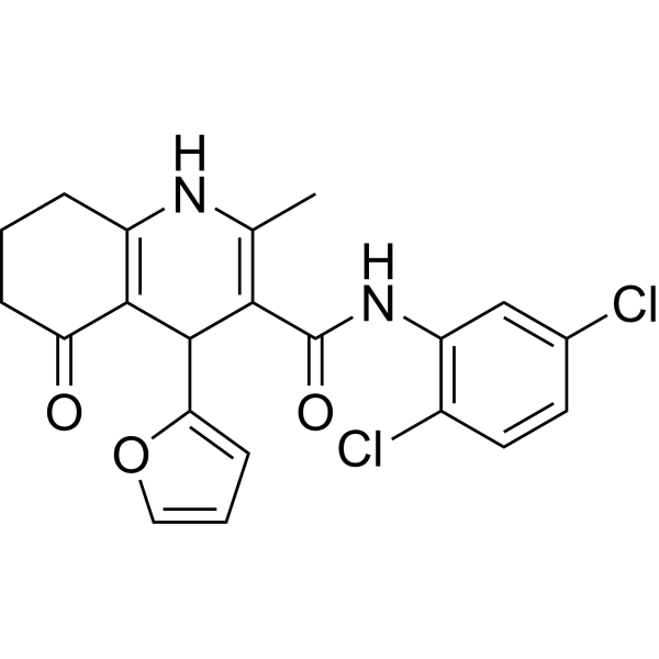
AR420626 is a selective agonist of free fatty acid receptor 3 (FFAR3) (IC50=117 nM). AR420626 has anti-inflammatory, anticancer and antidiabetic activities. AR420626 improves neurogenic diarrhea by inhibiting nAChR mediated neural pathways. AR420626 inhibits the growth of HepG2 xenografts and inhibits the proliferation of hepatoma cells by inducing apoptosis. AR420626 also suppresses allergic asthma and eczema and has the ability to activate GPR41 to increase Ca2+ signal-mediated glucose uptake and improve diabetes.
For research use only. We do not sell to patients.
- Purity : 98.40%
- CAS No.: 1798310-55-0
- Formula: C21H18Cl2N2O3
- Molecular Weight:417.29
-
Storage:Powder -20°C, 3 years , 4°C, 2 years ; In solvent -80°C, 6 months , -20°C, 1 month

To place orders, for customer services and technical support, please contact: MedChemExpress USA
Tel: 609-228-6898 E-mail: [email protected] [email protected]


-
 Biological Activity
-
Chemical Information
-
Solvent & Solubility
- Protocols
- Purity & Documentation
- References
-
 Help & FAQs


-
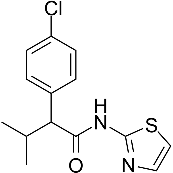
HY-P1125
4-CMTBFFA2 Agonist -
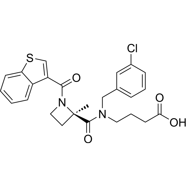
HY-12940
GLPG0974FFA2/GPR43 Antagonist -
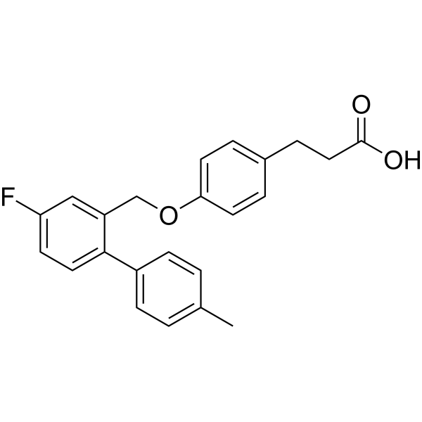
HY-100881
TUG-891FFA4/GPR120 Agonist -
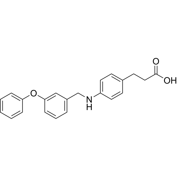
HY-15589
GW9508GPR40 Agonist -
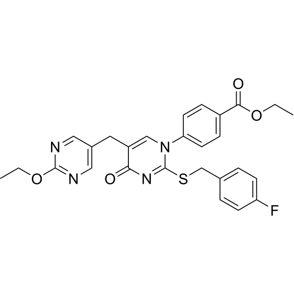
HY-50691
GW-1100Free Fatty Acid Receptor Antagonist -
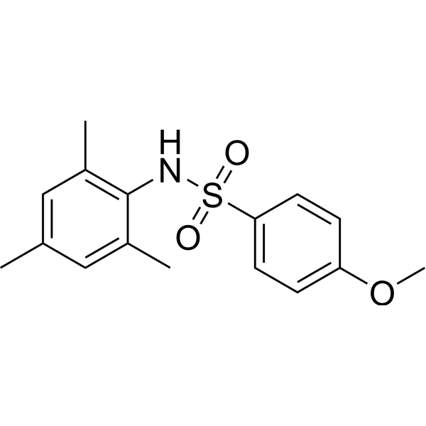
HY-19995
GSK137647AFFA4 Agonist -
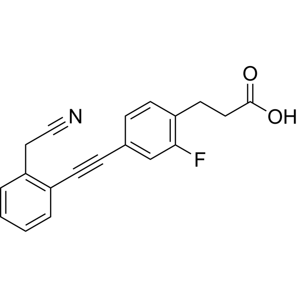
HY-15697
TUG-770GPR40/FFA1 Agonist -
HY-112813
TUG-1375FFA2/GPR43 Agonist -
HY-B0143
NiacinB3 vitamin -
HY-12764
6-OAUGPR84 Agonist -
HY-103252
Monomethyl fumarateGPR109A Agonist -
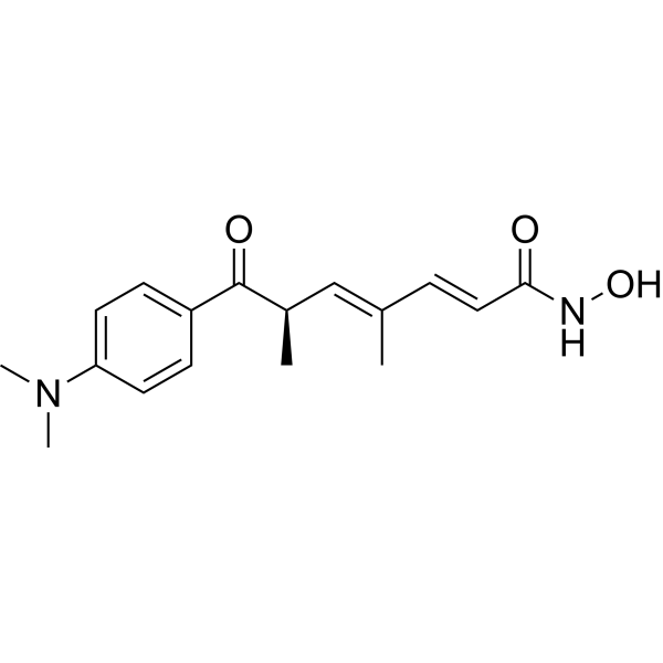
HY-15144
Trichostatin AHDAC Class I/II Inhibitor
Publications Citing Use of MedChemExpress (MCE) AR420626
MoreAll Histone Methyltransferase Isoforms
More

Biological Activity
Description
In Vitro
AR420626 (10 μM) inhibits 100 μM nicotine-induced contraction and 10 μM nicotine-induced relaxation of rat proximal colon circular muscle strips[1]. AR420626 (10 μM) reverses NO-mediated relaxation of circular muscles induced by 5-HT (100 μM)[1]. AR420626 (10 and 25 µM; 48 h) inhibits the proliferation of HLE cells and HepG2 cells at a concentration of 25 µM for 48 h, and induces apoptosis of HepG2 cells and HLE cells by inhibiting HDAC-induced TNF-α[2]. AR420626 (25 µM; 1, 3, 12 and 24 h) induces mTOR phosphorylation at the 1 h time point and for 24 h in HepG2 and HLE cells [2]. AR420626 (0.25, 0.5 and 1 µM; 1 h) increases Ca2+ influx and GLUT4 translocation via activation of CaMKII, CREB and p38 in C2C12 myotubes, thereby enhancing basal and insulin-stimulated glucose uptake[4].
MedChemExpress (MCE) has not independently confirmed the accuracy of these methods. They are for reference only. Further protocols information, click here.
-
Cell Line:HepG2 cells and HLE cells
-
Concentration:10, 25 µM
-
Incubation Time:24, 48 and 72 h
-
Result:Inhibited the proliferation of HepG2 cells at 25 µM for 24 h, 10 µM and 25 µM for 48 h and 72 h, and inhibited the proliferation of HLE cells at 25 µM for 48 h, 10 µM and 25 µM for 72 h.
-
Cell Line:HCC cells
-
Concentration:10, 25 µM
-
Incubation Time:48 h
-
Result:Increased the expression of cleaved caspase-3 in a dose-dependent manner. Increased cleaved caspase-8 in HCC cells in a dose-dependent manner and slightly increased cleaved caspase-9 in HepG2 cells at 25 µM.
-
Cell Line:HCC cells
-
Concentration:25 µM
-
Incubation Time:1, 3, 12 and 24 h
-
Result:Reduced the levels of HDAC 4-7 at 1 h and gradually decreased to 24h, reduced the levels of HDAC 1-3 at 24h, and reduced the levels of HDAC8 at 1h and gradually decreased to 24h in HepG2 cells. Upregulated histone H3 acetylation level at 3 h and gradually increased to 24 h in HepG2 cells.
-
Cell Line:HCC cells
-
Concentration:10, 25 µM
-
Incubation Time:1, 3, 12 and 24 h
-
Result:Increased the TNF-α mRNA level in HepG2 cells from 1 to 24 hours, reaching a peak at around 24 hours at a dose of 25µM, the TNF-α mRNA level in HLE cells from 1 to 24 hours was dose-dependent, reaching a peak at around 3 hours.
In Vivo
MedChemExpress (MCE) has not independently confirmed the accuracy of these methods. They are for reference only.
-
Animal Model:HepG2 xenograft model in male SHO nude mice [2]
-
Dosage:0.1 mg/kg on days 0-4 after tumors reach 500-1000 mm3
0.2 mg/kg on days 7-11 after tumors reach 500-1000 mm3 -
Administration:Intraperitoneal injection (i.p.)
-
Result:Inhibited the growth of HepG2 xenograft mice, and still had an inhibitory effect on the weight of mice after stopping administration.
-
Animal Model:Ovalbumin (OVA)-induced asthma induction in BALB/c mice [3]
-
Dosage:0.1 mg/kg
-
Administration:Intraperitoneal injection 30 min before OVA sensitization (D0 and D14) or 30 min before OVA challenge (D28, D29, and D30)
-
Result:Suppressed the increased IL-4 and IL-17A levels. Suppressed the increase in the number of eosinophils and total immune cells in bronchoalveolar lavage fluid (BALF). Inhibited the increase of cytokine (IL-4, IL-13, IFN-γ and IL-17a) levels.
-
Animal Model:1-chloro-2, 4-dinitrobenzene (DNCB)-induced eczema model in BALB/c mice[3]
-
Dosage:0.1 mg/kg
-
Administration:Intraperitoneal injection 30 min before the DNCB challenge from day 19
-
Result:Reduced the number of accumulated immune cells and mast cells, inhibited acanthosis and epidermal thickening and mast cell accumulation, and inhibited IL-17A, TSLP and IL-8 levels. Inhibited immune responses in the cervical lymph nodes, size enlargement, and expression of inflammatory cytokines and chemokines, but did not enhance the proportion of CD4+FoxP3+ Treg cells.


Chemical Information
-
CAS No. 1798310-55-0
-
Appearance Solid
-
Molecular Weight 417.29
-
Formula C21H18Cl2N2O3
-
Color Off-white to light yellow
-
SMILES
ClC1=CC(NC(C2=C(NC(CCC3)=C(C2C4=CC=CO4)C3=O)C)=O)=C(C=C1)Cl
-
Shipping
Room temperature in continental US; may vary elsewhere.
-
Storage
Powder -20°C 3 years 4°C 2 years In solvent -80°C 6 months -20°C 1 month


Publications (4)
-
Journal Impact Factor
-
Most Recent
-
Cancer Cell
Fusobacterium nucleatum facilitates anti-PD-1 therapy in microsatellite stable colorectal cancer. [Abstract]2024 Oct 14;42(10):1729-1746.e8. PMID: 39303724 -
-
ISME J
Ginsenoside Rg3 enriches SCFA-producing commensal bacteria to confer protection against enteric viral infection via the cGAS-STING-type I IFN axis. [Abstract]2023 Dec;17(12):2426-2440. PMID: 37950067 -
J Anim Sci Biotechnol
2023 Aug 5;14(1):111. PMID: 37542282


Solvent & Solubility
In Vitro:
DMSO : 20 mg/mL (47.93 mM; ultrasonic and warming and heat to 60°C; Hygroscopic DMSO has a significant impact on the solubility of product, please use newly opened DMSO)
Please refer to the solubility information to select the appropriate solvent. Once prepared, please aliquot and store the solution to prevent product inactivation from repeated freeze-thaw cycles.
Storage method and period of stock solution: -80°C, 6 months; -20°C, 1 month. When stored at -80°C, please use it within 6 months. When stored at -20°C, please use it within 1 month.
Please refer to the solubility information to select the appropriate solvent. Once prepared, please aliquot and store the solution to prevent product inactivation from repeated freeze-thaw cycles.
Storage method and period of stock solution: -80°C, 6 months; -20°C, 1 month. When stored at -80°C, please use it within 6 months. When stored at -20°C, please use it within 1 month.
Concentration (start) × Volume (start) = Concentration (final) × Volume (final)
In Vivo:
Select the appropriate dissolution method based on your experimental animal and administration route.
- For the following dissolution methods, please ensure to first prepare a clear stock solution using an In Vitro approach and then sequentially add co-solvents:
- To ensure reliable experimental results, the clarified stock solution can be appropriately stored based on storage conditions. As for the working solution for In Vivo experiments, it is recommended to prepare freshly and use it on the same day.
- The percentages shown for the solvents indicate their volumetric ratio in the final prepared solution. If precipitation or phase separation occurs during preparation, heat and/or sonication can be used to aid dissolution.
Add each solvent one by one: 10% DMSO 90% (20% SBE-β-CD in Saline)
Solubility: 2 mg/mL (4.79 mM); Suspended solution; Need ultrasonic
This protocol yields a suspended solution of 2 mg/mL. Suspended solution can be used for oral and intraperitoneal injection.
Taking 1 mL working solution as an example, add 100 μL DMSO stock solution (20.0 mg/mL) to 900 μL 20% SBE-β-CD in Saline, and mix evenly.
Preparation of 20% SBE-β-CD in Saline (4°C, storage for one week): 2 g SBE-β-CD powder is dissolved in 10 mL Saline, completely dissolve until clear.
In Vivo Dissolution Calculator
Please enter the basic information of animal experiments:
-
-
-
-
Recommended: Prepare an additional quantity of animals to account for potential losses during experiments.
Please enter your animal formula composition:
-
%DMSO +
Recommended: Keep the proportion of DMSO in working solution below 2% if your animal is weak.
-
%+
-
+%Tween-80 + +
-
%Saline +
The co-solvents required include: DMSO, . All of co-solvents are available by MedChemExpress (MCE). , Tween 80. All of co-solvents are available by MedChemExpress (MCE).
Working solution concentration: 0.22 mg/mL
Method for preparing stock solution: mg drug dissolved in μL DMSO. Stock solution concentration: mg/mL.
1. Take μL DMSO stock solution;
2. Add μL .
μL , mix evenly;
3. Then add μL Tween 80, mix evenly;
4. Then add μL
Please ensure that the stock solution in the first step is dissolved to a clear state, and add co-solvents in sequence. You can use ultrasonic heating (ultrasonic cleaner, recommended frequency 20-40 kHz), vortexing, etc. to assist dissolution.

Protocols
-
Apoptosis
Apoptosis, also called programmed cell death, is generally characterized by distinct morphological characteristics.
-
TUNEL staining for apoptotic DNA fragmentation
TUNEL staining detects DNA strand breaks by using terminal deoxynucleotidyl transferase to add labeled nucleotides to exposed 3′-OH DNA termini, generating either microscopic staining in fixed cells or tissue sections, or fluorescence/cytometric signal in cell suspensions. TUNEL positivity reflects DNA fragmentation but should not be interpreted alone as definitive apoptosis, because TUNEL can also label necrotic, autolytic, mechanically damaged, or DNA-repair-associated DNA breaks.
-
Cell Cytotoxicity Assay
Cytotoxicity assays are usually based on the assessment of cell membrane damage, which can also be indirectly detected by measuring cell viability. Detection methods include MTT assay, CKK-8 assay, LDH assay and ATP assay, etc.
-
Annexin V plus membrane-impermeant dye apoptosis staining
Annexin V-based apoptosis assays rely on the detection of phosphatidylserine (PS) externalization from the inner leaflet of the plasma membrane to the outer leaflet, an early biochemical hallmark of apoptosis. Fluorescently labeled Annexin V binds PS in a calcium-dependent manner, enabling identification of early apoptotic cells by flow cytometry or fluorescence microscopy. When combined with a membrane-impermeant DNA-binding dye (e. g. , propidium iodide), this approach allows discrimination between viable (Annexin V−/dye−), early apoptotic (Annexin V+/dye−), and late apoptotic or necrotic (Annexin V+/dye+) cell populations by assessing membrane integrity and PS exposure.
-
Ovalbumin-Induced Allergic Airway Inflammation
Ovalbumin-induced allergic airway inflammation is a mouse model in which systemic sensitization to ovalbumin, usually with aluminum hydroxide adjuvant, is followed by airway ovalbumin challenge to induce allergic airway inflammation, eosinophil recruitment, mucus production, serum antigen-specific IgE, Th2 cytokine responses, and airway hyperresponsiveness to methacholine. The model is used to study allergen-driven airway inflammation and asthma-like immune responses, but it does not reproduce every feature of human asthma. The main readouts are bronchoalveolar lavage fluid cellularity, lung histopathology, airway hyperresponsiveness, serum OVA-specific IgE, and cytokines such as IL-4, IL-5, and IL-13 in bronchoalveolar lavage fluid or lung samples. Eosinophilia and Th2 cytokines reflect allergic type 2 inflammation, while methacholine responsiveness provides a functional airway-reactivity endpoint.
-
Apoptosis Solutions
Apoptosis is a regulated, generally non-lytic cell-death pathway that removes unwanted, damaged, infected, or abnormal cells through coordinated morphological changes, caspase activation, DNA fragmentation, and membrane remodeling. The intrinsic apoptosis pathway is controlled mainly by mitochondrial outer membrane permeabilization, BCL-2 family proteins, cytochrome c release, apoptosome formation, caspase-9 activation, and downstream executioner caspase-3/7 activation. The extrinsic apoptosis pathway is initiated by death receptors such as Fas, TNFR, and TRAIL receptors, which recruit adaptor proteins and activate caspase-8 before engaging executioner caspases or mitochondrial amplification through BID cleavage. Apoptosis is linked to many phenotypes, including cancer cell killing, tissue homeostasis, immune regulation, neurodegeneration, infection response, and treatment-induced cytotoxicity; unresolved questions include how apoptosis interacts with necroptosis, pyroptosis, ferroptos
-
Research Protocol for Inflammation-related Diseases
The NLRP3 inflammasome is a cytosolic innate immune signaling platform that integrates priming signals and danger-signal activation to promote caspase-1 activation, maturation of IL-1β and IL-18, and gasdermin D-mediated pyroptotic cell death. The core experimental logic is to determine whether inflammatory disease phenotypes are driven by increased NLRP3 expression, ASC-containing inflammasome assembly, caspase-1 cleavage, GSDMD cleavage, and extracellular release of IL-1β/IL-18 rather than by nonspecific cell injury alone. The pathway is strongly linked to inflammation-related disease phenotypes because monosodium urate crystals activate NALP3/NLRP3 inflammasome signaling in gout-like crystal inflammation, cholesterol crystals activate NLRP3 inflammasomes in atherogenesis models, and DSS-induced intestinal inflammation has been reported to involve NLRP3 inflammasome activity. However, experimental colitis studies also show context-dependent protective effects of NLRP3 inflammasome co
-
Liver Cancer Modeling
Liver cancer can be classified into primary liver cancer and secondary liver cancer. Secondary liver cancer is the metastatic liver cancer. Primary liver cancer includes hepatocellular carcinoma (HCC), intrahepatic cholangiocarcinoma (ICC) and fibrolamellar HCC, of which HCC is the most common form, accounting for approximately 90% of primary liver cancers[1]. HCC mouse models include chemical agent-induced models, transplanted tumor models, and genetic engineered models.
-
Ca2+ Staining Technique
Ca2+ staining is an experimental technique that utilizes specific fluorescent probes (such as Fluo-4 AM, Fura-2, etc.) to qualitatively or quantitatively detect dynamic changes in intracellular Ca2+ concentrations; this is achieved by monitoring the changes in fluorescent signals generated when these probes bind to free intracellular calcium ions. The underlying principle relies primarily on the presence of chelating groups within the probe's molecular structure that possess high affinity for calcium ions.
-
Research Protocol for Metabolic Diseases
AMP-activated protein kinase, AMPK, is a conserved cellular energy sensor that responds to reduced cellular energy status and coordinates metabolism by increasing ATP-generating catabolic pathways while suppressing ATP-consuming anabolic processes. In metabolic disease research, the AMPK pathway is experimentally relevant because it regulates hepatic lipid synthesis, fatty acid oxidation, glucose production, skeletal-muscle glucose disposal, mTORC1-linked biosynthesis, autophagy, mitochondrial homeostasis, and whole-body energy balance. The central pathway logic is that energy stress, metformin, exercise-like stimulation, or direct AMPK activators increase AMPKα Thr172 phosphorylation and downstream substrate phosphorylation, including ACC and RAPTOR. Phosphorylation of ACC suppresses lipogenesis and supports fatty acid oxidation, whereas phosphorylation of RAPTOR suppresses mTORC1 signaling and links cellular energy status to growth and protein synthesis control. The pathway is linked
-
Cell Viability Determination by MTT Colorimetric Assay
The following protocol uses the MTT colorimetric assay as a classic literature-established method for assessing cell viability/metabolic activity in cultured mammalian cells. MTT[3-(4,5-dimethylthiazol-2-yl)-2,5-diphenyltetrazolium bromide] is reduced by metabolically active cells to a colored formazan product; the amount of formazan is quantified spectrophotometrically and provides an indirect measure of metabolically active viable cells. Importantly, MTT reduction reflects cellular oxidoreductase/metabolic activity rather than an absolute direct count of living cells, so changes in cellular metabolism can alter the signal independently of cell number.


Purity & Documentation
-
Data Sheet (287 KB)
-
SDS (394 KB)
- English - EN (394 KB)
- Français - FR (394 KB)
- Deutsch - DE (394 KB)
- Norwegian - NO (394 KB)
- Español - ES (394 KB)
- Swedish - SV (394 KB)
- Italian - IT (394 KB)
- Korean - KR (394 KB)
- Portuguese - PT (394 KB)
-
Handling Instructions (2659 KB)
References
[1]. Kaji I, et al. Free fatty acid receptor 3 activation suppresses neurogenic motility in rat proximal colon. Neurogastroenterol Motil. 2018 Jan;30(1):10.1111/nmo.13157. [Content Brief]
[2]. Mikami, et al. AR420626, a selective agonist of GPR41/FFA3, suppresses growth of hepatocellular carcinoma cells by inducing apoptosis via HDAC inhibition. Therapeutic Advances in Medical Oncology 12 (2020): 1758835920913432. [Content Brief]
[3]. Ye-Ji Lee, et al. Free fatty acid 3 receptor agonist AR420626 reduces allergic responses in asthma and eczema in mice. International Immunopharmacology 127 (2024): 111428. [Content Brief]
[4]. Do‐Hyung, et al. Gαi‐coupled GPR41 activation increases Ca2+ influx in C2C12 cells and shows a therapeutic effect in diabetic animals. Obesity 31.7 (2023): 1871-1883. [Content Brief]


Complete Stock Solution Preparation Table
Please refer to the solubility information to select the appropriate solvent. Once prepared, please aliquot and store the solution to prevent product inactivation from repeated freeze-thaw cycles.
Storage method and period of stock solution: -80°C, 6 months; -20°C, 1 month. When stored at -80°C, please use it within 6 months. When stored at -20°C, please use it within 1 month.
| Optional Solvent | Concentration Solvent Mass | 1 mg | 5 mg | 10 mg | 25 mg |
|---|---|---|---|---|---|
| DMSO | 1 mM | 2.3964 mL | 11.9821 mL | 23.9641 mL | 59.9104 mL |
| 5 mM | 0.4793 mL | 2.3964 mL | 4.7928 mL | 11.9821 mL | |
| 10 mM | 0.2396 mL | 1.1982 mL | 2.3964 mL | 5.9910 mL | |
| 15 mM | 0.1598 mL | 0.7988 mL | 1.5976 mL | 3.9940 mL | |
| 20 mM | 0.1198 mL | 0.5991 mL | 1.1982 mL | 2.9955 mL | |
| 25 mM | 0.0959 mL | 0.4793 mL | 0.9586 mL | 2.3964 mL | |
| 30 mM | 0.0799 mL | 0.3994 mL | 0.7988 mL | 1.9970 mL | |
| 40 mM | 0.0599 mL | 0.2996 mL | 0.5991 mL | 1.4978 mL |


AR420626 Related Classifications
HY-116522 Related Classifications
 Powered by Bioz