



Hordenine-d6

Based on 1 Customer Validation
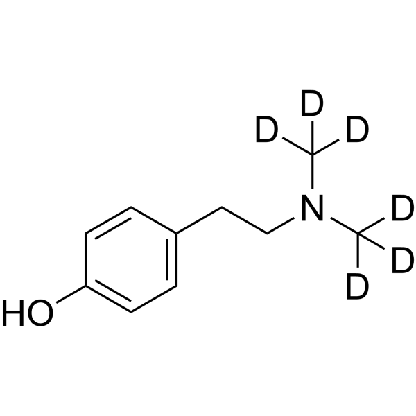
Hordenine-d6 (Ordenina-d6; Peyocactine-d6) is the deuterated-labeled Hordenine (HY-N0113). Hordenine (Ordenina; Peyocactine) is a multifunctional alkaloid with oral activity and blood-brain barrier penetration. Hordenine inhibits LPS-induced phosphorylation of p38, JNK, ERK1/2, p65, IκB, and AKT, prevents p65 nuclear translocation, and suppresses inflammatory cytokines and mediators. Hordenine promotes M2 macrophage polarization, restores blood-milk barrier integrity, alleviates oxidative stress by reducing ROS and MDA, and attenuates LPS-induced lung injury and pulmonary edema. Hordenine inhibits cAMP production, CREB phosphorylation, and MITF expression, thereby suppressing melanin synthesis in melanocytes and reconstructed epidermis. Hordenine activates the Wnt/β-catenin signaling pathway, promotes dermal papilla cell proliferation and hair shaft elongation, and accelerates hair regeneration. Hordenine activates DRD2, acts as a D3R partial agonist, α2A-AR full agonist, and 5-HT2A-R antagonist, and inhibits SERT and DAT. Hordenine inhibits α-synuclein accumulation and ameliorates motor deficits in Parkinson's disease models. Hordenine limits alcohol intake, reduces relapse drinking behavior, and modulates alcohol-induced conditioned place preference. Hordenine can be used for research on mastitis, skin pigmentation, acute lung injury, foodborne diseases, hair loss, Parkinson's disease, bacterial infections, and alcohol use disorder.
For research use only. We do not sell to patients.
- Purity : 98.03%
- CAS No.: 1346598-66-0
- Formula: C10H9D6NO
- Molecular Weight:171.27
-
Storage:
4°C, stored under nitrogen
* In solvent : -80°C, 6 months; -20°C, 1 month (stored under nitrogen)

To place orders, for customer services and technical support, please contact: MedChemExpress USA
Tel: 609-228-6898 E-mail: [email protected] [email protected]


-
 Biological Activity
-
Chemical Information
-
Solvent & Solubility
- Protocols
- Purity & Documentation
- References
-
 Help & FAQs


All Dopamine Receptor Isoforms
MoreAll Adrenergic Receptor Isoforms
MoreAll 5-HT Receptor Isoforms
MoreAll α-synuclein Isoforms
More

Biological Activity
Description
Application
1. This compound can be used as a tracer
2. This compound can be used as an internal standard for quantitative analysis by NMR, GC-MS, or LC-MS.


Chemical Information
-
CAS No. 1346598-66-0
-
Unlabeled CAS 539-15-1
-
Appearance Solid
-
Molecular Weight 171.27
-
Formula C10H9D6NO
-
Color White to off-white
-
SMILES
[2H]C([2H])([2H])N(C([2H])([2H])[2H])CCC1=CC=C(C=C1)O
-
Synonyms
Ordenina-d6; Peyocactine-d6
-
Shipping
Room temperature in continental US; may vary elsewhere.
-
Storage
4°C, stored under nitrogen
* In solvent : -80°C, 6 months; -20°C, 1 month (stored under nitrogen)


Solvent & Solubility
In Vitro:
DMF : ≥ 25 mg/mL (145.97 mM)
DMSO : ≥ 20 mg/mL (116.77 mM; Hygroscopic DMSO has a significant impact on the solubility of product, please use newly opened DMSO)
Ethanol : ≥ 5 mg/mL (29.19 mM)
* "≥" means soluble, but saturation unknown.
Please refer to the solubility information to select the appropriate solvent. Once prepared, please aliquot and store the solution to prevent product inactivation from repeated freeze-thaw cycles.
Storage method and period of stock solution: -80°C, 6 months; -20°C, 1 month (stored under nitrogen). When stored at -80°C, please use it within 6 months. When stored at -20°C, please use it within 1 month.
Please refer to the solubility information to select the appropriate solvent. Once prepared, please aliquot and store the solution to prevent product inactivation from repeated freeze-thaw cycles.
Storage method and period of stock solution: -80°C, 6 months; -20°C, 1 month (stored under nitrogen). When stored at -80°C, please use it within 6 months. When stored at -20°C, please use it within 1 month.
Concentration (start) × Volume (start) = Concentration (final) × Volume (final)

Protocols
-
RNA extraction experimental
By lysing cells, releasing RNA, and removing impurities such as proteins and DNA, high-purity RNA products are finally obtained. The commonly used traditional method is the guanidine isothiocyanate/phenol/chloroform method (Trizol), which is suitable for a variety of animal materials including animal tissues, microorganisms, cultured cells, etc., and most plant materials.
-
Somatic Cell Culture
A method of simulating the in vivo environment in vitro to maintain the cell growth, differentation and main functions.
-
Phagocytosis Functional Assay
A phagocytosis functional assay measures the ability of phagocytic cells, such as neutrophils, macrophages, monocytes, or microglia/macrophages, to bind and internalize particulate targets including bacteria, yeast particles, beads, or myelin particles. Fluorescent flow-cytometry assays detect target uptake as fluorescence associated with gated phagocytes, while pH-sensitive dyes such as pHrodo increase signal in acidic phagosomal compartments and therefore preferentially report internalized particles rather than particles remaining outside the cell. Microscopy or high-content imaging can be used to confirm intracellular localization and, in some protocols, to follow uptake kinetics.
-
Cytoplasmic-Nuclear Fractionated Protein Extraction
Cytoplasmic-nuclear fractionated protein extraction separates soluble cytoplasmic proteins from nuclear-enriched proteins by mild plasma-membrane permeabilization, differential centrifugation, washing of nuclei, and extraction of nuclear proteins for downstream immunoblotting or related molecular analysis. The readout is the relative abundance of a protein in cytoplasmic and nuclear fractions, commonly assessed by western blotting together with compartment markers such as tubulin or pyruvate kinase for cytoplasm and lamin, nucleoporin, hnRNP, H2AX, or Lamin B for nuclear fractions.
-
Transepithelial/transendothelial electrical resistance assay
TEER measures electrical resistance across epithelial or endothelial monolayers cultured on permeable supports, and the readout reflects ionic conductance through the cell barrier, especially the paracellular pathway regulated by junctional integrity. TEER can be measured without destroying the monolayer and is commonly used before or during transport, permeability, barrier-disruption, and barrier-maturation experiments. TEER values are influenced by biological maturation and technical conditions; reported factors include temperature, medium formulation, passage number, electrode geometry, membrane properties, and junctional length during early monolayer maturation. Therefore, TEER should be interpreted with blank-insert subtraction, area normalization, repeated readings, and, when possible, orthogonal barrier readouts such as FITC-dextran flux or tight-junction staining.
-
Inhalation Toxicity Study
Inhalation toxicity studies expose rodents to a controlled aerosol, vapor, gas, or smoke atmosphere and assess respiratory and systemic toxicity using exposure-atmosphere characterization, clinical observations, body and organ weights, bronchoalveolar lavage fluid, histopathology, blood chemistry, hematology, and, when included, molecular endpoints such as transcriptomics, proteomics, lipidomics, or tissue burden analysis. The primary biological readouts are airway irritation, pulmonary inflammation, cytotoxicity, altered surfactant or lipid homeostasis, impaired particle clearance, and tissue remodeling, reflected by BALF cell differentials, BALF protein, LDH, phosphatase activities, cytokines, lung weight, microscopic respiratory-tract lesions, and retained lung burden.
-
MTT Cell Proliferation Assay
The MTT assay is a colorimetric endpoint assay for estimating viable cell number, cell growth, cytotoxicity, or cell activation in cultured mammalian cells. Living cells reduce the yellow tetrazolium salt MTT into purple/blue formazan, while dead cells do not generate the same signal; the resulting color can be quantified with a multiwell spectrophotometer. MTT reduction is commonly interpreted as a readout of metabolic activity that often correlates with viable cell number, but it should not be treated as a direct cell-counting method unless the assay is optimized for the cell type and experimental condition. Studies show that MTT reduction can involve mitochondrial and non-mitochondrial reducing systems, and formazan may accumulate in intracellular lipid droplets rather than simply marking mitochondria.
-
Western Blot
Western blotting (WB) is a commonly used experimental method in molecular biology, biochemistry, and immunogenetics for identifying and quantifying target proteins. It combines gel electrophoresis with immunoassay, enabling researchers to analyze protein expression, post-translational modifications, and molecular weight.
-
ROS/oxidative-stress fluorescent staining
ROS/oxidative-stress fluorescent staining uses cell-permeant fluorogenic probes that become fluorescent after oxidation inside cells or tissues; commonly used examples include DCFH-DA/DCFDA for broad cellular oxidant detection, DHE for superoxide-related signal detection, MitoSOX for mitochondrial superoxide-related signal detection, and CellROX probes for oxidative-stress-associated fluorescence readouts. The assay detects probe oxidation rather than a single ROS species unless the probe and analysis method have been chemically validated for that species. DCFH-DA enters cells, is deacetylated by intracellular esterases to DCFH, and produces fluorescent DCF after oxidation, so the readout is used as an operational measure of total cellular oxidative stress rather than a species-specific ROS measurement. DHE and MitoSOX can report superoxide-related oxidation, but red fluorescence alone can include non-specific ethidium-like oxidation products; HPLC or optimized spectral approaches are
-
BrdU Incorporation Assay
Bromodeoxyuridine (BrdU) incorporation assay is based on the principle that BrdU, a thymidine analog, is incorporated into newly synthesized DNA during the S phase of the cell cycle, thereby serving as a marker of DNA replication and cellular proliferation. Incorporated BrdU can be detected using anti-BrdU antibodies following DNA denaturation, enabling visualization or quantification of proliferating cells through immunochemical detection methods such as immunofluorescence or immunohistochemistry.
-
CFSE Dye Dilution Proliferation Assay
The CFSE (carboxyfluorescein diacetate succinimidyl ester) dye dilution proliferation assay is based on the covalent labeling of intracellular proteins by a cell-permeant fluorescent dye that becomes fluorescent upon intracellular ester cleavage and then is stably retained within cells. As labeled cells divide, the dye is partitioned equally between daughter cells, resulting in a stepwise halving of fluorescence intensity that can be quantified by flow cytometry to determine the number of cell divisions undergone by each cell population. This fluorescence dilution approach enables quantitative tracking of lymphocyte proliferation at the single-cell level over multiple rounds of division. CFSE-based proliferation analysis has been widely applied to measure antigen-driven lymphocyte expansion in vitro, where discrete fluorescence peaks correspond to successive cell divisions and allow reconstruction of proliferative history within heterogeneous populations.
-
Dye-dilution cell tracking and proliferation staining
Dye-dilution cell tracking assays quantify cell proliferation by covalently labeling intracellular proteins with a stable fluorescent dye that is equally partitioned between daughter cells during mitosis, resulting in stepwise halving of fluorescence intensity with each cell division as measured by flow cytometry histograms. Carboxyfluorescein diacetate succinimidyl ester (CFSE) is a prototypical dye that diffuses into cells, is enzymatically converted into a fluorescent compound, and then covalently binds intracellular amine groups, producing long-lived fluorescence suitable for tracking multiple rounds of division in vitro and in vivo. Successive generations of dividing cells form discrete peaks of decreasing fluorescence intensity, enabling estimation of proliferation history, precursor frequency, and division index within heterogeneous populations. Alternative dyes such as CellTrace Violet (CTV) and far-red membrane dyes (e. g. , PKH26) follow the same dilution principle but differ
-
CCK-8/WST-8 Cell Proliferation Assay
The CCK-8/WST-8 assay is based on the reduction of the water-soluble tetrazolium salt WST-8 to a water-soluble formazan product by cellular dehydrogenases in metabolically active cells, where the generated formazan amount is proportional to the number of living cells and is quantified by measuring absorbance in the visible range, providing a colorimetric readout for cell viability and proliferation assessment. This class of tetrazolium-based assays improves upon earlier MTT-based systems by producing a water-soluble formazan, eliminating the need for organic solubilization steps and enabling direct spectrophotometric measurement in culture medium.
-
Protocol for Cell Cycle
Cell-cycle analysis by flow cytometry measures DNA content in single cells to estimate the fraction of cells in G0/G1, S, and G2/M phases. Propidium iodide intercalates into DNA, and after RNA removal with RNase, fluorescence intensity reflects cellular DNA content: 2N cells are assigned to G0/G1, cells between 2N and 4N to S phase, and 4N cells to G2/M. DNA-content analysis alone cannot reliably separate G0 from G1 or G2 from M. Ki-67 can distinguish quiescent G0 cells from cycling cells, EdU or BrdU incorporation marks active DNA synthesis in S phase, and phospho-histone H3 staining identifies mitotic cells within the 4N population.
-
Cell Counting-Based Growth Curve Assay
Cell counting-based growth curve assays quantify cell proliferation by directly measuring changes in viable cell number over time using manual or automated counting methods such as hemocytometer-based counting or instrument-assisted cell enumeration, enabling construction of growth curves that reflect population expansion dynamics in response to culture conditions. A widely used approach is trypan blue exclusion with hemocytometer counting, where membrane-compromised (non-viable) cells take up the dye, allowing discrimination between viable and non-viable cells while simultaneously enabling total cell number quantification. Repeated sampling across time points allows estimation of proliferation rate, growth phases, and comparative growth kinetics between experimental conditions.
-
Colony Formation (Clonogenic) Assay
The clonogenic (colony formation) assay measures the ability of a single cell to retain reproductive viability and form a macroscopic colony, typically defined as a cluster derived from one progenitor cell after a defined growth period. This assay is widely used to evaluate cell survival after exposure to ionizing radiation or cytotoxic treatments and is considered a standard method in radiation biology for generating dose-response relationships of reproductive cell death. Colony formation reflects long-term proliferative capacity rather than short-term metabolic activity, and survival is quantified by comparing treated versus untreated conditions based on colony number and derived survival fractions.
-
Cell Viability Determination by MTT Colorimetric Assay
The following protocol uses the MTT colorimetric assay as a classic literature-established method for assessing cell viability/metabolic activity in cultured mammalian cells. MTT[3-(4,5-dimethylthiazol-2-yl)-2,5-diphenyltetrazolium bromide] is reduced by metabolically active cells to a colored formazan product; the amount of formazan is quantified spectrophotometrically and provides an indirect measure of metabolically active viable cells. Importantly, MTT reduction reflects cellular oxidoreductase/metabolic activity rather than an absolute direct count of living cells, so changes in cellular metabolism can alter the signal independently of cell number.
-
Kinase activity and phosphorylation assays
Kinase activity assays measure the ability of kinases to transfer phosphate groups from ATP to specific substrates, while phosphorylation assays detect the presence and levels of phosphorylated proteins. Common methods include radiolabeled ATP incorporation (e. g. ,), ADP release detection via bioluminescence (e. g. ,[3]), enzyme-linked immunosorbent assays (ELISA) for phospho-specific epitopes (e. g. ,[6]), and microtiter-based formats for high-throughput screening (e. g. ,[8]). The ADP-Glo assay quantifies kinase activity by measuring ADP produced during phosphorylation using a luciferase-based system. Radiometric assays involve autoradiography or scintillation counting after incorporation of 32P-labeled ATP into substrate proteins. ELISA-based approaches rely on phospho-specific antibodies to detect activated kinases in cell lysates or purified samples.
-
EdU Incorporation Assay (Click Chemistry-Based DNA Synthesis Measurement)
The EdU incorporation assay measures DNA synthesis by adding the thymidine analog 5-ethynyl-2′-deoxyuridine to cells or tissues, where it is incorporated into newly synthesized DNA during S phase. Incorporated EdU is detected by copper-catalyzed azide-alkyne cycloaddition, in which a fluorescent azide covalently reacts with the ethynyl group on EdU, allowing S-phase cells to be detected by fluorescence microscopy, flow cytometry, or high-content imaging. EdU detection does not require DNA denaturation or anti-BrdU antibody access, which preserves sample structure and improves compatibility with immunostaining and multiparameter cytometry compared with BrdU-based detection. EdU can be cytotoxic in a cell-type- and exposure-dependent manner, so pulse duration, concentration, and continuous-labeling designs should be validated for each cell type.
-
Ki-67 Immunostaining Proliferation Assay
Ki-67 immunostaining measures the growth fraction of a cell population by detecting Ki-67, a nuclear antigen present in proliferating cells and absent in quiescent G0 cells. The readout is the percentage of Ki-67-positive nuclei among total counted cells, commonly called the Ki-67 labeling index or proliferation index.
-
LPS-Induced Endotoxemia/Systemic Inflammation
Lipopolysaccharide (LPS)-induced endotoxemia is a widely used in vivo model of acute systemic inflammation in which LPS, a Gram-negative bacterial endotoxin, activates innate immune signaling primarily through TLR4, leading to rapid and transient induction of pro-inflammatory cytokines such as TNF-α, IL-6, and IL-1β in circulation and tissues. This cytokine surge is commonly used as a measurable readout of systemic inflammatory activation and immune dysregulation, and is typically assessed within hours after intraperitoneal LPS administration in mouse models of endotoxemia. The model captures key features of systemic inflammatory response syndrome, including cytokine release, immune cell activation, and downstream tissue responses, and has been used to evaluate anti-inflammatory interventions such as cytokine modulation, lipid mediators, and immune cell-targeting therapies.
-
PCNA Immunodetection Proliferation Assay
PCNA immunodetection measures proliferative activity by detecting proliferating cell nuclear antigen, a nuclear protein associated with DNA polymerase δ function and DNA replication. The assay readout is the proportion of PCNA-positive nuclei among total counted cells, but PCNA labeling is not identical to BrdU labeling because PCNA can mark late G1/early S-associated replication competence and may persist beyond active DNA synthesis depending on fixation and extraction conditions.
-
Protocol for Cell Counting and Cell Density Analysis
Cell counting and cell-density analysis estimate the number of cells in a known volume or field area. Manual hemocytometer counting uses a chamber of defined geometry to convert counted cells into cells/mL, while automated counters and image-analysis workflows detect cell objects from optical, brightfield, fluorescence, impedance, or digital-image features. Trypan blue viability counting is based on dye exclusion: viable cells with intact membranes exclude dye, while non-viable cells with compromised membranes stain blue. The readout is total cell density, viable-cell density, dead-cell density, and percent viability. Cell density can also be estimated from microscopy images by counting objects per image area, from flow cytometry using calibrated volume or reference particles, or from in situ microscopy in bioreactors after calibration against reference methods such as hemocytometer or flow cytometry.
-
Human pluripotent stem cell midbrain dopaminergic neuron differentiation
Human pluripotent stem cells are directed toward midbrain dopaminergic neurons by first inducing a neural floor-plate-like progenitor state, then patterning cells with ventralizing SHH signaling and midbrain/WNT-FGF cues, and finally maturing progenitors into neurons expressing dopaminergic markers such as TH, NURR1/NR4A2, PITX3, DAT/SLC6A3, VMAT2/SLC18A2, GIRK2/KCNJ6, FOXA2, LMX1A, and EN1. The main readouts are loss of pluripotency, acquisition of FOXA2+/LMX1A+ midbrain floor-plate progenitors, emergence of βIII-tubulin+/MAP2+ neurons, and production of TH+ dopaminergic neurons with molecular, dopamine-release, and electrophysiological features of midbrain dopaminergic identity.
-
Primary monocyte-to-macrophage differentiation
Primary human monocytes can be differentiated ex vivo into monocyte-derived macrophages by culturing purified blood monocytes for approximately 5-7 days in macrophage-supporting cytokine conditions; M-CSF commonly yields CD14^high/CD163^high macrophages, while GM-CSF yields a phenotypically distinct macrophage population, so the cytokine condition should be chosen according to the downstream model. The readout of successful differentiation is a combined change in morphology, adherence, surface phenotype, and function: differentiated macrophages become adherent, enlarge, acquire macrophage-associated markers such as CD14, CD68, CD163, CD206, or HLA-DR depending on culture condition, and show increased phagocytic capacity compared with starting monocytes.
-
Protocol for Kinase activity and phosphorylation assays
Kinase activity assays measure transfer of phosphate from ATP to a protein or peptide substrate, generating phosphorylated substrate, ADP, or incorporated radiolabeled phosphate as the readout; phosphorylation assays measure site-specific phosphorylation in cells or tissues as a proxy for kinase-pathway activation, inhibition, or substrate regulation. Phosphorylation can be detected by phospho-specific Western blot, immunoprecipitation kinase assay, phospho-immunofluorescence, phospho-flow cytometry, luminescent ADP detection, radiolabeled ATP incorporation, or reporter-based pathway assays, and these readouts can be applied to cancer cells, primary neurons, mouse tumors, organoids, inflammatory macrophages, ferroptosis studies, and mitophagy studies when the kinase target is biologically relevant.


Purity & Documentation
-
Data Sheet (275 KB)
-
SDS (252 KB)
- English - EN (252 KB)
- Français - FR (252 KB)
- Deutsch - DE (252 KB)
- Norwegian - NO (252 KB)
- Español - ES (252 KB)
- Swedish - SV (252 KB)
- Italian - IT (252 KB)
- Korean - KR (252 KB)
- Portuguese - PT (252 KB)
-
Handling Instructions (2659 KB)
References
[2]. Kim SC, et al. Hordenine, a single compound produced during barley germination, inhibits melanogenesis in human melanocytes. Food chemistry. 2013 Nov 01;141(1):174-81. [Content Brief]
[6]. Li H, et al. Hordenine improves Parkinsonian-like motor deficits in mice and nematodes by activating dopamine D2 receptor-mediated signaling. Phytotherapy research : PTR. 2023 Aug;37(8):3296-3308. [Content Brief]


Complete Stock Solution Preparation Table
Please refer to the solubility information to select the appropriate solvent. Once prepared, please aliquot and store the solution to prevent product inactivation from repeated freeze-thaw cycles.
Storage method and period of stock solution: -80°C, 6 months; -20°C, 1 month (stored under nitrogen). When stored at -80°C, please use it within 6 months. When stored at -20°C, please use it within 1 month.
| Optional Solvent | Concentration Solvent Mass | 1 mg | 5 mg | 10 mg | 25 mg |
|---|---|---|---|---|---|
| Ethanol / DMSO / DMF | 1 mM | 5.8387 mL | 29.1937 mL | 58.3873 mL | 145.9684 mL |
| 5 mM | 1.1677 mL | 5.8387 mL | 11.6775 mL | 29.1937 mL | |
| 10 mM | 0.5839 mL | 2.9194 mL | 5.8387 mL | 14.5968 mL | |
| 15 mM | 0.3892 mL | 1.9462 mL | 3.8925 mL | 9.7312 mL | |
| 20 mM | 0.2919 mL | 1.4597 mL | 2.9194 mL | 7.2984 mL | |
| 25 mM | 0.2335 mL | 1.1677 mL | 2.3355 mL | 5.8387 mL | |
| DMSO / DMF | 30 mM | 0.1946 mL | 0.9731 mL | 1.9462 mL | 4.8656 mL |
| 40 mM | 0.1460 mL | 0.7298 mL | 1.4597 mL | 3.6492 mL | |
| 50 mM | 0.1168 mL | 0.5839 mL | 1.1677 mL | 2.9194 mL | |
| 60 mM | 0.0973 mL | 0.4866 mL | 0.9731 mL | 2.4328 mL | |
| 80 mM | 0.0730 mL | 0.3649 mL | 0.7298 mL | 1.8246 mL | |
| 100 mM | 0.0584 mL | 0.2919 mL | 0.5839 mL | 1.4597 mL |


Hordenine-d6 Related Classifications
HY-N0113S Related Classifications
 Powered by Bioz
Keywords
- Hordenine-d6
- 1346598-66-0
- Ordenina-d6
- Peyocactine-d6
- Isotope-Labeled Compounds
- Bacterial
- p38 MAPK
- JNK
- ERK
- Akt
- Interleukin Related
- TNF Receptor
- NO Synthase
- COX
- Reactive Oxygen Species (ROS)
- Microphthalmia Associated Transcription Factor (MITF)
- Wnt
- β-catenin
- Dopamine Receptor
- Adrenergic Receptor
- 5-HT Receptor
- Serotonin Transporter
- Dopamine Transporter
- α-synuclein
- TRP-1
- tyrosinase
- MITF
- cAMP
- Nrf2
- HO-1
- TLR4
- NF-κB
- AMPK
- MAPK
- Inhibitor
- inhibitor
- inhibit