



Gamma-glutamylcysteine
Based on 4 publication(s) in Google Scholar
Gamma-glutamylcysteine (γ-Glu-Cys) is an orally active, blood-brain barrier permeable dipeptide. Gamma-glutamylcysteine activates AMPK, SIRT1, IL-4/STAT6, AC/cAMP/PI3K, IGF-1R/IRS1/PI3K, and Nrf2 signaling pathways; it inhibits NF-κB, JAK1/STAT1/3, MAPKs, cadmium-induced p38 MAPK, JNK, and PI3K/Akt signaling pathways. Gamma-glutamylcysteine regulates macrophage polarization, modulates the trafficking of CD36 and GLUT4, induces glutathione synthesis, improves metabolic dysfunction, reduces lipid deposition, ameliorates glucose homeostasis, inhibits apoptosis (Apoptosis), stabilizes mitochondria, suppresses lipid peroxidation, iron accumulation and ferroptosis (Ferroptosis), reduces ds-HMGB1 levels, reverses mechanical hyperalgesia, and alleviates hepatic lipid droplet formation. Gamma-glutamylcysteine is applicable to research related to inflammatory bowel disease, type 2 diabetes, cadmium-induced neurotoxicity, Alzheimer's disease, cerebral ischemia/reperfusion injury, neuropathy, and alcoholic liver disease.
For research use only. We do not sell to patients.
- Purity : 98.53%
- CAS No.: 636-58-8
- Formula: C8H14N2O5S
- Molecular Weight:250.27
-
Storage:
-20°C, sealed storage, away from moisture
* In solvent : -80°C, 6 months; -20°C, 1 month (sealed storage, away from moisture)

To place orders, for customer services and technical support, please contact: MedChemExpress USA
Tel: 609-228-6898 E-mail: [email protected] [email protected]


-
 Biological Activity
-
Chemical Information
-
Solvent & Solubility
- Protocols
- Purity & Documentation
- References
-
 Help & FAQs


-
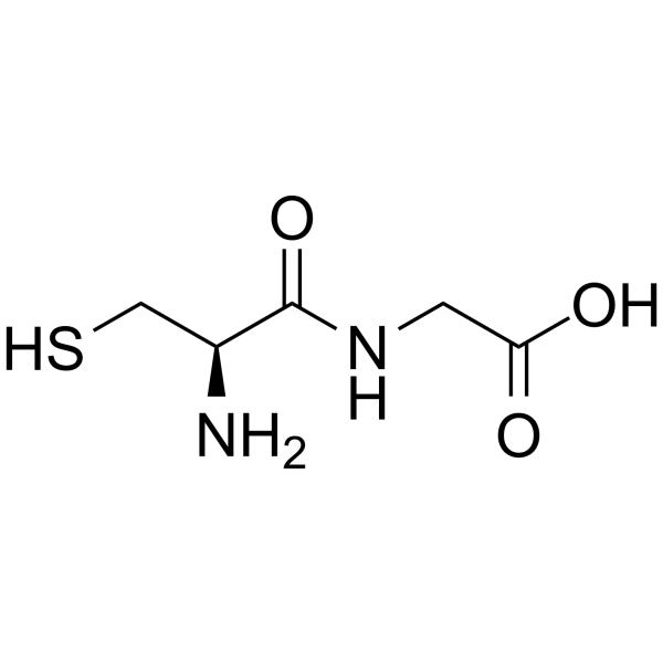
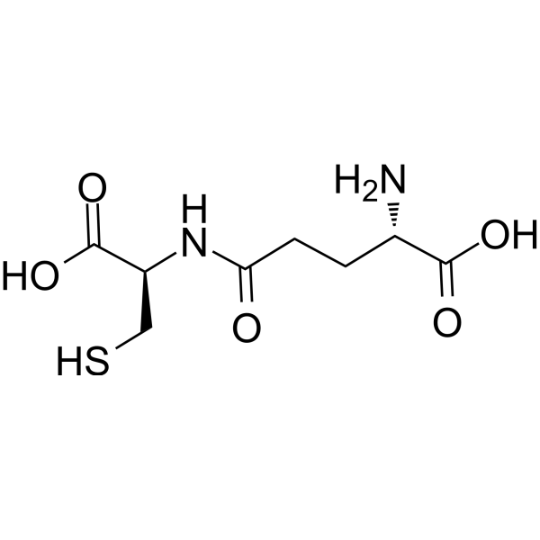
HY-113110
CysteinylglycineEndogenous Metabolite -
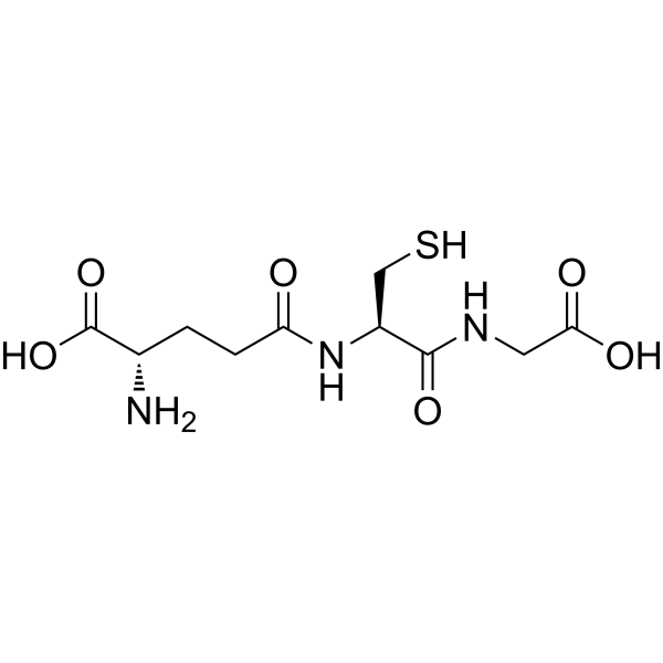
HY-D0187
L-Glutathione reducedAntioxidant -

HY-113402R
Gamma-glutamylcysteine (Standard)GSH Precursor -
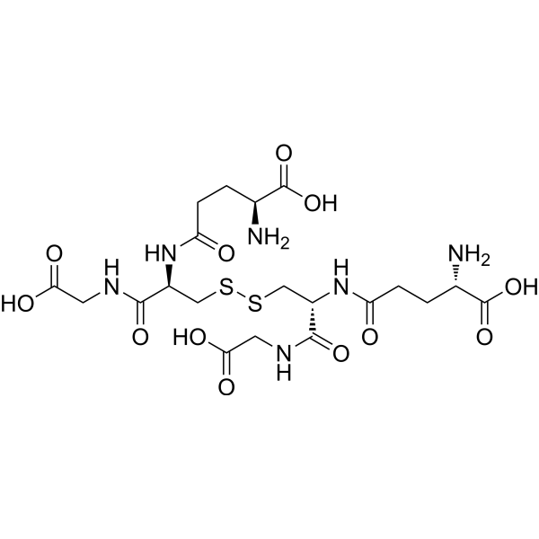
HY-D0844
Glutathione oxidizedAntioxidant -
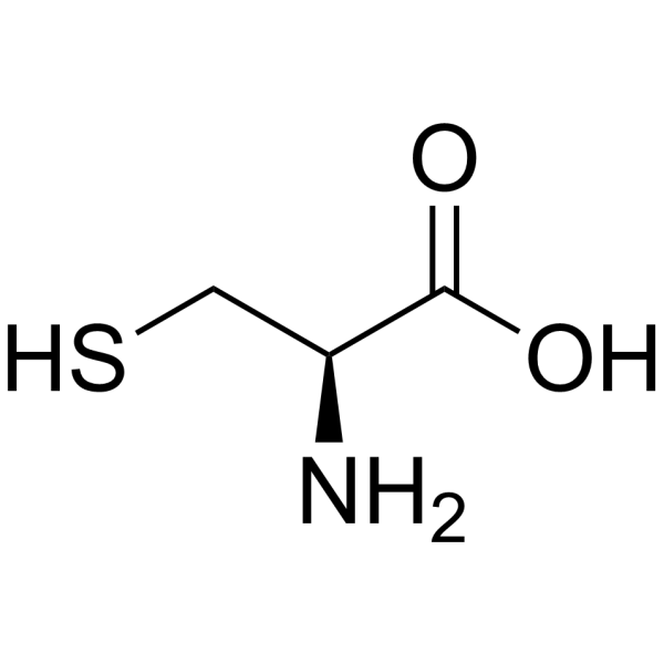
HY-Y0337
L-CysteineAnorectic Agent -
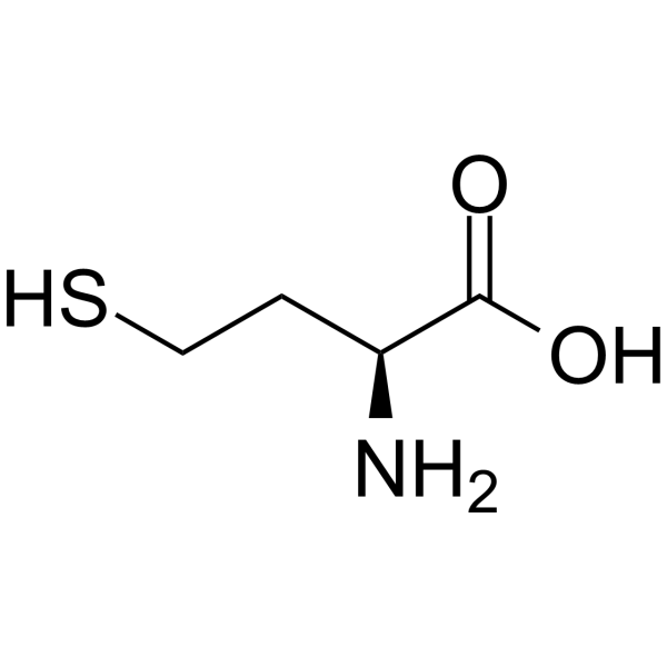
HY-W010347
L-HomocysteineAmino Acid -

HY-B0215
AcetylcysteineMucolytic Agent -

HY-10583
Y-27632 dihydrochlorideROCK Inhibitor -
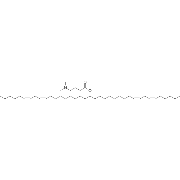
HY-112251
D-Lin-MC3-DMAsiRNA Delivery Vehicle -
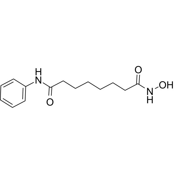
HY-10221
VorinostatHDAC Inhibitor -

HY-15763
ErastinFerroptosis Inducer -
HY-B0152
AdenineNucleobase
Publications Citing Use of MedChemExpress (MCE) Gamma-glutamylcysteine
More Customer Validation & Images

-
WB
-
In Vivo Efficacy Study
-
Bio/Physico-chemical Assay
All Endogenous Metabolite Isoforms
MoreAll AMPK Isoforms
More

Biological Activity
Description
IC50 & Target
|
Human Endogenous Metabolite |
JNK1 |
SIRT3 |
SIRT1 |
STAT6 |
IL-4 |
In Vitro
Gamma-glutamylcysteine (0-80 μM; 24 h) inhibits M1 polarization of Raw264.7 macrophages by suppressing the JAK1/STAT1/3, AKT, MAPKs and NF-κB signaling pathways, while activating the AMPK/SIRT1 axis[1].
Gamma-glutamylcysteine (0-80 μM; 24 h) promotes M2 polarization of Raw264.7 macrophages, and drives their repolarization from M1 to M2 via activation of the IL-4/STAT6 and AMPK/SIRT1 signaling pathways[1].
Gamma-glutamylcysteine (20-80 μM; 24 h) induces the polarization of human Jurcat CD4+ T lymphocytes toward Th2 by promoting IL-4 secretion[1].
Gamma-glutamylcysteine (2 mM; 6 h) activates the Nrf2 signaling pathway via nuclear translocation and upregulates the expression of antioxidant target genes and proteins in primary intestinal epithelial cells (IECs) of chicken embryos[2].
Gamma-glutamylcysteine (2 mM; 6 h) inhibits oxidative stress induced by Salmonella typhimurium in primary wild-type chicken embryonic intestinal epithelial cells (IECs) by maintaining Nrf2 activation[2].
Gamma-glutamylcysteine (2 mM; 6 h) protects primary intestinal epithelial cells (IECs) from wild-type chicken embryos against intestinal barrier disruption and inflammatory injury induced by Salmonella typhimurium, and this protective effect depends on the Nrf2 signaling pathway[2].
Gamma-glutamylcysteine (20-80 μM; administered for 24 h after 24 h of pre-treatment with insulin + PA) dose-dependently activates the IGF-1R/IRS1/PI3K/Akt signaling pathway in insulin-resistant HepG2 cells, primary mouse hepatocytes and C2C12 myotubes induced by Insulin (HY-P701240) + Palmitic acid (HY-N0830) (PA), by increasing the phosphorylation levels of key pathway proteins[3].
Gamma-glutamylcysteine (2-4 mM; 2 h pretreatment followed by 12 h cadmium exposure) inhibits the activation of JNK/p38 MAPK and PI3K/Akt signaling pathways in cadmium-treated cells by downregulating the expression of pro-apoptotic markers and normalizing the Bax/Bcl-2 ratio. This consequently suppresses cadmium-induced changes in apoptosis-related proteins, inhibits cadmium-induced cell apoptosis, and prevents cadmium-induced mitochondrial transmembrane potential depolarization in PC12 cells[4].
Gamma-glutamylcysteine (2-4 mM; 2 h pretreatment followed by 12 h cadmium exposure) dose-dependently inhibits cadmium-induced oxidative stress in PC12 cells by reducing ROS and lipid peroxidation levels, restoring antioxidant enzyme activity, and maintaining intracellular GSH homeostasis[4].
Gamma-glutamylcysteine (0.25-16 mM; 24 h) shows no toxicity to BV-2 cells at concentrations up to 4 mM after 24 h of incubation, while higher concentrations (8, 16 mM) reduce cell viability in a dose-dependent manner[5].
Gamma-glutamylcysteine (2-4 mM; 30 min pretreatment, 24 h AβO exposure) dose-dependently inhibits the release of proinflammatory mediators (TNF-α, IL-1β, NO) and the expression of proinflammatory proteins (iNOS, COX-2) induced by AβO in BV-2 cells, suppresses AβO-induced oxidative stress, and restores the antioxidant capacity of cells[5].
Gamma-glutamylcysteine (4 mM; 30 min pretreatment, followed by 6-24 h of AβO exposure) inhibits AβO-induced activation of the NF-κB signaling pathway in BV-2 cells, upregulates and maintains the expression of Nurr1 mRNA and protein, and thereby exerts anti-inflammatory effects by suppressing the binding of NF-κB p65 to the iNOS promoter induced by AβO in BV-2 cells[5].
Gamma-glutamylcysteine (2-4 mM; 30 min pretreatment, followed by 24 h AβO exposure) dose-dependently inhibits the release of proinflammatory mediators (TNF-α, IL-1β, NO) induced by AβO in primary mouse microglia, suppresses oxidative stress, and restores the antioxidant capacity of cells[5].
Gamma-glutamylcysteine (0.25-2 mM; 12 h) increases GSH levels, GSH/GSSG ratio, GPX activity, and cell viability in primary cortical neurons treated with OGD/R, with the strongest effect observed at 2 mM for 12 h[6].
Gamma-glutamylcysteine (0.85-7 mM; 12 h) increases GSH level, GSH/GSSG ratio, GPX activity and cell viability in PC12 cells treated with oxygen-glucose deprivation/reoxygenation (OGD/R)[6].
Gamma-glutamylcysteine (1.7-7 mM; 0-12 h, time-course 0-12 h) regulates the mRNA and protein levels of GSS in OGD/R-treated PC12 cells, promotes nuclear translocation of Nrf2 in cells, reduces the interaction between Nrf2 and Keap1, inhibits the upregulation of Keap1, decreases MDA and Fe2+ levels, improves cell viability, and thereby inhibits ferroptosis in cells[6].
Gamma-glutamylcysteine (3.5 mM; 12 h, time-course 0-12 h) activates Nrf2 in OGD/R-treated PC12 cells by increasing the level of phosphorylated PKC-ε[6].
Gamma-glutamylcysteine (200 μM; 15 min pre-incubation, 24 h co-incubation with oligomeric Aβ40) protects primary human astrocytes against oligomeric Aβ40-induced cytotoxicity, apoptosis, oxidative stress, neuroinflammation, and dysregulated metalloproteinase activity, while restoring antioxidant status and GSH levels[7].
Gamma-glutamylcysteine (20-80 μM; 2 h pretreatment followed by 24 h ethanol exposure; 400 μM; 24 h single treatment) protects human L02 hepatocytes against ethanol-induced injury by dose-dependently increasing cell viability, reducing hepatic enzyme release, inhibiting cell apoptosis, suppressing oxidative stress and mitochondrial damage, and attenuating the activation of pro-inflammatory signaling pathways[9].
MedChemExpress (MCE) has not independently confirmed the accuracy of these methods. They are for reference only. Further protocols information, click here.
-
Cell Line:insulin + palmitic acid-induced insulin-resistant HepG2 cells, primary mouse hepatocytes, C2C12 mouse skeletal muscle myotubes
-
Concentration:0, 20, 40, 80 μM
-
Incubation Time:24 h (following 24 h insulin + PA pre-treatment)
-
Result:Significantly increased phosphorylation of p-IGF-1R (Tyr1135), p-IRS1 (Tyr612), p-PI3K, and p-Akt (Ser473) in a dose-dependent manner relative to insulin + PA-only treated cells across all three cell types, with significant increases observed at 20, 40, and 80 μM.
-
Cell Line:PC12 cells
-
Concentration:4 mM
-
Incubation Time:2 h pretreatment + 12 h cadmium exposure
-
Result:Significantly reduced the number of TUNEL-positive PC12 cells.
Decreased the percentage of apoptotic cells induced by cadmium.
-
Cell Line:PC12 cells
-
Concentration:4 mM
-
Incubation Time:2 h pretreatment + 12 h cadmium exposure
-
Result:Down-regulated the ratio of Bax/Bcl-2 in cadmium-treated PC12 cells.
Reduced the protein levels of cytosolic cytopigment c, cleaved-caspase-9, cleaved-caspase-3, and cleaved-PARP in cadmium-treated PC12 cells.
In Vivo
Gamma-glutamylcysteine (250-500 mg/kg; p.o.; daily; 8 weeks) dose-dependently improves glycemic control, insulin sensitivity, β-cell function and hepatic steatosis, while alleviating diabetes-related organ damage in db/db mice[3].
Gamma-glutamylcysteine (100-400 mg/kg/d; p.o.; daily; 20 days) dose-dependently inhibits AβO-induced neuroinflammation in male ICR mice[5].
Gamma-glutamylcysteine (688 mg/kg; p.o.; single dose) upregulates GSH by activating the PKC-ε/Nrf2/GSS pathway, and significantly reduces cerebral infarction volume, neurological dysfunction, and neuronal ferroptosis induced by cerebral ischemia/reperfusion injury in male Sprague-Dawley rats[6].
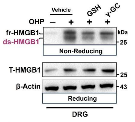
Gamma-glutamylcysteine (600 mg/kg; p.o.; once) significantly reduces the level of ds-HMGB1 in DRG of OIPN mice and reverses oxaliplatin-induced mechanical hyperalgesia[8].
Gamma-glutamylcysteine (700-1200 mg/kg; p.o.; daily; 7 days) alleviates acute ethanol-induced hepatotoxicity in male C57BL/6JNifdc mice in a dose-dependent manner by reducing hepatic enzyme release, restoring hepatic antioxidant levels, alleviating histopathological damage, and inhibiting inflammatory signaling pathways[9].
MedChemExpress (MCE) has not independently confirmed the accuracy of these methods. They are for reference only.
-
Animal Model:BALB/c (8-week-old male, 20-22 g, TNBS-induced colitis)[1]
-
Dosage:600 mg/kg; 1200 mg/kg
-
Administration:p.o.; daily; ~3 days
-
Result:Conferred significant protection against lethality.
Reversed TNBS-induced body weight loss.
Reduced the increase in Disease Activity Index (DAI).
Mitigated colon shortening, bleeding, and ulcerations.
Lowered histological injury scores.
Significantly reduced colon tissue mRNA levels of M1 markers (Inos, Il-1β).
Significantly increased colon tissue mRNA levels of M2 markers (Cd206, Arg1).
Significantly reduced serum TNF-α levels relative to the TNBS model group.
Confirmed reduced M1 marker (iNOS, IL-1β) and increased M2 marker (CD206, ARG1) protein levels in colon tissue via Western blot and immunohistochemistry.
Significantly reduced TNBS-induced phosphorylation of JAK1, STAT1, STAT3, AKT, JNK, ERK, p38, IKKα/β, and IkBα in colon lamina propria mononuclear cells.
Increased phosphorylation of STAT6 and serum IL-4 levels.
-
Animal Model:C57BL/6J mice (male, 6 weeks old, 20-22 g); db/db mice (male, 6 weeks old, 30-35 g, spontaneous leptin receptor mutation model)[3]
-
Dosage:250 mg/kg; 500 mg/kg
-
Administration:p.o.; daily; 8 weeks
-
Result:Significantly decreased food intake, food efficiency, and water intake in db/db mice at 500 mg/kg dose.
Decreased water intake in db/db mice at 250 mg/kg dose.
Dose-dependently decreased fasting blood glucose, serum HbA1c, serum insulin levels, and HOMA-IR index, while increased HOMA-β index in db/db mice.
Dose-dependently improved glucose tolerance (reduced oral glucose tolerance test AUC) and insulin sensitivity (reduced insulin tolerance test AUC) in db/db mice.
Dose-dependently improved organ coefficients (reduced liver coefficient, increased cardiac and kidney coefficients) and reduced tissue damage (cardiomyocyte degeneration, kidney glomerular/tubular damage, hepatocyte ballooning, pancreatic islet loss) in db/db mice, with 500 mg/kg treatment more effective than metformin.
Dose-dependently reduced urinary MAU/ALB levels and increased hepatic glycogen content in db/db mice.
Dose-dependently reduced subcutaneous adipose tissue (SAT) and visceral adipose tissue (VAT) weight, and decreased adipocyte size in db/db mice.
Dose-dependently reduced hepatic triglyceride (TG), total cholesterol (TC), and serum TG, TC, LDL-C levels, while increased serum HDL-C levels in db/db mice; 500 mg/kg treatment reduced hepatic TG more effectively than metformin.
Dose-dependently reduced hepatic lipid droplet accumulation (Oil Red O staining) and decreased serum ALT and AST levels in db/db mice.
Dose-dependently reduced hepatic CD36, PPARα, and CPT1A protein expression, and increased hepatic p-IGF-1R, p-IRS1, p-PI3K, and p-Akt protein expression in db/db mice.
Showed no significant effect on body weight, fasting blood glucose, organ coefficients, or tissue histology in C57BL/6J mice at both doses.
-
Animal Model:ICR mice (male, 22-25 g, Alzheimer's disease model induced by intracerebroventricular administration of 2 μg AβO)[5]
-
Dosage:100 mg/kg/d; 400 mg/kg/d
-
Administration:p.o.; daily; 20 days
-
Result:Suppressed AβO-induced microglial activation (reduced Iba1 fluorescence intensity in the hippocampus), and inhibited upregulation of hippocampal COX-2, iNOS, and Iba1 protein expression (100 mg/kg/d dose).
Suppressed AβO-induced microglial activation with greater reduction in Iba1 fluorescence intensity than the 100 mg/kg/d dose, and exhibited stronger inhibition of AβO-induced upregulation of hippocampal COX-2, iNOS, and Iba1 protein expression compared to the 100 mg/kg/d dose (400 mg/kg/d dose).
-
Animal Model:Sprague-Dawley (SD) (male, 260-300 g, transient focal cerebral ischemia induced by 90-minute middle cerebral artery occlusion followed by reperfusion)[6]
-
Dosage:688 mg/kg
-
Administration:p.o.; single dose (administered 1.5 hours after MCAO)
-
Result:Significantly reduced cerebral infarction volume.
Significantly lowered neurological deficit scores.
Significantly increased the number of Nissl-positive neurons in the ipsilateral cerebral cortex.
Significantly reduced the number of FJB+/NeuN+ (dying) neurons in the cerebral cortex.
Alleviated MCAO/R-induced neuronal mitochondrial damage, including reduced loss of mitochondrial cristae and outer membrane rupture.
Significantly decreased cortical H2O2, malondialdehyde (MDA), and Fe2+ levels, and reduced 4-HNE-positive neuronal counts.
Restored MCAO/R-induced alterations in ferroptosis-related protein and mRNA levels: increased FTH1 and GPX4, and decreased ACSL4 and TF (no effect on SLC7A11).
Significantly elevated cortical GSH levels, GSH/GSSG ratio, and glutathione peroxidase (GPX) activity.
Inhibited MCAO/R-induced reduction of cortical GSS mRNA and protein levels, and increased GSS/NeuN-positive neuron counts.
Increased total Nrf2 and phosphorylated Nrf2 protein levels in cortical tissue, and elevated Nrf2/NeuN and p-Nrf2/NeuN-positive neuron counts.
Increased phosphorylated PKC protein levels in cerebral cortical tissue.
-
Animal Model:C57BL/6J (male, 8-10 weeks old, 22-26 g, SPF grade, oxaliplatin-induced peripheral neuropathy model)[8]
-
Dosage:600 mg/kg
-
Administration:p.o.; 2 hours prior to each weekly oxaliplatin injection
-
Result:Reduced ds-HMGB1 levels in the DRG to ~2.0 relative units.
Produced a smaller reduction in serum ds-HMGB1 to ~1.6 relative units.
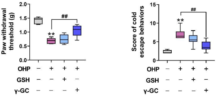
Increased paw withdrawal thresholds to ~1.2 g, reversing oxaliplatin-induced mechanical allodynia.
Reduced cold escape behavior scores to ~4.0, modestly improving cold sensitivity.
Exhibited greater efficacy at reducing DRG ds-HMGB1 levels and improving pain responses than equimolar glutathione.
-
Animal Model:C57BL/6JNifdc (male, 6-8 weeks old, acute ethanol-induced hepatotoxicity model)[9]
-
Dosage:700 mg/kg; 1200 mg/kg
-
Administration:p.o.; daily; 7 days
-
Result:Reduced ethanol-induced elevations in serum ALT, AST, and TG levels in a dose-dependent manner.
Reversed ethanol-induced depletion of hepatic GSH levels.
Reduced hepatic lipid droplet formation and inflammatory cell infiltration.
Decreased ethanol-induced increases in hepatic protein levels of iNOS, p-p65, p-IKKα/β, and p-IκBα in a dose-dependent manner.


Chemical Information
-
CAS No. 636-58-8
-
Appearance Solid
-
Molecular Weight 250.27
-
Formula C8H14N2O5S
-
Color White to off-white
-
SMILES
SC[C@@H](C(O)=O)NC(CC[C@H](N)C(O)=O)=O
-
Synonyms
γ-Glu-Cys
-
Structure Classification
-
Initial Source
-
Shipping
Room temperature in continental US; may vary elsewhere.
-
Storage
-20°C, sealed storage, away from moisture
* In solvent : -80°C, 6 months; -20°C, 1 month (sealed storage, away from moisture)


Publications (4)
-
Journal Impact Factor
-
Most Recent
-
Cell Rep
Microglial macrophage-derived ds-HMGB1 in DRG orchestrates neuropathic pain through immune-neural signaling. [Abstract]2025 Dec 10;44(12):116671. PMID: 41385369
Gamma-glutamylcysteine purchased from MedChemExpress. Usage Cited in: Cell Rep. 2025 Dec 10;44(12):116671. [Abstract]
Administration of glutathione (GSH) or Gamma-glutamylcysteine (γ-GC, a cell-permeable GSH analog) (600 mg/kg; p.o.; single dose) targeted to the DRG during OIPN. Western blots showing that oral γ-GC (which elevates intracellular GSH) significantly decreases DRG ds-HMGB1 compared to untreated OIPN controls (and more effectively than equimolar GSH).
Gamma-glutamylcysteine purchased from MedChemExpress. Usage Cited in: Cell Rep. 2025 Dec 10;44(12):116671. [Abstract]
Behavioral assessments demonstrating that Gamma-glutamylcysteine (γ-GC, 600 mg/kg; p.o.; single dose) treatment mitigates pain: mechanical allodynia thresholds and cold-plate latencies/scores in OIPN mice at week 1. γ-GC reverses oxaliplatin-induced hypersensitivity, whereas GSH has a more modest effect.
-
Probiotics Antimicrob Proteins
Lactobacillus crispatus 7-4 Mitigates Salmonella typhimurium-Induced Enteritis via the γ‑Glutamylcysteine-Mediated Nrf2 Pathway. [Abstract]2024 Jun 3. PMID: 38829566 -
Food Chem (Oxf)
Glutathione as a taste modulator: molecular mechanisms of interaction with umami and sweet taste receptors. [Abstract]2025 Oct 26:11:100319. PMID: 41246561
Gamma-glutamylcysteine purchased from MedChemExpress. Usage Cited in: Food Chem (Oxf). 2025 Oct 26:11:100319. [Abstract]
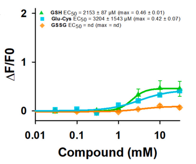
Dose-response curves of hTAS1R1/rTAS1R3 stimulated with GSH, Gamma-glutamylcysteine (Glu-Cys), and GSSG. The results showed that the application of Glu-Cys produced weak agonist activity, with an EC50 value of 3204 ± 1543 μM and a max ∆F/F0 of 0.42 ± 0.07.
-
Structure
Structural insights into the convergent evolution of sulfoxide synthase EgtB-IV, an ergothioneine-biosynthetic homolog of ovothiol synthase OvoA. [Abstract]2024 Nov 7;32(11):2013-2022.e5. PMID: 39216472


Solvent & Solubility
In Vitro:
H2O : 100 mg/mL (399.57 mM; Need ultrasonic)
Please refer to the solubility information to select the appropriate solvent. Once prepared, please aliquot and store the solution to prevent product inactivation from repeated freeze-thaw cycles.
Storage method and period of stock solution: -80°C, 6 months; -20°C, 1 month (sealed storage, away from moisture). When stored at -80°C, please use it within 6 months. When stored at -20°C, please use it within 1 month.
* Note: If you choose water as the stock solution, please dilute it to the working solution, then filter and sterilize it with a 0.22 μm filter before use.
Please refer to the solubility information to select the appropriate solvent. Once prepared, please aliquot and store the solution to prevent product inactivation from repeated freeze-thaw cycles.
Storage method and period of stock solution: -80°C, 6 months; -20°C, 1 month (sealed storage, away from moisture). When stored at -80°C, please use it within 6 months. When stored at -20°C, please use it within 1 month.
* Note: If you choose water as the stock solution, please dilute it to the working solution, then filter and sterilize it with a 0.22 μm filter before use.
Concentration (start) × Volume (start) = Concentration (final) × Volume (final)
In Vivo:
For the following dissolution methods, please prepare the working solution directly:
It is recommended to prepare fresh solutions and use them promptly within a short period of time.
The percentages shown for the solvents indicate their volumetric ratio in the final prepared solution. If precipitation or phase separation occurs during preparation, heat and/or sonication can be used to aid dissolution.
Add each solvent one by one: PBS
Solubility: 100 mg/mL (399.57 mM); Clear solution; Need ultrasonic

Protocols
-
Neurotoxicity Study
This protocol assesses in vitro neurotoxicity by combining neuronal viability, mitochondrial/metabolic activity, neurite outgrowth, and optional neuronal network function readouts. Calcein-AM or resazurin/PrestoBlue readouts estimate viable or metabolically active cells; βIII-tubulin immunofluorescence detects neuronal morphology and neurite networks; TMRE detects mitochondrial membrane potential; and MEA recordings detect functional changes in neuronal network activity.
-
Apoptosis
Apoptosis, also called programmed cell death, is generally characterized by distinct morphological characteristics.
-
TUNEL staining for apoptotic DNA fragmentation
TUNEL staining detects DNA strand breaks by using terminal deoxynucleotidyl transferase to add labeled nucleotides to exposed 3′-OH DNA termini, generating either microscopic staining in fixed cells or tissue sections, or fluorescence/cytometric signal in cell suspensions. TUNEL positivity reflects DNA fragmentation but should not be interpreted alone as definitive apoptosis, because TUNEL can also label necrotic, autolytic, mechanically damaged, or DNA-repair-associated DNA breaks.
-
Annexin V plus membrane-impermeant dye apoptosis staining
Annexin V-based apoptosis assays rely on the detection of phosphatidylserine (PS) externalization from the inner leaflet of the plasma membrane to the outer leaflet, an early biochemical hallmark of apoptosis. Fluorescently labeled Annexin V binds PS in a calcium-dependent manner, enabling identification of early apoptotic cells by flow cytometry or fluorescence microscopy. When combined with a membrane-impermeant DNA-binding dye (e. g. , propidium iodide), this approach allows discrimination between viable (Annexin V−/dye−), early apoptotic (Annexin V+/dye−), and late apoptotic or necrotic (Annexin V+/dye+) cell populations by assessing membrane integrity and PS exposure.
-
DSS-Induced Colitis
Dextran sulfate sodium (DSS)-induced colitis is generated by administering DSS in mouse drinking water, producing epithelial injury, barrier disruption, weight loss, diarrhea, fecal blood, colon shortening, histologic mucosal damage, and inflammatory mediator changes; the model is mainly used to study acute or chronic intestinal inflammation resembling selected features of ulcerative colitis. DSS injury is interpreted through clinical and tissue readouts rather than a single molecular endpoint: daily body weight, stool consistency, and bleeding are combined into a disease activity index, while colon length, histology, cytokines, myeloperoxidase activity, intestinal permeability, and tight-junction markers provide complementary measures of inflammation and barrier damage.
-
TNBS-Induced Colitis
TNBS-induced colitis is produced by intrarectal delivery of 2,4,6-trinitrobenzene sulfonic acid in ethanol, where ethanol disrupts the mucosal barrier and TNBS haptenates colonic proteins, generating immune-mediated colonic inflammation with weight loss, diarrhea, ulceration, transmural injury, inflammatory-cell infiltration, and cytokine responses. The model is used as an experimental intestinal inflammation model with Crohn’s disease–like features, especially when Th1-type responses, IL-12–dependent inflammation, chronic relapsing inflammation, or fibrosis-related endpoints are studied.
-
Primary monocyte-to-macrophage differentiation
Primary human monocytes can be differentiated ex vivo into monocyte-derived macrophages by culturing purified blood monocytes for approximately 5-7 days in macrophage-supporting cytokine conditions; M-CSF commonly yields CD14^high/CD163^high macrophages, while GM-CSF yields a phenotypically distinct macrophage population, so the cytokine condition should be chosen according to the downstream model. The readout of successful differentiation is a combined change in morphology, adherence, surface phenotype, and function: differentiated macrophages become adherent, enlarge, acquire macrophage-associated markers such as CD14, CD68, CD163, CD206, or HLA-DR depending on culture condition, and show increased phagocytic capacity compared with starting monocytes.
-
Apoptosis Solutions
Apoptosis is a regulated, generally non-lytic cell-death pathway that removes unwanted, damaged, infected, or abnormal cells through coordinated morphological changes, caspase activation, DNA fragmentation, and membrane remodeling. The intrinsic apoptosis pathway is controlled mainly by mitochondrial outer membrane permeabilization, BCL-2 family proteins, cytochrome c release, apoptosome formation, caspase-9 activation, and downstream executioner caspase-3/7 activation. The extrinsic apoptosis pathway is initiated by death receptors such as Fas, TNFR, and TRAIL receptors, which recruit adaptor proteins and activate caspase-8 before engaging executioner caspases or mitochondrial amplification through BID cleavage. Apoptosis is linked to many phenotypes, including cancer cell killing, tissue homeostasis, immune regulation, neurodegeneration, infection response, and treatment-induced cytotoxicity; unresolved questions include how apoptosis interacts with necroptosis, pyroptosis, ferroptos
-
Research Protocol for Inflammation-related Diseases
The NLRP3 inflammasome is a cytosolic innate immune signaling platform that integrates priming signals and danger-signal activation to promote caspase-1 activation, maturation of IL-1β and IL-18, and gasdermin D-mediated pyroptotic cell death. The core experimental logic is to determine whether inflammatory disease phenotypes are driven by increased NLRP3 expression, ASC-containing inflammasome assembly, caspase-1 cleavage, GSDMD cleavage, and extracellular release of IL-1β/IL-18 rather than by nonspecific cell injury alone. The pathway is strongly linked to inflammation-related disease phenotypes because monosodium urate crystals activate NALP3/NLRP3 inflammasome signaling in gout-like crystal inflammation, cholesterol crystals activate NLRP3 inflammasomes in atherogenesis models, and DSS-induced intestinal inflammation has been reported to involve NLRP3 inflammasome activity. However, experimental colitis studies also show context-dependent protective effects of NLRP3 inflammasome co
-
Ferroptosis Solutions
Ferroptosis is an iron-dependent, non-apoptotic form of regulated cell death characterized by lethal lipid peroxidation and sensitivity to suppression by iron chelators or lipophilic radical-trapping antioxidants. The core pathway links cystine uptake through system Xc−, glutathione availability, GPX4-dependent detoxification of phospholipid hydroperoxides, iron-dependent oxidative reactions, and polyunsaturated-phospholipid metabolism into a cell-death program that is biochemically and morphologically distinct from apoptosis, necrosis, and autophagy. The ferroptosis pathway is experimentally linked to phenotype through chemical and genetic perturbation. Erastin induces ferroptosis by inhibiting cystine uptake through system Xc− and weakening antioxidant defenses, while GPX4 inhibition or depletion causes lipid peroxide accumulation and ferroptotic cancer-cell death. ACSL4 and oxidizable arachidonoyl- or adrenoyl-containing phosphatidylethanolamines shape ferroptosis sensitivity by con
-
Research Protocol for Metabolic Diseases
AMP-activated protein kinase, AMPK, is a conserved cellular energy sensor that responds to reduced cellular energy status and coordinates metabolism by increasing ATP-generating catabolic pathways while suppressing ATP-consuming anabolic processes. In metabolic disease research, the AMPK pathway is experimentally relevant because it regulates hepatic lipid synthesis, fatty acid oxidation, glucose production, skeletal-muscle glucose disposal, mTORC1-linked biosynthesis, autophagy, mitochondrial homeostasis, and whole-body energy balance. The central pathway logic is that energy stress, metformin, exercise-like stimulation, or direct AMPK activators increase AMPKα Thr172 phosphorylation and downstream substrate phosphorylation, including ACC and RAPTOR. Phosphorylation of ACC suppresses lipogenesis and supports fatty acid oxidation, whereas phosphorylation of RAPTOR suppresses mTORC1 signaling and links cellular energy status to growth and protein synthesis control. The pathway is linked
-
Alzheimer’s Disease Modeling
Alzheimer’s Disease (AD) is a neurodegenerative disorder characterized by a progressive decline in cognitive functions and loss of specific types of neurons and synapses. Alzheimer's symptoms can be simulated in mice by injecting drugs (such as Aβ) or genetically modified.


Purity & Documentation
-
Data Sheet (300 KB)
-
SDS (762 KB)
- English - EN (762 KB)
- Français - FR (762 KB)
- Deutsch - DE (762 KB)
- Norwegian - NO (762 KB)
- Español - ES (762 KB)
- Swedish - SV (762 KB)
- Italian - IT (762 KB)
- Korean - KR (762 KB)
- Portuguese - PT (762 KB)
-
Handling Instructions (2659 KB)
References
[1]. Zhou J, et al. γ-Glutamylcysteine rescues mice from TNBS-driven inflammatory bowel disease through regulating macrophages polarization. Inflamm Res. 2023 Mar;72(3):603-621. [Content Brief]
[2]. the γ‑Glutamylcysteine-Mediated Nrf2 Pathway. Probiotics Antimicrob Proteins. 2025;17(5):3378-3391. [Content Brief]
[3]. Zhou J, et al. γ-glutamylcysteine alleviates insulin resistance and hepatic steatosis by regulating adenylate cyclase and IGF-1R/IRS1/PI3K/Akt signaling pathways. J Nutr Biochem. 2023 Sep;119:109404. [Content Brief]
[4]. Bi A, et al. γ-glutamylcysteine suppresses cadmium-induced apoptosis in PC12 cells via regulating oxidative stress. Toxicology. 2022;465:153029. [Content Brief]
[5]. Bi A, et al. γ-Glutamylcysteine attenuates amyloid-β oligomers-induced neuroinflammation in microglia via blocking NF-κB signaling pathway. Chem Biol Interact. 2022;363:110019. [Content Brief]
[6]. Zhang R, et al. γ-Glutamylcysteine Exerts Neuroprotection Effects against Cerebral Ischemia/Reperfusion Injury through Inhibiting Lipid Peroxidation and Ferroptosis. Antioxidants (Basel). 2022;11(9):1653. Published 2022 Aug 25. [Content Brief]
[8]. Yang Y, et al. Microglial macrophage-derived ds-HMGB1 in DRG orchestrates neuropathic pain through immune-neural signaling. Cell Rep. 2025;44(12):116671. [Content Brief]
[9]. Liu J, et al. γ-Glutamylcysteine alleviates ethanol-induced hepatotoxicity via suppressing oxidative stress, apoptosis, and inflammation. J Food Biochem. 2022;46(10):e14318. [Content Brief]


Complete Stock Solution Preparation Table
Please refer to the solubility information to select the appropriate solvent. Once prepared, please aliquot and store the solution to prevent product inactivation from repeated freeze-thaw cycles.
Storage method and period of stock solution: -80°C, 6 months; -20°C, 1 month (sealed storage, away from moisture). When stored at -80°C, please use it within 6 months. When stored at -20°C, please use it within 1 month.
| Optional Solvent | Concentration Solvent Mass | 1 mg | 5 mg | 10 mg | 25 mg |
|---|---|---|---|---|---|
| H2O | 1 mM | 3.9957 mL | 19.9784 mL | 39.9568 mL | 99.8921 mL |
| 5 mM | 0.7991 mL | 3.9957 mL | 7.9914 mL | 19.9784 mL | |
| 10 mM | 0.3996 mL | 1.9978 mL | 3.9957 mL | 9.9892 mL | |
| 15 mM | 0.2664 mL | 1.3319 mL | 2.6638 mL | 6.6595 mL | |
| 20 mM | 0.1998 mL | 0.9989 mL | 1.9978 mL | 4.9946 mL | |
| 25 mM | 0.1598 mL | 0.7991 mL | 1.5983 mL | 3.9957 mL | |
| 30 mM | 0.1332 mL | 0.6659 mL | 1.3319 mL | 3.3297 mL | |
| 40 mM | 0.0999 mL | 0.4995 mL | 0.9989 mL | 2.4973 mL | |
| 50 mM | 0.0799 mL | 0.3996 mL | 0.7991 mL | 1.9978 mL | |
| 60 mM | 0.0666 mL | 0.3330 mL | 0.6659 mL | 1.6649 mL | |
| 80 mM | 0.0499 mL | 0.2497 mL | 0.4995 mL | 1.2487 mL | |
| 100 mM | 0.0400 mL | 0.1998 mL | 0.3996 mL | 0.9989 mL |
* Note: If you choose water as the stock solution, please dilute it to the working solution, then filter and sterilize it with a 0.22 μm filter before use.


Gamma-glutamylcysteine Related Classifications
HY-113402 Related Classifications
 Powered by Bioz
Keywords
- Gamma-glutamylcysteine
- 636-58-8
- γ-Glu-Cys
- Endogenous Metabolite
- Interleukin Related
- TNF Receptor
- AMPK
- Sirtuin
- STAT
- PI3K
- NF-κB
- JAK
- p38 MAPK
- JNK
- Akt
- Apoptosis
- Ferroptosis
- Raw264.7 murine macrophages
- PC12 cells
- BV-2 cells
- dipeptide
- inflammatory bowel disease
- type 2 diabetes
- cadmium-induced neurotoxicity
- Alzheimer's disease
- cerebral ischemia/reperfusion injury
- neuropathy
- alcoholic liver disease
- Inhibitor
- inhibitor
- inhibit