



Bicyclol

Based on 1 Customer Validation
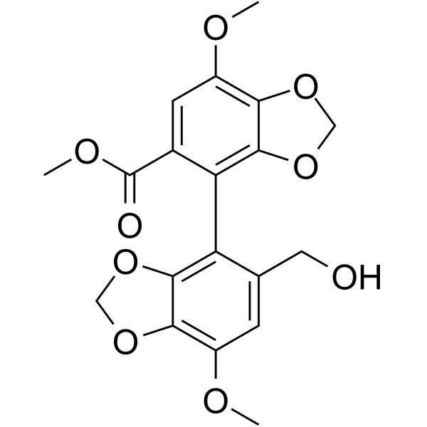
Bicyclol (SY801) is an orally active derivative of the traditional Chinese medicine Schisandra chinensis, which has antiviral, anti-inflammatory, immunomodulatory, antioxidant, anti-steatosis, anti-fibrotic and anti-tumor activities. Bicyclol regulates the expression of heat shock proteins and plays an anti-apoptosis role in hepatocytes. Bicyclol reduces the activation of NF-κB and the levels of inflammatory factors in hepatocytes infected with hepatitis C virus (HCV) by inhibiting the activation of the ROS-MAPK-NF-κB pathway, and prevents ferroptosis in acute liver injury. Bicyclol can change the expression of Mdr-1, GSH/GST and Bcl-2, increase the intracellular concentration of anticancer drugs, and sensitize drug-resistant cells to anticancer drugs. Bicyclol inhibits the proliferation of human malignant hepatoma cells by regulating the PI3K/AKT pathway and the Ras/Raf/MEK/ERK pathway. Bicyclol can be used in the study of chronic hepatitis, acute liver injury, nonalcoholic fatty liver disease, liver fibrosis and hepatocellular carcinoma.
연구목적의 판매만을 진행합니다. 환자를 대상으로 한 판매는 하지 않습니다.
- Purity : 99.91%
- CAS No.: 118159-48-1
- 화학식: C19H18O9
- 분자량:390.34
-
보관:Powder -20°C, 3 years , 4°C, 2 years ; In solvent -80°C, 2 years , -20°C, 1 year

To place orders, for customer services and technical support, please contact: MedChemExpress USA
Tel: 609-228-6898 E-mail: [email protected] [email protected]


-
 Biological Activity
-
Chemical Information
-
용액&용해도
- Protocol
- 순도&문서
- References
-
 Help & FAQs


-
HY-N7073
SilymarinMpro Inhibitor -
HY-N0693
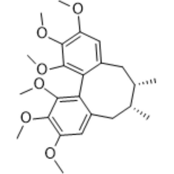
Schisandrin ACytochrome P450 Inhibitor -
HY-W018791
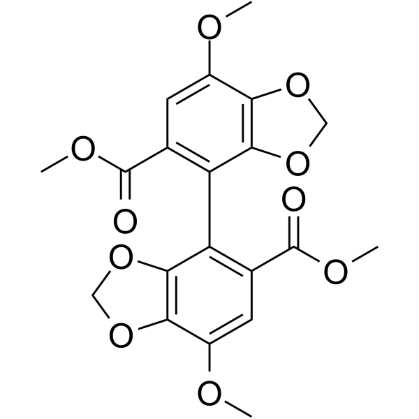
BifendateAntioxidant, Anti-HBV Agent -
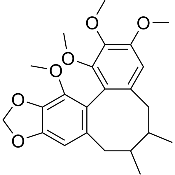
HY-N0089
Schisandrin BAntioxidant -
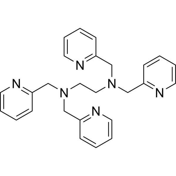
HY-100202
TPENHeavy Metal Chelator -
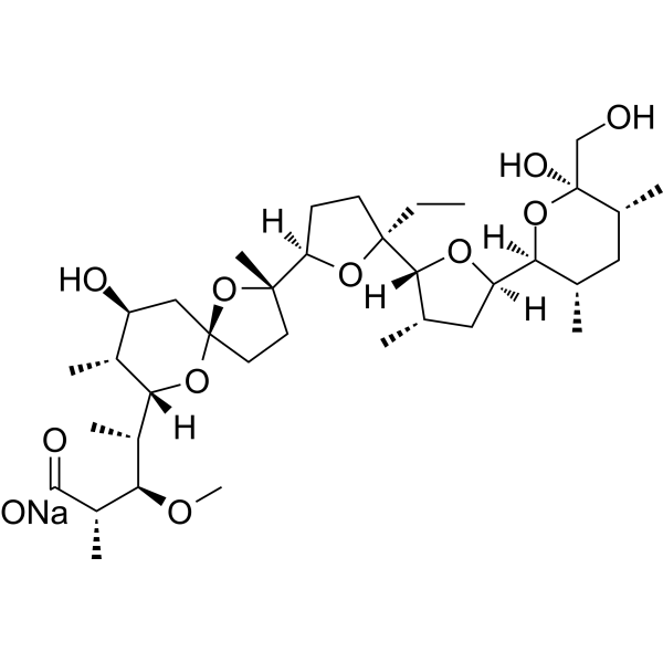
HY-N0150
Monensin sodiumAntibiotic -

HY-N0150R
Monensin sodium (Standard)Antibiotic -

HY-100202R
TPEN (Standard)Heavy Metal Chelator


Biological Activity
제품 설명
In Vitro
Bicyclol (0.1-1 mmol/L, 3 h) inhibits the release of ALT and AST, as well as the production of MDA, in CCl4-induced hepatotoxic Wistar rat hepatocytes in a dose-dependent manner[1]. Bicyclol (40 and 80 μM; 1 h) reduces palmitic acid (HY-N0830)-induced apoptosis in human hepatocyte HL-7702 cells and inhibits palmitic acid (HY-N0830)-induced activation of MAPKs and NF-κB in macrophages[2]. Bicyclol (200 μM; 10 h) reduces the level of intracellular oxygen free radicals in HCV-infected Huh7.5 cells and inhibits the phosphorylation of p38, ERK, JNK, and NF-κB[4]. Bicyclol (10 μM; 30 min) alleviates serrata-induced hepatocyte inactivation, destruction, and lipid peroxidation. Bicyclol (0-500 μM; 0-48 h) effectively inhibits the proliferation of HepG2 cells in a dose-dependent and time-dependent manner[5]. Bicyclol (0-100 μM; 24 h) reverses vincristine sulfate (HY-N0488) resistance in VinRKB cells by 2.8, 7.3, and 20.7 times, respectively[6]. Bicyclol (50 and 100 μM; 12-72 h) alters the intracellular drug concentrations in vincristine sulfate (HY-N0488)-resistant human epidermoid carcinoma VinRKB and doxorubicin (HY-15142A)-resistant human breast cancer AdrRMCF-7 cells, sensitizing the resistant cells to anticancer drugs[8].
MedChemExpress (MCE) has not independently confirmed the accuracy of these methods. They are for reference only.
-
Cell Line:CCL4-induced intoxication of Wistar rat hepatocytes
-
Concentration:10-4-10-3 mol/L
-
Incubation Time:3 h
-
Result:Improved rat liver cell viability and reduced cell surface damage such as microvilli aggregation and shedding.
-
Cell Line:HepG2 cells
-
Concentration:50, 100, 200 and 500 μM
-
Incubation Time:24 and 48 h
-
Result:Inhibited the phosphorylation of Akt and ERK, and downregulates the expression of cyclin D1, cyclin E2, CDK2, CDK4, p-Rb, and p-mTOR.
-
Cell Line:Palmitic acid induces human hepatocyte HL-7702 cells
-
Concentration:40 and 80 μM
-
Incubation Time:1 h
-
Result:Inhibited the expression of IL-6 and TNF-α induced by PA.
-
Cell Line:Vincristine-resistant human epidermoid carcinoma VinRKB and doxorubicin-resistant human breast cancer AdrRMCF-7
-
Concentration:50 and 100 μM
-
Incubation Time:24-72 h
-
Result:Reduced the level of p-glycoprotein encoded by the MDR-1 gene in VinRKB and AdrRMCF-7 cells to the level of drug-sensitive cells. The increase of GSH content in virrkb and AdrRMCF-7 cells increased the GST activity of AdrRMCF-7 cells, and the overexpression of Bcl-2 protein.
In Vivo
MedChemExpress (MCE) has not independently confirmed the accuracy of these methods. They are for reference only.
-
Animal Model:CCl4, D-Galactosamine, Acetaminophen-Induced Liver Injury in Mice [1]
-
Dosage:50, 100 and 200 mg/kg
-
Administration:Oral gavage (p.o.) Single dose administration
-
Result:Reduced the elevated serum ALT and AST levels in a dose-dependent manner, and also ameliorated the liver lesions.
-
Animal Model:Acetaminophen-Induced Liver Injury in Mice [1]
-
Dosage:50 and 150 mg/kg
-
Administration:Oral gavage (p.o.); Once a day for 3 days
-
Result:Reduced mitochondrial ultrastructural damage, mitochondrial AST release, mitochondrial fluidity and mitochondrial swelling, and mitochondrial cytochrome C release.
-
Animal Model:ConA and acetaminophen-Induced Liver Injury in Mice [1]
-
Dosage:300 mg/kg
-
Administration:Oral gavage (p.o.); Once a day for 3 days
-
Result:Induced the expression of HSP70/27 mRNA and protein in mouse liver and activated heat shock factor-1 (HSF1) in mouse liver.
-
Animal Model:HFD-induced liver inflammation in mice[2]
-
Dosage:25 and 50 mg/kg
-
Administration:Oral gavage (p.o.); once every other day for the last four weeks of the 16-week HFD induction
-
Result:Inhibited the expression levels of pro-inflammatory genes IL-6, TNF-α, IL-1β, COX-2, ICAM-1, VCAM-1, and MCP-1.
-
Animal Model:CCl4-induced ALI mice[6].
-
Dosage:200 mg/kg
-
Administration:Intraperitoneal injection (i.p.); 3 times a day for 2 days
-
Result:Inhibited iron accumulation, excessive production of reactive oxygen species, enhanced lipid peroxidation, altered mitochondrial morphology, and decreased GPx4 and xCT protein levels in mouse liver.
- Research Protocol for Infectious Diseases
- RNA extraction experimental
- Collagen: Sirius Red Staining
- Annexin V plus membrane-impermeant dye apoptosis staining
- ROS/oxidative-stress fluorescent staining
- Connective Tissue: Masson's Trichrome/Collagen Trichrome Staining
- Research Protocol for Inflammation-related Diseases
- Liver Cancer Modeling
- Research Protocol for Metabolic Diseases
- Liver Histomorphometry
- Cell Viability Determination by MTT Colorimetric Assay
- Fibrosis/Collagen Morphometry
- Detection of Apoptosis
- TUNEL staining for apoptotic DNA fragmentation
- Apoptosis Solutions
- Ferroptosis Solutions
Clinical Trial
| NCT Number | Sponsor | Condition | Start Date |
Phase

|
|---|---|---|---|---|
| NCT01329991 | 2011-05 | PHASE1 |


Chemical Information
-
CAS No. 118159-48-1
-
Appearance Solid
-
분자량 390.34
-
화학식 C19H18O9
-
Color White to off-white
-
SMILES
O=C(C1=CC(OC)=C(OCO2)C2=C1C3=C4OCOC4=C(OC)C=C3CO)OC
-
Synonyms
SY801
-
선적
Room temperature in continental US; may vary elsewhere.
-
보관
Powder -20°C 3 years 4°C 2 years In solvent -80°C 2 years -20°C 1 year


용액&용해도
In Vitro:
DMSO : 100 mg/mL (256.19 mM; Need ultrasonic; Hygroscopic DMSO has a significant impact on the solubility of product, please use newly opened DMSO)
Please refer to the solubility information to select the appropriate solvent. Once prepared, please aliquot and store the solution to prevent product inactivation from repeated freeze-thaw cycles.
Storage method and period of stock solution: -80°C, 2 years; -20°C, 1 year. When stored at -80°C, please use it within 2 years. When stored at -20°C, please use it within 1 year.
Please refer to the solubility information to select the appropriate solvent. Once prepared, please aliquot and store the solution to prevent product inactivation from repeated freeze-thaw cycles.
Storage method and period of stock solution: -80°C, 2 years; -20°C, 1 year. When stored at -80°C, please use it within 2 years. When stored at -20°C, please use it within 1 year.
Concentration (start) × Volume (start) = Concentration (final) × Volume (final)
In Vivo:
Select the appropriate dissolution method based on your experimental animal and administration route.
- For the following dissolution methods, please ensure to first prepare a clear stock solution using an In Vitro approach and then sequentially add co-solvents:
- To ensure reliable experimental results, the clarified stock solution can be appropriately stored based on storage conditions. As for the working solution for In Vivo experiments, it is recommended to prepare freshly and use it on the same day.
- The percentages shown for the solvents indicate their volumetric ratio in the final prepared solution. If precipitation or phase separation occurs during preparation, heat and/or sonication can be used to aid dissolution.
Add each solvent one by one: 10% DMSO 40% PEG300 5% Tween-80 45% Saline
Solubility: ≥ 2.5 mg/mL (6.40 mM); Clear solution
This protocol yields a clear solution of ≥ 2.5 mg/mL (saturation unknown).
Taking 1 mL working solution as an example, add 100 μL DMSO stock solution (25.0 mg/mL) to 400 μL PEG300, and mix evenly; then add 50 μL Tween-80 and mix evenly; then add 450 μL Saline to adjust the volume to 1 mL.
Preparation of Saline: Dissolve 0.9 g sodium chloride in ddH₂O and dilute to 100 mL to obtain a clear Saline solution.
Add each solvent one by one: 10% DMSO 90% (20% SBE-β-CD in Saline)
Solubility: 2.5 mg/mL (6.40 mM); Suspended solution; Need ultrasonic
This protocol yields a suspended solution of 2.5 mg/mL. Suspended solution can be used for oral and intraperitoneal injection.
Taking 1 mL working solution as an example, add 100 μL DMSO stock solution (25.0 mg/mL) to 900 μL 20% SBE-β-CD in Saline, and mix evenly.
Preparation of 20% SBE-β-CD in Saline (4°C, storage for one week): 2 g SBE-β-CD powder is dissolved in 10 mL Saline, completely dissolve until clear.
For the following dissolution methods, please prepare the working solution directly:
It is recommended to prepare fresh solutions and use them promptly within a short period of time.
The percentages shown for the solvents indicate their volumetric ratio in the final prepared solution. If precipitation or phase separation occurs during preparation, heat and/or sonication can be used to aid dissolution.
Add each solvent one by one: 15% Cremophor EL 85% Saline
Solubility: 2 mg/mL (5.12 mM); Clear solution; Need ultrasonic
In Vivo Dissolution Calculator
Please enter the basic information of animal experiments:
-
-
-
-
Recommended: Prepare an additional quantity of animals to account for potential losses during experiments.
Please enter your animal formula composition:
-
%DMSO +
Recommended: Keep the proportion of DMSO in working solution below 2% if your animal is weak.
-
%+
-
+%Tween-80 + +
-
%Saline +
The co-solvents required include: DMSO, . All of co-solvents are available by MedChemExpress (MCE). , Tween 80. All of co-solvents are available by MedChemExpress (MCE).
Working solution concentration: 0.22 mg/mL
Method for preparing stock solution: mg drug dissolved in μL DMSO. Stock solution concentration: mg/mL.
1. Take μL DMSO stock solution;
2. Add μL .
μL , mix evenly;
3. Then add μL Tween 80, mix evenly;
4. Then add μL
Please ensure that the stock solution in the first step is dissolved to a clear state, and add co-solvents in sequence. You can use ultrasonic heating (ultrasonic cleaner, recommended frequency 20-40 kHz), vortexing, etc. to assist dissolution.


순도&문서
-
Data Sheet (293 KB)
-
SDS (398 KB)
- English - EN (398 KB)
- Français - FR (398 KB)
- Deutsch - DE (398 KB)
- Norwegian - NO (398 KB)
- Español - ES (398 KB)
- Swedish - SV (398 KB)
- Italian - IT (398 KB)
- Korean - KR (398 KB)
- Portuguese - PT (398 KB)
-
Handling Instructions (2659 KB)
References
[1]. Walsh, S.W., Y. Wang, and A. Killian, AA-2414, an antioxidant and thromboxane receptor blocker, completely inhibits peroxide-induced vasoconstriction in the human placenta. J Pharmacol Exp Ther, 1999. 290(1): p. 220-6. [Content Brief]
[2]. Weixin Zhao, et al. "Bicyclol ameliorates nonalcoholic fatty liver disease in mice via inhibiting MAPKs and NF-κB signaling pathways." Biomedicine & Pharmacotherapy 141 (2021): 111874. [Content Brief]
[3]. Hui-Juan Dai, et al. "Induction of heat shock protein 27 by bicyclol attenuates d-galactosamine/lipopolysaccharide-induced liver injury." European Journal of Pharmacology 791 (2016): 482-490. [Content Brief]
[4]. Li Hu, et al. "Bicyclol attenuates liver inflammation induced by infection of hepatitis C virus via repressing ROS-mediated activation of MAPK/NF-κB signaling pathway." Frontiers in Pharmacology 9 (2018): 1438. [Content Brief]
[5]. Tianming Zhao, et al. "Regulating Nrf2-GPx4 axis by bicyclol can prevent ferroptosis in carbon tetrachloride-induced acute liver injury in mice." Cell death discovery 8.1 (2022): 380. [Content Brief]
[6]. Yu Wang, et al. "Bicyclol induces cell cycle arrest and autophagy in HepG2 human hepatocellular carcinoma cells through the PI3K/AKT and Ras/Raf/MEK/ERK pathways." BMC cancer 16 (2016): 1-15. [Content Brief]
[7]. Tianming Zhao, et al. "Therapeutic potential of bicyclol in liver diseases: lessons from a synthetic drug based on herbal derivative in traditional Chinese medicine." International Immunopharmacology 91 (2021): 107308. [Content Brief]
[8]. Bing Zhu, et al. "Chemosensitizing multiple drug resistance of human carcinoma by Bicyclol involves attenuated P-glycoprotein, GST-P and Bcl-2." Cancer biology & therapy 5.5 (2006): 536-543. [Content Brief]


Complete Stock Solution Preparation Table
Please refer to the solubility information to select the appropriate solvent. Once prepared, please aliquot and store the solution to prevent product inactivation from repeated freeze-thaw cycles.
Storage method and period of stock solution: -80°C, 2 years; -20°C, 1 year. When stored at -80°C, please use it within 2 years. When stored at -20°C, please use it within 1 year.
| Optional Solvent | Concentration Solvent Mass | 1 mg | 5 mg | 10 mg | 25 mg |
|---|---|---|---|---|---|
| DMSO | 1 mM | 2.5619 mL | 12.8093 mL | 25.6187 mL | 64.0467 mL |
| 5 mM | 0.5124 mL | 2.5619 mL | 5.1237 mL | 12.8093 mL | |
| 10 mM | 0.2562 mL | 1.2809 mL | 2.5619 mL | 6.4047 mL | |
| 15 mM | 0.1708 mL | 0.8540 mL | 1.7079 mL | 4.2698 mL | |
| 20 mM | 0.1281 mL | 0.6405 mL | 1.2809 mL | 3.2023 mL | |
| 25 mM | 0.1025 mL | 0.5124 mL | 1.0247 mL | 2.5619 mL | |
| 30 mM | 0.0854 mL | 0.4270 mL | 0.8540 mL | 2.1349 mL | |
| 40 mM | 0.0640 mL | 0.3202 mL | 0.6405 mL | 1.6012 mL | |
| 50 mM | 0.0512 mL | 0.2562 mL | 0.5124 mL | 1.2809 mL | |
| 60 mM | 0.0427 mL | 0.2135 mL | 0.4270 mL | 1.0674 mL | |
| 80 mM | 0.0320 mL | 0.1601 mL | 0.3202 mL | 0.8006 mL | |
| 100 mM | 0.0256 mL | 0.1281 mL | 0.2562 mL | 0.6405 mL |


Bicyclol Related Classifications
HY-B0766 Related Classifications
 Powered by Bioz