



Loliolide

Based on 1 Customer Validation
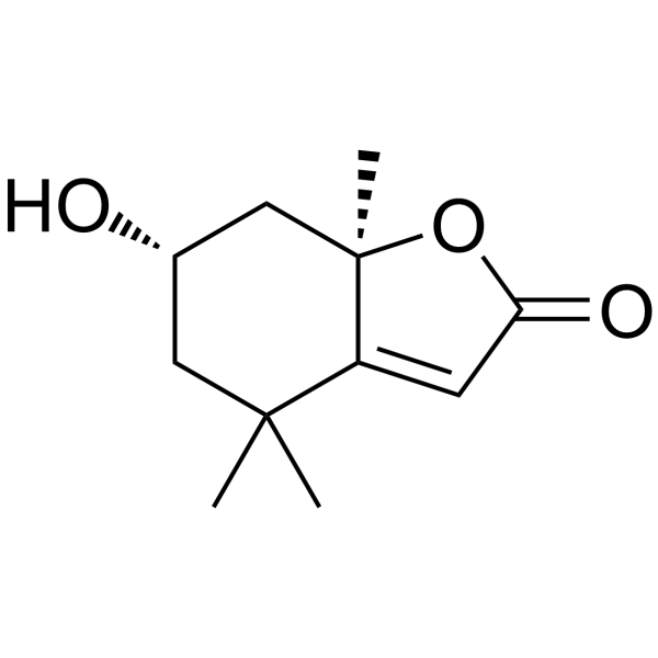
Loliolide (Loliolid) is a β-carotene metabolite. Loliolide reduces caspase 3, 8, 9 expression, enhances PI3K, AKT, SIRT1, inhibits ROS, apoptosis, and blocks NF-κB p65 nuclear translocation. Loliolide protects mitochondria, reduces oxidative stress, and increases cell viability in neuroblastoma cells. Loliolide can be used for the research of UV-induced skin damage and Parkinson’s disease.
For research use only. We do not sell to patients.
- Purity : 99.92%
- CAS No.: 5989-02-6
- Formula: C11H16O3
- Molecular Weight:196.24
-
Storage:
4°C, protect from light
* In solvent : -80°C, 6 months; -20°C, 1 month (protect from light)

To place orders, for customer services and technical support, please contact: MedChemExpress USA
Tel: 609-228-6898 E-mail: [email protected] [email protected]


-
 Biological Activity
-
Chemical Information
-
Solvent & Solubility
- Protocols
- Purity & Documentation
- References
-
 Help & FAQs


-
HY-N0761
Isoferulic acidAnti-glycation Agent -
HY-W028393
Indole-3-pyruvic acidAHR Agonist -

HY-W028393R
Indole-3-pyruvic acid (Standard)Reference Standard -
HY-12687S
Tizoxanide-d4Stable Isotope -
HY-N9182
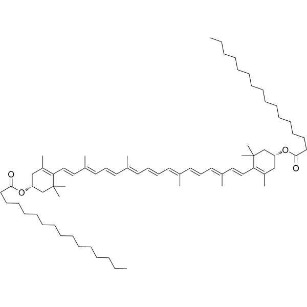
Zeaxanthin dipalmitatecarotenoid -
HY-N6635
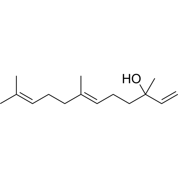
trans-NerolidolAnti-fungal Agent -
HY-W011641
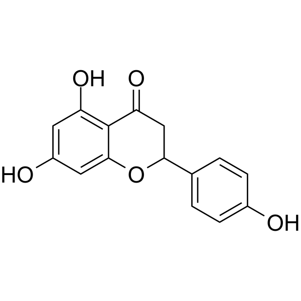
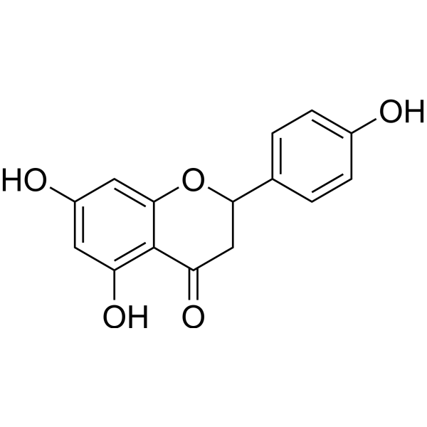
(±)-NaringeninAnti-inflammatory Agents -
HY-121038
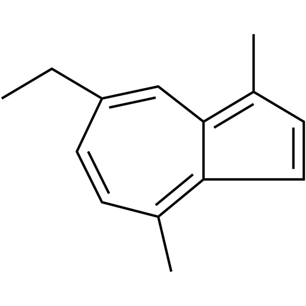
ChamazuleneLeukotriene B4 Inhibitor -

HY-N10544
δ-CadineneApoptosis Inducer -
HY-W011641R
(±)-Naringenin (Standard) -

HY-N6635R
trans-Nerolidol (Standard)Anti-fungal Agent


Biological Activity
Description
IC50 & Target
[1]|
SIRT1 |
MMP-9 |
Caspase 3 |
Caspase 8 |
Caspase 9 |
Cellular Effect
|
Cell Line

|
Type | Value | Description | References |
|---|---|---|---|---|
| A549 | IC50 |
>10 μM
Compound: 19
|
Antiproliferative activity against human A549 cells assessed as reduction in cell viability after 72 hrs by SRB assay
Antiproliferative activity against human A549 cells assessed as reduction in cell viability after 72 hrs by SRB assay
|
[PMID: 32946697] |
| HepG2 | IC50 |
>20 μg/mL
Compound: loliolide
|
Growth inhibition of HepG2 cells
Growth inhibition of HepG2 cells
|
[PMID: 17547458] |
| HT-29 | IC50 |
>10 μM
Compound: S11
|
Cytotoxicity against human HT-29 cells assessed as reduction in cell viability after 72 hrs by CellTiter 96 aqueous one solution assay
Cytotoxicity against human HT-29 cells assessed as reduction in cell viability after 72 hrs by CellTiter 96 aqueous one solution assay
|
[PMID: 30057155] |
| HT-29 | IC50 |
>10 μM
Compound: S11
|
Inhibition of mitochondrial membrane potential in human HT-29 cells after 3 hrs by JC-1 staining based fluorescence assay
Inhibition of mitochondrial membrane potential in human HT-29 cells after 3 hrs by JC-1 staining based fluorescence assay
|
[PMID: 30057155] |
| KB | IC50 |
>10 μM
Compound: 19
|
Antiproliferative activity against human KB cells assessed as reduction in cell viability after 72 hrs by SRB assay
Antiproliferative activity against human KB cells assessed as reduction in cell viability after 72 hrs by SRB assay
|
[PMID: 32946697] |
| MCF7 | IC50 |
>10 μM
Compound: 19
|
Antiproliferative activity against human MCF7 cells assessed as reduction in cell viability after 72 hrs by SRB assay
Antiproliferative activity against human MCF7 cells assessed as reduction in cell viability after 72 hrs by SRB assay
|
[PMID: 32946697] |
| MDA-MB-231 | IC50 |
>10 μM
Compound: 19
|
Antiproliferative activity against human MDA-MB-231 cells assessed as reduction in cell viability after 72 hrs by SRB assay
Antiproliferative activity against human MDA-MB-231 cells assessed as reduction in cell viability after 72 hrs by SRB assay
|
[PMID: 32946697] |
| MDA-MB-231 | IC50 |
>10 μM
Compound: S11
|
Cytotoxicity against human MDA-MB-231 cells assessed as reduction in cell viability after 72 hrs by CellTiter 96 aqueous one solution assay
Cytotoxicity against human MDA-MB-231 cells assessed as reduction in cell viability after 72 hrs by CellTiter 96 aqueous one solution assay
|
[PMID: 30057155] |
| MDA-MB-435 | IC50 |
>10 μM
Compound: S11
|
Cytotoxicity against human MDA-MB-435 cells assessed as reduction in cell viability after 72 hrs by CellTiter 96 aqueous one solution assay
Cytotoxicity against human MDA-MB-435 cells assessed as reduction in cell viability after 72 hrs by CellTiter 96 aqueous one solution assay
|
[PMID: 30057155] |
| OVCAR-3 | IC50 |
>10 μM
Compound: S11
|
Cytotoxicity against human OVCAR3 cells assessed as reduction in cell viability after 72 hrs by CellTiter 96 aqueous one solution assay
Cytotoxicity against human OVCAR3 cells assessed as reduction in cell viability after 72 hrs by CellTiter 96 aqueous one solution assay
|
[PMID: 30057155] |
| RAW264.7 | CC50 |
>100 μM
Compound: 11
|
Cytotoxicity against mouse RAW264.7 cells assessed as reduction in cell viability measured after 24 hrs by MTT assay
Cytotoxicity against mouse RAW264.7 cells assessed as reduction in cell viability measured after 24 hrs by MTT assay
|
[PMID: 33667099] |
| RAW264.7 | IC50 |
>0.4 μM
Compound: loliolide
|
Antiinflammatory activity against mouse RAW264.7 cells assessed as inhibition of LPS-induced nitric oxide release after 24 hrs
Antiinflammatory activity against mouse RAW264.7 cells assessed as inhibition of LPS-induced nitric oxide release after 24 hrs
|
[PMID: 20192236] |
| RAW264.7 | IC50 |
>50 μM
Compound: 11
|
Inhibition of LPS-induced IL-1beta production in mouse RAW264.7 cells after 24 hrs by ELISA
Inhibition of LPS-induced IL-1beta production in mouse RAW264.7 cells after 24 hrs by ELISA
|
[PMID: 24963714] |
| RAW264.7 | IC50 |
>50 μM
Compound: 11
|
Inhibition of LPS-induced NO production in mouse RAW264.7 cells after 24 hrs by Griess reagent based assay
Inhibition of LPS-induced NO production in mouse RAW264.7 cells after 24 hrs by Griess reagent based assay
|
[PMID: 24963714] |
| RAW264.7 | IC50 |
56.7 μM
Compound: 11
|
Inhibition of LPS induced NO production in mouse RAW264.7 cells measured after 24 hrs by Griess reagent based assay
Inhibition of LPS induced NO production in mouse RAW264.7 cells measured after 24 hrs by Griess reagent based assay
|
[PMID: 33667099] |
| WI-38 | IC50 |
>20 μg/mL
Compound: loliolide
|
Growth inhibition of WI38 cells
Growth inhibition of WI38 cells
|
[PMID: 17547458] |
| WI-38 VA13 | IC50 |
>20 μg/mL
Compound: loliolide
|
Growth inhibition of VA13 cells
Growth inhibition of VA13 cells
|
[PMID: 17547458] |
In Vitro
Loliolide (25-100 μM; 24 h) is non-toxic to HaCaT human keratinocytes at concentrations up to 100 μM and dose-dependently recovers UVB-reduced HaCaT cell viability[1].
Loliolide (25-100 μM; 24 h) dose-dependently inhibits UVB-induced reactive oxygen species generation and DNA damage in HaCaT cells[1].
Loliolide (50-100 μM; 24 h) reduces UVB-induced apoptosis in HaCaT cells[1].
Loliolide (25-100 μM; 24 h) dose-dependently inhibits UVB-induced activation of caspase 3, 8, and 9 to reduce apoptosis in HaCaT cells[1].
Loliolide (25-200 μM; 24 h) dose-dependently inhibits UVB-induced MMP-1, MMP-2, MMP-3, and MMP-9 expression and restores UVB-reduced SIRT1 expression in HaCaT cells[1].
Loliolide (100 μM; 8-24 h) significantly accelerates scratch wound closure via increased cell migration in HaCaT cells[1].
Loliolide (25-100 μM; 24-72 h) dose-dependently increases proliferation in HaCaT cells[1].
Loliolide (25-100 μM; 24 h) dose-dependently upregulates migration factors, growth factor KGF, and wound-healing cytokines, while downregulating inflammation-related genes in scratched HaCaT cells[1].
Loliolide (25-100 μM; 24 h) dose-dependently activates the PI3K/AKT EGFR signaling pathway in scratched HaCaT human keratinocytes, with maximum activity at 100 μM[1].
Loliolide (1-100 μM; 24 h) exhibits no cytotoxicity and inhibits 6-hydroxydopamin (6-OHDA)-induced cytotoxicity in SH-SY5Y cells[2].
Loliolide (50-100 μM; 6 h) reduces 6-OHDA-induced ROS production and Caspase-3 activity, stimulates catalase activity, prevents 6-OHDA-induced mitochondrial membrane depolarization in SH-SY5Y cells[2].
Loliolide (50-100 μM; 24 h) prevents 6-OHDA-induced nuclear fragmentation and condensation in SH-SY5Y cells[2].
Loliolide (50-100 μM; 6 h) inhibits 6-OHDA-induced NF-κB p65 translocation from the cytoplasm to the nucleus in SH-SY5Y cells[2].
Loliolide (50-100 μM; 24 h) is non-cytotoxic to RAW 264.7 cells and does not induce NO production in RAW 264.7 cells[2].
Loliolide (50-100 μM; 24 h) reduces LPS-induced NO production and reduces LPS (HY-D1056)-induced production of TNF-α and IL-6 in RAW 264.7 cells[2].
Loliolide (300 μM; 24 h leaf treatment, 5 d mite incubation) reduces the survival rate of Tetranychus urticae and egg deposition in Solanum lycopersicum cv. Micro-Tom leaves [3].
MedChemExpress (MCE) has not independently confirmed the accuracy of these methods. They are for reference only. Further protocols information, click here.
-
Cell Line:HaCaT cells
-
Concentration:25; 50; 100 μM
-
Incubation Time:24 h
-
Result:Showed no effect on HaCaT cell viability at concentrations up to 100 μM under normal culture conditions.
Recovered UVB-reduced cell viability in a dose-dependent manner, with 100 μM producing the strongest statistically significant recovery.
-
Cell Line:HaCaT cells
-
Concentration:25; 50; 100 μM
-
Incubation Time:24 h
-
Result:Dose-dependently reduced UVB-induced expression of cleaved caspase 3, 8, and 9 in HaCaT cells.
Dose-dependently increased phosphorylation of PI3K and AKT in scratched HaCaT cells.
Showed the strongest activation at 100 μM, while total PI3K and AKT levels remained unchanged.
-
Cell Line:HaCaT cells
-
Concentration:25; 50; 100; 200 μM
-
Incubation Time:24 h
-
Result:Dose-dependently reduced UVB-induced mRNA expression of MMP-1, MMP-2, MMP-3, and MMP-9 in HaCaT cells.
Restored UVB-reduced SIRT1 mRNA expression in HaCaT cells.
Dose-dependently increased mRNA expression of migration factors K6, K16, K17, growth factor KGF, and cytokines IL-1, IL-17, IL-22 in scratched HaCaT cells.
Dose-dependently reduced mRNA expression of inflammation-related genes TNF-α and MMP-1 in scratched HaCaT cells.
-
Cell Line:HaCaT cells
-
Concentration:25; 50; 100 μM
-
Incubation Time:8, 16, 24 h
-
Result:Significantly increased HaCaT cell migration and scratch wound closure at 8, 16, and 24 h post-scratching.
Showed statistically significant differences (p<0.01 compared to untreated scratched control) at all time points.
-
Cell Line:HaCaT cells
-
Concentration:25-100 μM
-
Incubation Time:24, 48, 72 h
-
Result:Dose-dependently increased HaCaT cell proliferation over 72 h.
Produced statistically significant increases at 72 h for all concentrations, and at 48 h for 100 μM.
-
Cell Line:SH-SY5Y cells
-
Concentration:1; 5; 10; 50;100 μM
-
Incubation Time:24 h
-
Result:Did not induce cytotoxicity at any tested concentration.
Showed cell viability remained close to vehicle control levels.\nIncreased cell viability by 23.70% at 50 μM and 41.06% at 100 μM.
-
Cell Line:SH-SY5Y cells
-
Concentration:50; 100 μM
-
Incubation Time:6 h
-
Result:Downregulated nuclear NF-κB p65 and upregulated cytosolic levels, with significant inhibition of translocation observed at 100 μM compared to cells treated with 6-OHDA alone.
-
Cell Line:RAW 264.7 murine macrophage cells
-
Concentration:50; 100 μM
-
Incubation Time:24 h
-
Result:Reduced TNF-α production by 305% at 50 μM and 238% at 100 μM.
Reduced IL-6 production by 197% at 50 μM and 220% at 100 μM compared to cells stimulated with LPS alone.
Did not significantly affect IL-10 levels.
-
Cell Line:HaCaT cells
-
Concentration:50; 100 μM
-
Incubation Time:24 h
-
Result:Reduced UVB-induced apoptosis in HaCaT cells.
Showed stronger activity at 100 μM than 50 μM.


Chemical Information
-
CAS No. 5989-02-6
-
Appearance Solid
-
Molecular Weight 196.24
-
Formula C11H16O3
-
Color White to off-white
-
SMILES
C[C@]12C(C(C)(C[C@@H](C1)O)C)=CC(O2)=O
-
Synonyms
Loliolid; Digiprolactone
-
Structure Classification
-
Shipping
Room temperature in continental US; may vary elsewhere.
-
Storage
4°C, protect from light
* In solvent : -80°C, 6 months; -20°C, 1 month (protect from light)


Solvent & Solubility
In Vitro:
Methanol : 58.87 mg/mL (299.99 mM; Need ultrasonic and warming)
Please refer to the solubility information to select the appropriate solvent. Once prepared, please aliquot and store the solution to prevent product inactivation from repeated freeze-thaw cycles.
Storage method and period of stock solution: -80°C, 6 months; -20°C, 1 month (protect from light). When stored at -80°C, please use it within 6 months. When stored at -20°C, please use it within 1 month.
Please refer to the solubility information to select the appropriate solvent. Once prepared, please aliquot and store the solution to prevent product inactivation from repeated freeze-thaw cycles.
Storage method and period of stock solution: -80°C, 6 months; -20°C, 1 month (protect from light). When stored at -80°C, please use it within 6 months. When stored at -20°C, please use it within 1 month.
Concentration (start) × Volume (start) = Concentration (final) × Volume (final)

Protocols
-
RNA extraction experimental
By lysing cells, releasing RNA, and removing impurities such as proteins and DNA, high-purity RNA products are finally obtained. The commonly used traditional method is the guanidine isothiocyanate/phenol/chloroform method (Trizol), which is suitable for a variety of animal materials including animal tissues, microorganisms, cultured cells, etc., and most plant materials.
-
Mammalian live/dead viability and cytotoxicity staining
Live/dead viability and cytotoxicity staining assays are based on the simultaneous detection of intracellular esterase activity in metabolically active (viable) cells and membrane integrity loss in non-viable cells. In commonly used dual-staining approaches, membrane-permeant fluorogenic substrates are converted by intracellular esterases into fluorescent products in live cells, while impermeant DNA-binding dyes selectively enter cells with compromised plasma membranes and label nucleic acids in dead or dying cells, enabling discrimination between viable and non-viable populations by fluorescence microscopy or flow cytometry.
-
Annexin V plus membrane-impermeant dye apoptosis staining
Annexin V-based apoptosis assays rely on the detection of phosphatidylserine (PS) externalization from the inner leaflet of the plasma membrane to the outer leaflet, an early biochemical hallmark of apoptosis. Fluorescently labeled Annexin V binds PS in a calcium-dependent manner, enabling identification of early apoptotic cells by flow cytometry or fluorescence microscopy. When combined with a membrane-impermeant DNA-binding dye (e. g. , propidium iodide), this approach allows discrimination between viable (Annexin V−/dye−), early apoptotic (Annexin V+/dye−), and late apoptotic or necrotic (Annexin V+/dye+) cell populations by assessing membrane integrity and PS exposure.
-
ROS/oxidative-stress fluorescent staining
ROS/oxidative-stress fluorescent staining uses cell-permeant fluorogenic probes that become fluorescent after oxidation inside cells or tissues; commonly used examples include DCFH-DA/DCFDA for broad cellular oxidant detection, DHE for superoxide-related signal detection, MitoSOX for mitochondrial superoxide-related signal detection, and CellROX probes for oxidative-stress-associated fluorescence readouts. The assay detects probe oxidation rather than a single ROS species unless the probe and analysis method have been chemically validated for that species. DCFH-DA enters cells, is deacetylated by intracellular esterases to DCFH, and produces fluorescent DCF after oxidation, so the readout is used as an operational measure of total cellular oxidative stress rather than a species-specific ROS measurement. DHE and MitoSOX can report superoxide-related oxidation, but red fluorescence alone can include non-specific ethidium-like oxidation products; HPLC or optimized spectral approaches are
-
CCK-8/WST-8 Cell Proliferation Assay
The CCK-8/WST-8 assay is based on the reduction of the water-soluble tetrazolium salt WST-8 to a water-soluble formazan product by cellular dehydrogenases in metabolically active cells, where the generated formazan amount is proportional to the number of living cells and is quantified by measuring absorbance in the visible range, providing a colorimetric readout for cell viability and proliferation assessment. This class of tetrazolium-based assays improves upon earlier MTT-based systems by producing a water-soluble formazan, eliminating the need for organic solubilization steps and enabling direct spectrophotometric measurement in culture medium.
-
Research Protocol for Inflammation-related Diseases
The NLRP3 inflammasome is a cytosolic innate immune signaling platform that integrates priming signals and danger-signal activation to promote caspase-1 activation, maturation of IL-1β and IL-18, and gasdermin D-mediated pyroptotic cell death. The core experimental logic is to determine whether inflammatory disease phenotypes are driven by increased NLRP3 expression, ASC-containing inflammasome assembly, caspase-1 cleavage, GSDMD cleavage, and extracellular release of IL-1β/IL-18 rather than by nonspecific cell injury alone. The pathway is strongly linked to inflammation-related disease phenotypes because monosodium urate crystals activate NALP3/NLRP3 inflammasome signaling in gout-like crystal inflammation, cholesterol crystals activate NLRP3 inflammasomes in atherogenesis models, and DSS-induced intestinal inflammation has been reported to involve NLRP3 inflammasome activity. However, experimental colitis studies also show context-dependent protective effects of NLRP3 inflammasome co
-
Cell Viability Determination by MTT Colorimetric Assay
The following protocol uses the MTT colorimetric assay as a classic literature-established method for assessing cell viability/metabolic activity in cultured mammalian cells. MTT[3-(4,5-dimethylthiazol-2-yl)-2,5-diphenyltetrazolium bromide] is reduced by metabolically active cells to a colored formazan product; the amount of formazan is quantified spectrophotometrically and provides an indirect measure of metabolically active viable cells. Importantly, MTT reduction reflects cellular oxidoreductase/metabolic activity rather than an absolute direct count of living cells, so changes in cellular metabolism can alter the signal independently of cell number.
-
Cytoplasmic-Nuclear Fractionated Protein Extraction
Cytoplasmic-nuclear fractionated protein extraction separates soluble cytoplasmic proteins from nuclear-enriched proteins by mild plasma-membrane permeabilization, differential centrifugation, washing of nuclei, and extraction of nuclear proteins for downstream immunoblotting or related molecular analysis. The readout is the relative abundance of a protein in cytoplasmic and nuclear fractions, commonly assessed by western blotting together with compartment markers such as tubulin or pyruvate kinase for cytoplasm and lamin, nucleoporin, hnRNP, H2AX, or Lamin B for nuclear fractions.
-
Apoptosis
Apoptosis, also called programmed cell death, is generally characterized by distinct morphological characteristics.
-
TUNEL staining for apoptotic DNA fragmentation
TUNEL staining detects DNA strand breaks by using terminal deoxynucleotidyl transferase to add labeled nucleotides to exposed 3′-OH DNA termini, generating either microscopic staining in fixed cells or tissue sections, or fluorescence/cytometric signal in cell suspensions. TUNEL positivity reflects DNA fragmentation but should not be interpreted alone as definitive apoptosis, because TUNEL can also label necrotic, autolytic, mechanically damaged, or DNA-repair-associated DNA breaks.
-
Cell Cytotoxicity Assay
Cytotoxicity assays are usually based on the assessment of cell membrane damage, which can also be indirectly detected by measuring cell viability. Detection methods include MTT assay, CKK-8 assay, LDH assay and ATP assay, etc.
-
Apoptosis Solutions
Apoptosis is a regulated, generally non-lytic cell-death pathway that removes unwanted, damaged, infected, or abnormal cells through coordinated morphological changes, caspase activation, DNA fragmentation, and membrane remodeling. The intrinsic apoptosis pathway is controlled mainly by mitochondrial outer membrane permeabilization, BCL-2 family proteins, cytochrome c release, apoptosome formation, caspase-9 activation, and downstream executioner caspase-3/7 activation. The extrinsic apoptosis pathway is initiated by death receptors such as Fas, TNFR, and TRAIL receptors, which recruit adaptor proteins and activate caspase-8 before engaging executioner caspases or mitochondrial amplification through BID cleavage. Apoptosis is linked to many phenotypes, including cancer cell killing, tissue homeostasis, immune regulation, neurodegeneration, infection response, and treatment-induced cytotoxicity; unresolved questions include how apoptosis interacts with necroptosis, pyroptosis, ferroptos
-
MTT Cell Proliferation Assay
The MTT assay is a colorimetric endpoint assay for estimating viable cell number, cell growth, cytotoxicity, or cell activation in cultured mammalian cells. Living cells reduce the yellow tetrazolium salt MTT into purple/blue formazan, while dead cells do not generate the same signal; the resulting color can be quantified with a multiwell spectrophotometer. MTT reduction is commonly interpreted as a readout of metabolic activity that often correlates with viable cell number, but it should not be treated as a direct cell-counting method unless the assay is optimized for the cell type and experimental condition. Studies show that MTT reduction can involve mitochondrial and non-mitochondrial reducing systems, and formazan may accumulate in intracellular lipid droplets rather than simply marking mitochondria.


Purity & Documentation
-
Data Sheet (285 KB)
-
SDS (394 KB)
- English - EN (394 KB)
- Français - FR (394 KB)
- Deutsch - DE (394 KB)
- Norwegian - NO (394 KB)
- Español - ES (394 KB)
- Swedish - SV (394 KB)
- Italian - IT (394 KB)
- Korean - KR (394 KB)
- Portuguese - PT (394 KB)
-
Handling Instructions (2659 KB)
References
[1]. Park SH, et al. Loliolide Presents Antiapoptosis and Antiscratching Effects in Human Keratinocytes. Int J Mol Sci. 2019;20(3):651. Published 2019 Feb 2. [Content Brief]
[2]. Silva J, et al. Loliolide, a New Therapeutic Option for Neurological Diseases? In Vitro Neuroprotective and Anti-Inflammatory Activities of a Monoterpenoid Lactone Isolated from Codium tomentosum. Int J Mol Sci. 2021;22(4):1888. Published 2021 Feb 14. [Content Brief]
[3]. Murata M, et al. Loliolide, a Carotenoid Metabolite, Is a Potential Endogenous Inducer of Herbivore Resistance. Plant Physiol. 2019;179(4):1822-1833. [Content Brief]


Complete Stock Solution Preparation Table
Please refer to the solubility information to select the appropriate solvent. Once prepared, please aliquot and store the solution to prevent product inactivation from repeated freeze-thaw cycles.
Storage method and period of stock solution: -80°C, 6 months; -20°C, 1 month (protect from light). When stored at -80°C, please use it within 6 months. When stored at -20°C, please use it within 1 month.
| Optional Solvent | Concentration Solvent Mass | 1 mg | 5 mg | 10 mg | 25 mg |
|---|---|---|---|---|---|
| Methanol | 1 mM | 5.0958 mL | 25.4790 mL | 50.9580 mL | 127.3950 mL |
| 5 mM | 1.0192 mL | 5.0958 mL | 10.1916 mL | 25.4790 mL | |
| 10 mM | 0.5096 mL | 2.5479 mL | 5.0958 mL | 12.7395 mL | |
| 15 mM | 0.3397 mL | 1.6986 mL | 3.3972 mL | 8.4930 mL | |
| 20 mM | 0.2548 mL | 1.2740 mL | 2.5479 mL | 6.3698 mL | |
| 25 mM | 0.2038 mL | 1.0192 mL | 2.0383 mL | 5.0958 mL | |
| 30 mM | 0.1699 mL | 0.8493 mL | 1.6986 mL | 4.2465 mL | |
| 40 mM | 0.1274 mL | 0.6370 mL | 1.2740 mL | 3.1849 mL | |
| 50 mM | 0.1019 mL | 0.5096 mL | 1.0192 mL | 2.5479 mL | |
| 60 mM | 0.0849 mL | 0.4247 mL | 0.8493 mL | 2.1233 mL | |
| 80 mM | 0.0637 mL | 0.3185 mL | 0.6370 mL | 1.5924 mL | |
| 100 mM | 0.0510 mL | 0.2548 mL | 0.5096 mL | 1.2740 mL |


Loliolide Related Classifications
HY-118020A Related Classifications
 Powered by Bioz
Keywords
- Loliolide
- 5989-02-6
- Loliolid
- Digiprolactone
- Endogenous Metabolite
- Caspase
- PI3K
- Apoptosis
- Akt
- Sirtuin
- Reactive Oxygen Species (ROS)
- NF-κB
- MMP
- SH-SY5Y cells
- Solanum lycopersicum cv. Micro-Tom
- RAW 264.7 cells
- Arabidopsis thaliana
- Spodoptera litura
- Frankliniella occidentalis
- Parkinson’s disease
- HaCaT human keratinocytes
- NF-κB p65
- Tetranychus urticae
- Inhibitor
- inhibitor
- inhibit